
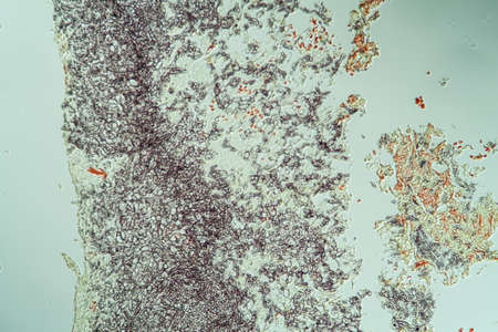
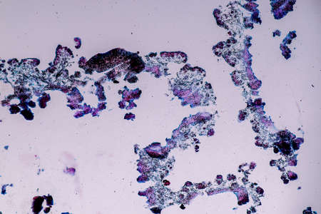
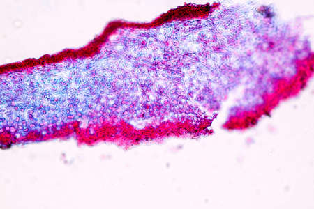
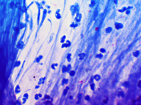
diseased ear tissue infected with Aspergillus 200x
Коллекция по умолчанию
Коллекция по умолчанию
Создать новую
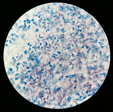
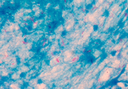
Mycobacterium tuberculosis positive (small red rod) in sputum smear, acid-fast stain, analyze by microscope 1000x
Коллекция по умолчанию
Коллекция по умолчанию
Создать новую
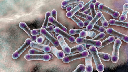
Scientific image of bacteria Corynebacterium showing their characteristic morphology, rod-shaped bacteria with wider ends and club shape appearance, 3D illustration. C. diphtheriae and other
Коллекция по умолчанию
Коллекция по умолчанию
Создать новую
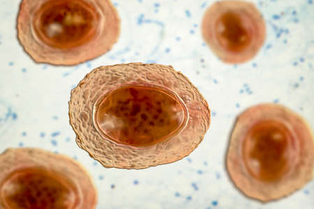
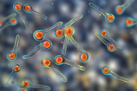
Ascaris lumbricoides, a large roundworm, unfertilized egg, 3D illustration
Коллекция по умолчанию
Коллекция по умолчанию
Создать новую

Mycobacterium tuberculosis undermicroscope 100x /AFB POSITIVE 3+
Коллекция по умолчанию
Коллекция по умолчанию
Создать новую
Methanobrevibacter smithii microorganisms, 3D illustration, the predominant archaeon in the microbiota of the human intestine, a methanogen, plays a role in the digestion of polysaccharides
Коллекция по умолчанию
Коллекция по умолчанию
Создать новую

Texture of nature - gem agate close - up
Коллекция по умолчанию
Коллекция по умолчанию
Создать новую
Mycobacterium leprae bacteria, the causative agent of leprosy, 3D illustration
Коллекция по умолчанию
Коллекция по умолчанию
Создать новую
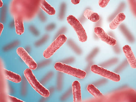
Dense Clumps of Rod-Shaped Bacteria (Bacilli) Under a Microscope
Коллекция по умолчанию
Коллекция по умолчанию
Создать новую
Bone marrow biopsy from myelodysplastic condition under the microscope view. Histology tissue of bone marrow biopsy.
Коллекция по умолчанию
Коллекция по умолчанию
Создать новую
Clostridium tetani bacteria, the causative agent of tetanus, 3D illustration
Коллекция по умолчанию
Коллекция по умолчанию
Создать новую
Probiotics Bacteria Vector illustration. Biology, Science background. Microscopic bacteria closeup.
Коллекция по умолчанию
Коллекция по умолчанию
Создать новую
Smear of Positive Acid Fast Bacilli AFB stained for MTB, under 100X light microscope.
Коллекция по умолчанию
Коллекция по умолчанию
Создать новую
Abstract watercolor vector background blue with yellow spots, backdrop, template, vertical. Vector illustration
Коллекция по умолчанию
Коллекция по умолчанию
Создать новую
Characteristics of Lichen, hyphae and Symbiotic algae under the microscope for education.
Коллекция по умолчанию
Коллекция по умолчанию
Создать новую
Niallia tiangongensis bacteria, 3D illustration of spaceborne bacterium found aboard China's Tiangong station, forming spores and biofilms in microgravity.
Коллекция по умолчанию
Коллекция по умолчанию
Создать новую

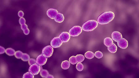
Leuconostoc bacteria, 3D illustration. Coccoid lactic acid bacteria, found on plants, used for production of fermented milk, can cause meningitis, bacteremia, urinary tract and pulmonary infections
Коллекция по умолчанию
Коллекция по умолчанию
Создать новую
Leuconostoc bacteria, 3D illustration. Coccoid lactic acid bacteria, found on plants, used for production of fermented milk, can cause meningitis, bacteremia, urinary tract and pulmonary infections
Коллекция по умолчанию
Коллекция по умолчанию
Создать новую
Bacillus gram positive stain under the microscope view. Bacillus is rod-shaped bacteria.
Коллекция по умолчанию
Коллекция по умолчанию
Создать новую
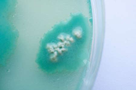
Yeast in petri dish, Microbiology for education in laboratories.
Коллекция по умолчанию
Коллекция по умолчанию
Создать новую
Bacterial culture growth on MacConkey agar (Gram Negative Bacilli)contains small light grains. Focus on all agar surface.
Коллекция по умолчанию
Коллекция по умолчанию
Создать новую

Mycobacterium leprae bacteria, the causative agent of leprosy, 3D illustration
Коллекция по умолчанию
Коллекция по умолчанию
Создать новую
Moderate bacteria cells with Gram stain method fide with microscope.
Коллекция по умолчанию
Коллекция по умолчанию
Создать новую
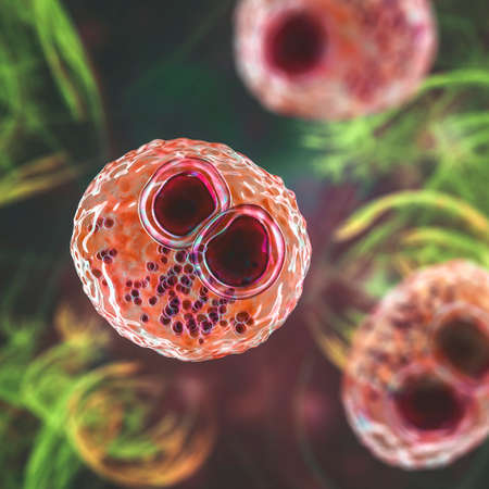
stool specimen shows Cryptosporidium eggs in red. They can cause diarrhea and more serious problems in children and adults who immune systems are suppressed.
Коллекция по умолчанию
Коллекция по умолчанию
Создать новую
Bifidobacterium bacteria, 3D illustration. Gram-positive anaerobic bacteria important for gut health, commonly used as probiotics and studied for their role in preventing gastrointestinal disorders.
Коллекция по умолчанию
Коллекция по умолчанию
Создать новую
Staphylococcus aureus: Gram-positive, to Gram-variable, nonmotile, Coccus,beta haemolysis, saprotrophic bacterium that belongs to the family Staphylococcus growth on blood agar.
Коллекция по умолчанию
Коллекция по умолчанию
Создать новую
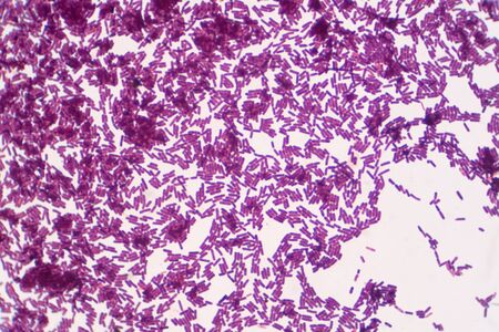
Microscopic Field of Dense, Clumped Bacilli (Rod-Shaped Bacteria) After Gram Staining
Коллекция по умолчанию
Коллекция по умолчанию
Создать новую
Erysipelothrix bacteria, 3D illustration. A species of pleomorphic rod-shaped bacteria causing the skin disease erysipeloid, particularly in individuals working with fish and animal products
Коллекция по умолчанию
Коллекция по умолчанию
Создать новую

Mycobacterium tuberculosis undermicroscope
Коллекция по умолчанию
Коллекция по умолчанию
Создать новую
Pathogenic yeast fungus Cryptococcus neoformans that causes cryptococcal meningoencephalitis and lung disease in immunocompromised patients with AIDS
Коллекция по умолчанию
Коллекция по умолчанию
Создать новую
Microscope of black fungus spore strain with Lactophenol cotton blue, molds or yeasts with macro 40x lens, contamination in air room, pollution aerosol environmental. Microbiology laboratory concepts.
Коллекция по умолчанию
Коллекция по умолчанию
Создать новую

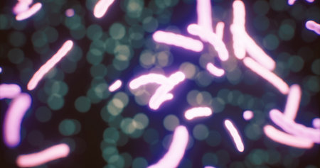
Abstract macro image of particles looking like bacteria, macro shot, microbiology theme
Коллекция по умолчанию
Коллекция по умолчанию
Создать новую
Vibrant shapes swirl and dance through a dark backdrop, representing the unseen world of microorganisms. Their movements create a mesmerizing visual experience full of life and energy.
Коллекция по умолчанию
Коллекция по умолчанию
Создать новую
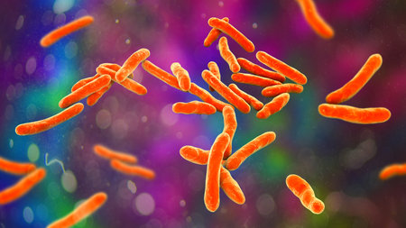
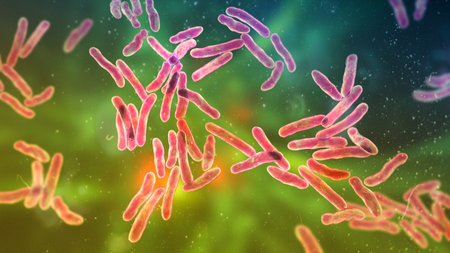
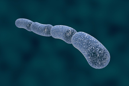
Bacteria which cause tuberculosis Mycobacterium tuberculosis, 3D illustration
Коллекция по умолчанию
Коллекция по умолчанию
Создать новую
Characteristics of Lichen, hyphae and Symbiotic algae under the microscope for education.
Коллекция по умолчанию
Коллекция по умолчанию
Создать новую
abstract background
Коллекция по умолчанию
Коллекция по умолчанию
Создать новую

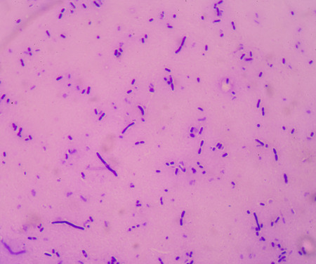
Bacteria gram staining
Коллекция по умолчанию
Коллекция по умолчанию
Создать новую
A very sharp and detailed cardboard texture
Коллекция по умолчанию
Коллекция по умолчанию
Создать новую
Sepsis or septicaemia is a life-threatening illness.
Коллекция по умолчанию
Коллекция по умолчанию
Создать новую
Nocardia bacteria, 3D illustration. Nocardia are rod-shaped Gram-positive bacteria that cause pulmonary infection nocardiosis, tropical infection of skin and bones mycetoma, nocardiosis in animals
Коллекция по умолчанию
Коллекция по умолчанию
Создать новую

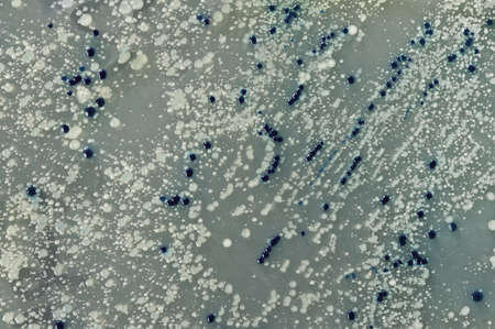
Black bacterial colonies of Salmonella species on Salmonella Shigella agar (SS agar, selective and differential medium) plate on white, medical background.
Коллекция по умолчанию
Коллекция по умолчанию
Создать новую
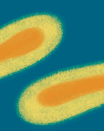
Bacteria Mycobacterium tuberculosis, the causative agent of tuberculosis, 3D illustration, can be used for M. leprae, M. avium complex and other mycobacteria
Коллекция по умолчанию
Коллекция по умолчанию
Создать новую
Bacteria Mycobacterium tuberculosis, the causative agent of tuberculosis, 3D illustration, can be used for M. leprae, M. avium complex and other mycobacteria
Коллекция по умолчанию
Коллекция по умолчанию
Создать новую

Mixed of bacteria colonies in Petri dish
Коллекция по умолчанию
Коллекция по умолчанию
Создать новую
Chromosomes Human under the microscope for education.
Коллекция по умолчанию
Коллекция по умолчанию
Создать новую

Blue spots from the dye in the white tub dissolves in water
Коллекция по умолчанию
Коллекция по умолчанию
Создать новую

Scientific image of bacteria Corynebacterium showing their characteristic morphology, rod-shaped bacteria with wider ends and club shape appearance, 3D illustration. C. diphtheriae and other
Коллекция по умолчанию
Коллекция по умолчанию
Создать новую
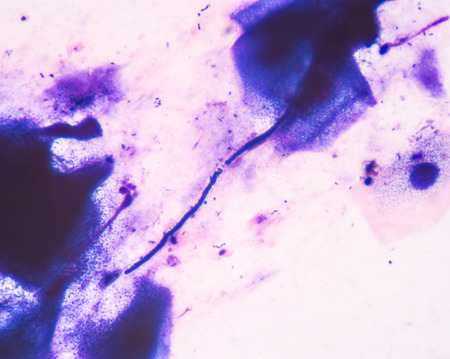
Human cervical squamous epithelial cells, pap smear, NILM (class II non specific chronic inflammation) under a microscope view for education histology. Human tissue.
Коллекция по умолчанию
Коллекция по умолчанию
Создать новую
Probiotic bacteria Bacillus clausii, 3D illustration. B. clausii is a rod-shaped Gram-positive aerobic bacterium used to restore microflora of intestine
Коллекция по умолчанию
Коллекция по умолчанию
Создать новую
blood smear is often used as a follow-up test to abnormal results on a complete blood count (CBC) to evaluate the different types of blood cells.
Коллекция по умолчанию
Коллекция по умолчанию
Создать новую
Hand in glove holding Petri dish with bacteria growing in it
Коллекция по умолчанию
Коллекция по умолчанию
Создать новую
Micro bacterium tuberculosis 3+ moderate red cells on blue background .Medical science background concept.
Коллекция по умолчанию
Коллекция по умолчанию
Создать новую
Niallia tiangongensis bacteria, 3D illustration of spaceborne bacterium found aboard China's Tiangong station, forming spores and biofilms in microgravity.
Коллекция по умолчанию
Коллекция по умолчанию
Создать новую
Cytomegalovirus CMV in a human cell, owl's eye inclusion in nucleus, multinucleated cell, 3D illustration. It is herpes virus, causes diseases in fetus, organ transplant patients, HIV infected people
Коллекция по умолчанию
Коллекция по умолчанию
Создать новую

Smear of Acid-Fast bacilli AFB stained from sputum specimen, under 100X light microscope.
Коллекция по умолчанию
Коллекция по умолчанию
Создать новую

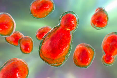
Budding probiotic yeasts
Коллекция по умолчанию
Коллекция по умолчанию
Создать новую

Bacteria Mycobacterium tuberculosis, the causative agent of tuberculosis, 3D illustration, can be used for M. leprae, M. avium complex and other mycobacteria
Коллекция по умолчанию
Коллекция по умолчанию
Создать новую
Bacterial colonies on agar surface, white ones carry a plasmid
Коллекция по умолчанию
Коллекция по умолчанию
Создать новую

Bacteria cells background. 3d render
Коллекция по умолчанию
Коллекция по умолчанию
Создать новую

Hands holding agar plate with bacterial colonies for plasmid vector cloning
Коллекция по умолчанию
Коллекция по умолчанию
Создать новую

Bacillus bacteria close up mycobacterium 3d illustration
Коллекция по умолчанию
Коллекция по умолчанию
Создать новую

Agar surface with bacterial colonies for plasmid
Коллекция по умолчанию
Коллекция по умолчанию
Создать новую
Yeast cells with epithelial tissue in Gram stain method.
Коллекция по умолчанию
Коллекция по умолчанию
Создать новую
Bacteria on blue background. 3d illustration. Escherichia coli rods.
Коллекция по умолчанию
Коллекция по умолчанию
Создать новую
Bacteria Corynebacterium diphtheriae
Коллекция по умолчанию
Коллекция по умолчанию
Создать новую
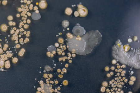
Backgrounds of Characteristics and Different shaped Colony of Bacteria and Mold growing on agar plates from Soil samples for education in Microbiology laboratory.
Коллекция по умолчанию
Коллекция по умолчанию
Создать новую
Two Petri dishes with bacteria growing in them
Коллекция по умолчанию
Коллекция по умолчанию
Создать новую

Staphylococcus speciesm( Staphylococcus aureus) to colonies of white bacteria culture on blood agar in microbiology department hospital.
Коллекция по умолчанию
Коллекция по умолчанию
Создать новую
Backgrounds of Characteristics and Different shaped Colony of Bacteria and Mold growing on agar plates from Soil samples for education in Microbiology laboratory.
Коллекция по умолчанию
Коллекция по умолчанию
Создать новую

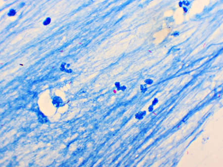
Slide sputum AFB.
Коллекция по умолчанию
Коллекция по умолчанию
Создать новую
Microscopic Section of Lymphoid Tissue Showing Contrasting Follicle Zones
Коллекция по умолчанию
Коллекция по умолчанию
Создать новую
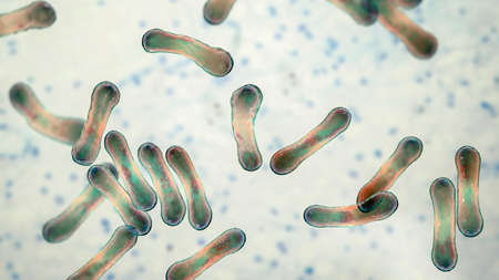
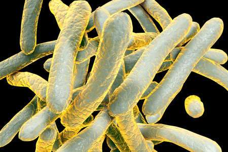
Bacillus subtilis, gram-positive bacteria, non-pathogenic for humans, used as fungicides on plants and in biotechnology for antibiotic production. 3D illustration
Коллекция по умолчанию
Коллекция по умолчанию
Создать новую
stool specimen shows Cryptosporidium eggs in red. They can cause diarrhea and more serious problems in children and adults who immune systems are suppressed.
Коллекция по умолчанию
Коллекция по умолчанию
Создать новую
Optochin subsensitivity test on blood agar plate contains small light grains for Streptococcus pneumoniae; Focus on all agar surface.
Коллекция по умолчанию
Коллекция по умолчанию
Создать новую

Backgrounds of Characteristics and Different shaped Colony of Bacteria and Mold growing on agar plates from Soil samples for education in Microbiology laboratory.
Коллекция по умолчанию
Коллекция по умолчанию
Создать новую

Klebsiella pneumoniae or Klebsiella spp.; bacterial culture growth on MacConkey agar contains small light grains. Focus on all agar surface.
Коллекция по умолчанию
Коллекция по умолчанию
Создать новую
Listeria, bacteria in a petri dish, closeup
Коллекция по умолчанию
Коллекция по умолчанию
Создать новую
Bacteria Coxiella burnetii, 3D illustration. Gram-negative bacteria that cause Q fever, are transmitted to humans from sheep, goats and cattle
Коллекция по умолчанию
Коллекция по умолчанию
Создать новую
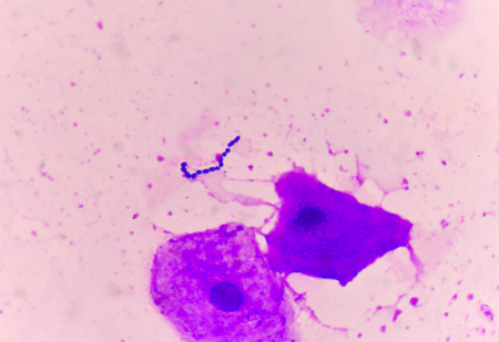
Mycobacterium tuberculosis positive (small red rod) in sputum smear, acid-fast stain, analyze by microscope 1000x
Коллекция по умолчанию
Коллекция по умолчанию
Создать новую
These Gram-negative rod-shaped bacteria have a single polar flagellum.They are the cause of cholera, an infection of the small intestine that is transmitted to humans via contaminated food or water
Коллекция по умолчанию
Коллекция по умолчанию
Создать новую

Ice texture background, ink in water pattern frost. Crystal winter design
Коллекция по умолчанию
Коллекция по умолчанию
Создать новую
360 degree panorama view of Porphyromonas gingivalis bacteria, 3D illustration. Bacteria that cause periodontal disease, bacterial vaginosis
Коллекция по умолчанию
Коллекция по умолчанию
Создать новую
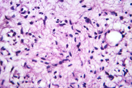
Photomicrograph of a neurofibroma tissue sample in neurofibromatosis genetic disease under a microscope, revealing spindle-shaped cells within a myxoid stroma and wavy nuclei.
Коллекция по умолчанию
Коллекция по умолчанию
Создать новую
medical treatment cell bacteria background
Коллекция по умолчанию
Коллекция по умолчанию
Создать новую
Microscopic Field of Purple Rod-Shaped Bacteria (Bacilli) in Scattered and Dense Colonies
Коллекция по умолчанию
Коллекция по умолчанию
Создать новую
Mycelium of mushrooms on agar in a petri dish Mushroom cultivation. Fungi culture on petri dish plate Macro. top view.
Коллекция по умолчанию
Коллекция по умолчанию
Создать новую
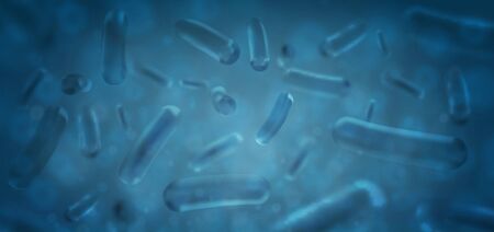
Three-dimensional drawing of rod-shaped bacteria in blue color, streptobacilli, Bacillus anthracis, antrax, model of bacteriarealistic illustration of microbes, microorganisms, bacteria
Коллекция по умолчанию
Коллекция по умолчанию
Создать новую
Haemophilus ducreyi bacteria, 3D illustration of gram-negative coccobacilli. Causes chancroid, a sexually transmitted disease with painful genital ulcers and lymphadenopathy.
Коллекция по умолчанию
Коллекция по умолчанию
Создать новую
Bacteria Sphingomonas, scientific 3D illustration. Gram-negative rod-shaped bacterium widely distributed in nature, and also was isolated in patients with peritonitis, septicemia, meningitis and other
Коллекция по умолчанию
Коллекция по умолчанию
Создать новую
Candida spp. - unicellular fungi. Top view
Коллекция по умолчанию
Коллекция по умолчанию
Создать новую
Microscopic Field of Cocci Bacteria Clustered in Tetrads and Small Groups
Коллекция по умолчанию
Коллекция по умолчанию
Создать новую
Flu of the lungs Diseased tissue 200x
Коллекция по умолчанию
Коллекция по умолчанию
Создать новую
Microscopic fungi Trichosporon, 3D illustration shows septate hyphae, pseudohyphae, blastoconidia singly or in short chains, arthroconidia. Cause white piedra, superficial and invasive infections
Коллекция по умолчанию
Коллекция по умолчанию
Создать новую
Scientific image of bacteria Bacteroides fragilis and other Bacteroides, Gram-negative anaerobic bacterium, one of the major components of normal microbiome of human intestine, 3D illustration
Коллекция по умолчанию
Коллекция по умолчанию
Создать новую
Petri dish close up. Bacteria culture.
Коллекция по умолчанию
Коллекция по умолчанию
Создать новую

Microscopic fungi Malassezia furfur, 3D illustration. They are naturally found on the skin surfaces and are also associated with dandruff, seborrhoeic dermatitis and tinea versicolor
Коллекция по умолчанию
Коллекция по умолчанию
Создать новую

red Mycobacterium tuberculosis on blue background.
Коллекция по умолчанию
Коллекция по умолчанию
Создать новую
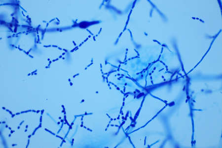
Budding yeast cells with pseudohyphae from sputum gram stain test, in laboratory,fine with microscope.
Коллекция по умолчанию
Коллекция по умолчанию
Создать новую
Metal sheet with spots of rust, painted green. Abstract texture. Close up.
Коллекция по умолчанию
Коллекция по умолчанию
Создать новую

3d rendering, infectious virus with surface details on blue background. Computer digital image.
Коллекция по умолчанию
Коллекция по умолчанию
Создать новую
Legion-Media
Создайте свои проекты на основе качественных стоковых фотографий и видео.


Copyright © Legion-Media.