
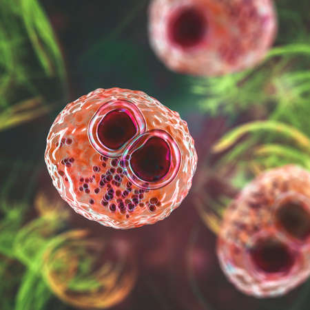
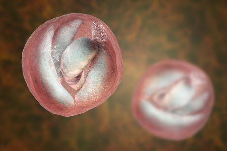
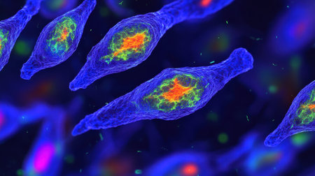
Cytomegalovirus CMV in a human cell, owl's eye inclusion in nucleus, multinucleated cell, 3D illustration. It is herpes virus, causes diseases in fetus, organ transplant patients, HIV infected people
Коллекция по умолчанию
Коллекция по умолчанию
Создать новую
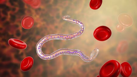
Ascaris lumbricoides, a large roundworm, unfertilized egg, 3D illustration
Коллекция по умолчанию
Коллекция по умолчанию
Создать новую

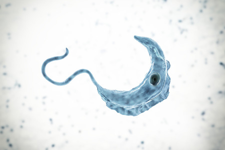
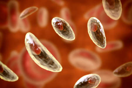
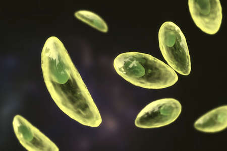
Trypanosoma brucei parasites, 3D illustration. A protozoan that is transmitted by tse-tse fly and causes African sleeping sickness
Коллекция по умолчанию
Коллекция по умолчанию
Создать новую
Trypanosoma brucei parasite, 3D illustration. A protozoan that is transmitted by tse-tse fly and causes African sleeping sickness
Коллекция по умолчанию
Коллекция по умолчанию
Создать новую
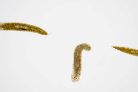
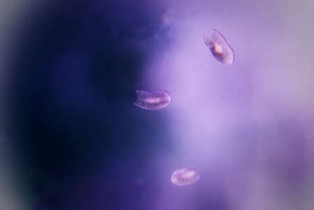
Planarian parasite (flatworm) under microscope view.
Коллекция по умолчанию
Коллекция по умолчанию
Создать новую
plasmodium falciparum ring form state , multiple in fection of red blood cells
Коллекция по умолчанию
Коллекция по умолчанию
Создать новую
Soothing Pink Abstract background with little impurities
Коллекция по умолчанию
Коллекция по умолчанию
Создать новую

Cytomegalovirus CMV in a human cell, owl's eye inclusion in nucleus, multinucleated cell, 3D illustration. It is herpes virus, causes diseases in fetus, organ transplant patients, HIV infected people
Коллекция по умолчанию
Коллекция по умолчанию
Создать новую
Fasciola hepatica, or liver fluke, 3D illustration. A parasitic trematode worm that causes fasciolosis, an infection of liver
Коллекция по умолчанию
Коллекция по умолчанию
Создать новую

Cancer Cell in human showing abnormal cells.Medical science background concept.
Коллекция по умолчанию
Коллекция по умолчанию
Создать новую

Activated and non-activated platelets, thrombocytes, 3D illustration. Activated thrombocytes have cell membrane projections on the surface, unactivated platelets are biconvex discoid, or lens-shaped
Коллекция по умолчанию
Коллекция по умолчанию
Создать новую
Eggs of parasitic roundworm Trichuris trichiura, or whipworm, the causative agent of trichuriasis, disease of a human large intestine, 3D illustration
Коллекция по умолчанию
Коллекция по умолчанию
Создать новую
Cryptosporidium parvum oocyst, 3D illustration. Cryptosporidium is a protozoan, microscopic parasite, the causative agent of the diarrheal disease cryptosporidiosis
Коллекция по умолчанию
Коллекция по умолчанию
Создать новую
bacteria under the microscope background, background bacteria
Коллекция по умолчанию
Коллекция по умолчанию
Создать новую
3D illustration of Phagocytosis. Neutrophe that uses its plasma membrane to engulf bacteria. From endocytosis to exocytosis. Digestion process in phagocytes. immune system, 3d render
Коллекция по умолчанию
Коллекция по умолчанию
Создать новую
Cell division process, micro
Коллекция по умолчанию
Коллекция по умолчанию
Создать новую

Fusobacterium, 3D illustration. An oral bacterium, causes periodontal diseases, periodontal plague formation, sore throat, Lemmieres syndrome. It is also associated with preterm birth and colon cancer
Коллекция по умолчанию
Коллекция по умолчанию
Создать новую
Ice texture background, ink in water pattern frost. Crystal winter design
Коллекция по умолчанию
Коллекция по умолчанию
Создать новую
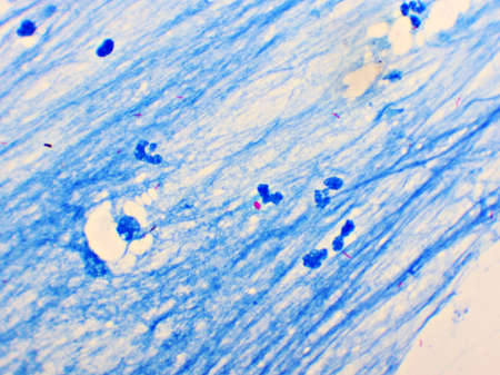
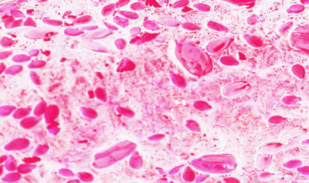
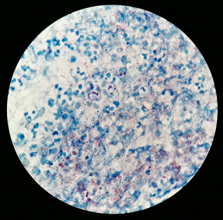
Mycobacterium tuberculosis positive (small red rod) in sputum smear, acid-fast stain, analyze by microscope 1000x
Коллекция по умолчанию
Коллекция по умолчанию
Создать новую
Signet ring cell carcinoma of the stomach, light micrograph, photo under microscope
Коллекция по умолчанию
Коллекция по умолчанию
Создать новую

Fusobacterium, 3D illustration. An oral bacterium, causes periodontal diseases, periodontal plague formation, sore throat, Lemmieres syndrome. It is also associated with preterm birth and colon cancer
Коллекция по умолчанию
Коллекция по умолчанию
Создать новую
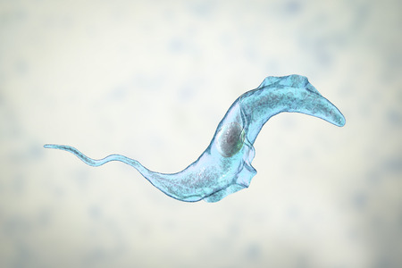
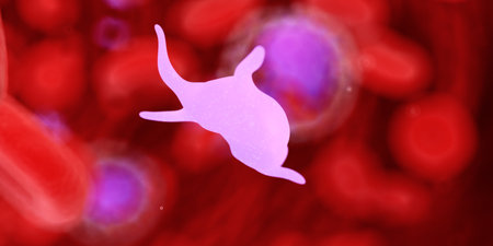
Trypanosoma cruzi parasite, 3D illustration. A protozoan that causes Chagas disease transmitted to humans by the bite of triatomine bug
Коллекция по умолчанию
Коллекция по умолчанию
Создать новую

Fusobacterium, 3D illustration. An oral bacterium, causes periodontal diseases, periodontal plague formation, sore throat, Lemmieres syndrome. It is also associated with preterm birth and colon cancer
Коллекция по умолчанию
Коллекция по умолчанию
Создать новую

Abstract macro image of particles looking like bacteria, macro shot, microbiology theme
Коллекция по умолчанию
Коллекция по умолчанию
Создать новую
Chromosomes Human under the microscope for education.
Коллекция по умолчанию
Коллекция по умолчанию
Создать новую
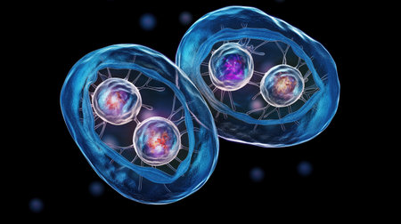
Babesia parasites inside red blood cell, the causative agent of babesiosis. 3D illustration showing classic tetrad-forms of Babesia merozoites so-called Maltese cross formation
Коллекция по умолчанию
Коллекция по умолчанию
Создать новую
Immature white blood cells in leukemia.Science concept.
Коллекция по умолчанию
Коллекция по умолчанию
Создать новую
Clostridium tetani bacteria, the causative agent of tetanus, 3D illustration
Коллекция по умолчанию
Коллекция по умолчанию
Создать новую

Test and sample virus crown covid virus 19
Коллекция по умолчанию
Коллекция по умолчанию
Создать новую

Blood vessel with flowing blood cells, 3D illustration. Small blood vessels, capillaries
Коллекция по умолчанию
Коллекция по умолчанию
Создать новую
Leech on the glass. Bloodsucking animal. subclass of ringworms from the belt-type class. Hirudotherapy.
Коллекция по умолчанию
Коллекция по умолчанию
Создать новую

Rows of microscope glass slide in the cells
Коллекция по умолчанию
Коллекция по умолчанию
Создать новую
Human egg cell, 3D illustration closeup
Коллекция по умолчанию
Коллекция по умолчанию
Создать новую

Realistic molecule bacterium illustration graphics 3d microbe
Коллекция по умолчанию
Коллекция по умолчанию
Создать новую

Blue spots from the dye in the white tub dissolves in water
Коллекция по умолчанию
Коллекция по умолчанию
Создать новую

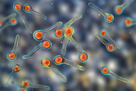
Promastigotes of Leishmania parasite which cause leishmaniasis, 3D illustration
Коллекция по умолчанию
Коллекция по умолчанию
Создать новую
3d rendered medically accurate illustration of cells
Коллекция по умолчанию
Коллекция по умолчанию
Создать новую
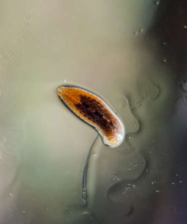
Rare image of Ghost flatworm - Maricola (Planarian) triclad flatworms in reef aquarium glass
Коллекция по умолчанию
Коллекция по умолчанию
Создать новую
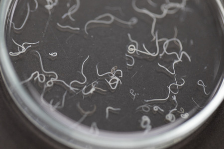
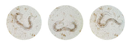
Ascariasis is a disease caused by the parasitic roundworm Ascaris lumbricoides for education in laboratories.
Коллекция по умолчанию
Коллекция по умолчанию
Создать новую
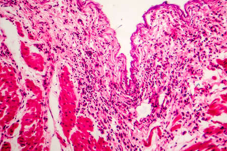
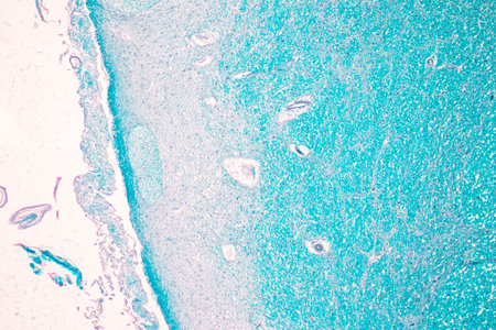
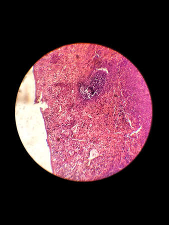
Histopathology of human liver under microscope view for medical education.
Коллекция по умолчанию
Коллекция по умолчанию
Создать новую
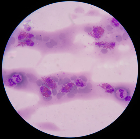
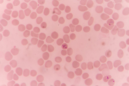
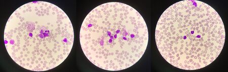
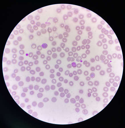
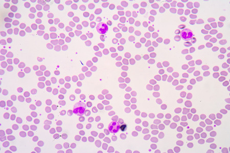
blood smear is often used as a follow-up test to abnormal results on a complete blood count (CBC) to evaluate the different types of blood cells.
Коллекция по умолчанию
Коллекция по умолчанию
Создать новую

Fusobacterium, 3D illustration. An oral bacterium, causes periodontal diseases, periodontal plague formation, sore throat, Lemmieres syndrome. It is also associated with preterm birth and colon cancer
Коллекция по умолчанию
Коллекция по умолчанию
Создать новую

Ovarian cancer, light micrograph, photo under microscope. Photograph shows a fragment of a cancerous tumor in the female ovary. Selective focus
Коллекция по умолчанию
Коллекция по умолчанию
Создать новую
Pancreas cancer cells, light micrograph for medical education.
Коллекция по умолчанию
Коллекция по умолчанию
Создать новую
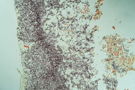
Characteristics of Lichen, hyphae and Symbiotic algae under the microscope for education.
Коллекция по умолчанию
Коллекция по умолчанию
Создать новую

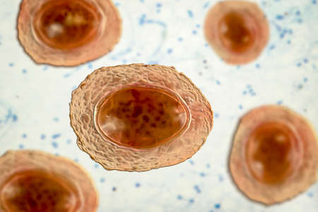
Parasitic protozoans Toxoplasma gondii, the causative agent of toxoplasmosis in tachyzoite stage, 3D illustration
Коллекция по умолчанию
Коллекция по умолчанию
Создать новую
Thyroid follicular carcinoma, light micrograph, photo under microscope
Коллекция по умолчанию
Коллекция по умолчанию
Создать новую
Dive into a stunning microscopic view showcasing vibrant microorganisms in an aquatic setting. This image reveals the intricacies of diverse life forms through scientific observation.
Коллекция по умолчанию
Коллекция по умолчанию
Создать новую

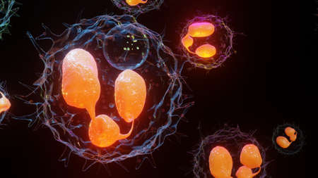
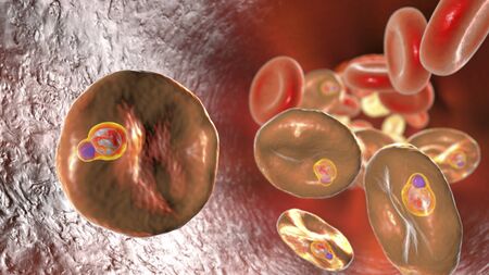
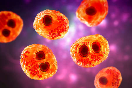
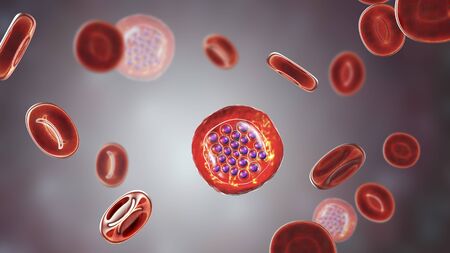
The malaria-infected red blood cells. 3D illustration showing malaria parasite Plasmodium falciparum in schizont stage inside red blood cells, the causative agent of tropical malaria
Коллекция по умолчанию
Коллекция по умолчанию
Создать новую

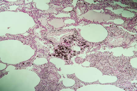
Histopathology of lung emphysema, light micrograph, photo under microscope showing enlargement of air spaces in lung tissue and destruction of alveolar septa
Коллекция по умолчанию
Коллекция по умолчанию
Создать новую
Tongue Tissue with taste buds across 200x
Коллекция по умолчанию
Коллекция по умолчанию
Создать новую
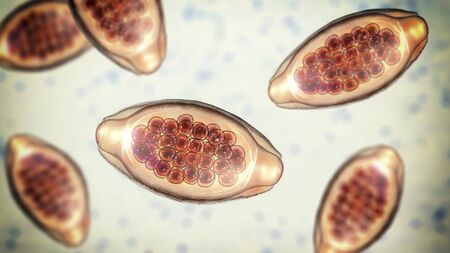
Wuchereria bancrofti, a roundworm nematode, one of the causative agents of lymphatic filariasis, 3D illustration showing presence of sheath around the worm and tail nuclei non-extending to tip
Коллекция по умолчанию
Коллекция по умолчанию
Создать новую

Meningococcal meningitis, cerebrospinal fluid smear containing neutrophils with and without bacteria Neisseria meningitidis, 3D illustration
Коллекция по умолчанию
Коллекция по умолчанию
Создать новую
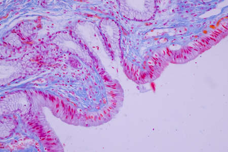
Columnar epithelium of human gall bladder under the microscope in Lab.
Коллекция по умолчанию
Коллекция по умолчанию
Создать новую
Acute pyelonephritis, light micrograph, photo under microscope
Коллекция по умолчанию
Коллекция по умолчанию
Создать новую

Columnar epithelium of human gall bladder under the microscope in Lab.
Коллекция по умолчанию
Коллекция по умолчанию
Создать новую

Slide sputum AFB.
Коллекция по умолчанию
Коллекция по умолчанию
Создать новую

Sediment sulfonamide crystal in urine.Urinalysis concept.
Коллекция по умолчанию
Коллекция по умолчанию
Создать новую
stool specimen shows Cryptosporidium eggs in red. They can cause diarrhea and more serious problems in children and adults who immune systems are suppressed.
Коллекция по умолчанию
Коллекция по умолчанию
Создать новую
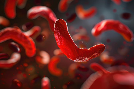
Multiple red blood cells float weightlessly in the air, An image representing the sickle-shaped blood cells found in sickle cell anemia, AI Generated
Коллекция по умолчанию
Коллекция по умолчанию
Создать новую

Bladder cat- cell nature background. Abstract- photo macro sections with high magnification with light microscope
Коллекция по умолчанию
Коллекция по умолчанию
Создать новую
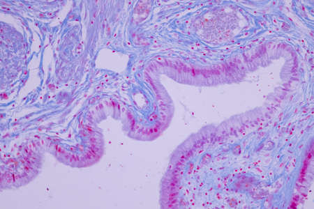
Columnar epithelium of human gall bladder under the microscope in Lab.
Коллекция по умолчанию
Коллекция по умолчанию
Создать новую
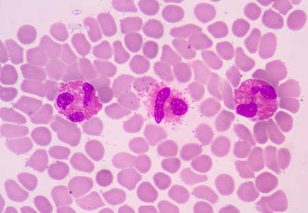
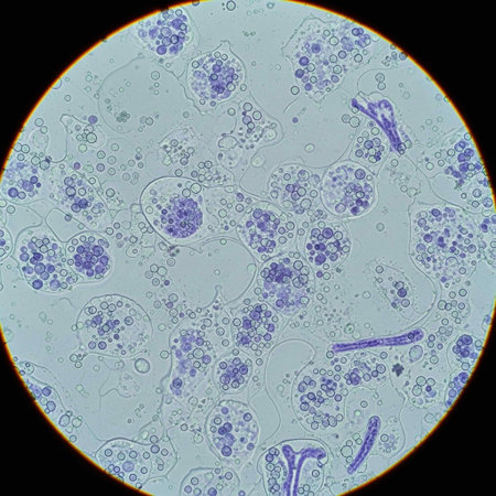
White blood cells of a human, Eosinophil photomicrograph panorama as seen under the microscope
Коллекция по умолчанию
Коллекция по умолчанию
Создать новую

Helminths Toxocara canis dog roundworm, the cause of toxocariasis in man, an infestation transmitted from material contaminated by eggs in dogs feces. 3D illustration of a first larval stage
Коллекция по умолчанию
Коллекция по умолчанию
Создать новую
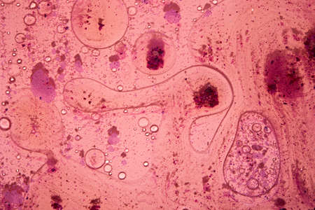
science aquaculture fish parasite hook clip worm micrograph
Коллекция по умолчанию
Коллекция по умолчанию
Создать новую
Parasitic protozoans Toxoplasma gondii, the causative agent of toxoplasmosis in tachyzoite stage, 3D illustration
Коллекция по умолчанию
Коллекция по умолчанию
Создать новую

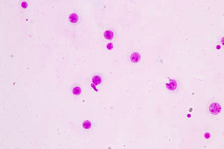
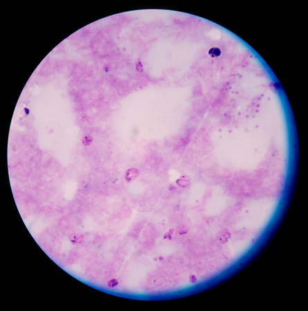
Abnormal cells pink cells with microscope.Medical science background concept.
Коллекция по умолчанию
Коллекция по умолчанию
Создать новую
Cytomegalovirus CMV in a human cell, owl's eye inclusion in nucleus, multinucleated cell, 3D illustration. It is herpes virus, causes diseases in fetus, organ transplant patients, HIV infected people
Коллекция по умолчанию
Коллекция по умолчанию
Создать новую
Test and sample virus crown covid virus 19
Коллекция по умолчанию
Коллекция по умолчанию
Создать новую
Strongyloides stercoralis in stool
Коллекция по умолчанию
Коллекция по умолчанию
Создать новую
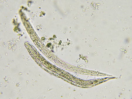
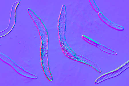
Microscopic view collection of adult free-living female Strongyloides stercoralis, a human pathogenic parasitic roundworm causing the disease strongyloidiasis.
Коллекция по умолчанию
Коллекция по умолчанию
Создать новую
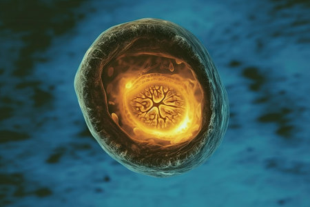
A microscopic image of a developing embryo with vessels sprouting and branching out to nourish and sustain the growing life inside
Коллекция по умолчанию
Коллекция по умолчанию
Создать новую
Lung tissue as dust lung under the microscope 100x
Коллекция по умолчанию
Коллекция по умолчанию
Создать новую
Fusobacterium, 3D illustration. An oral bacterium, causes periodontal diseases, periodontal plague formation, sore throat, Lemmieres syndrome. It is also associated with preterm birth and colon cancer
Коллекция по умолчанию
Коллекция по умолчанию
Создать новую
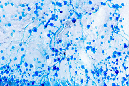
Smear of Positive Acid Fast Bacilli AFB stained for MTB, under 100X light microscope.
Коллекция по умолчанию
Коллекция по умолчанию
Создать новую
Malaria blood parasite infected red blood cells laboratory background.
Коллекция по умолчанию
Коллекция по умолчанию
Создать новую

Histological Uterus human, Uterine tube human, Placenta human and Umbilical cord Human under the microscope for education.
Коллекция по умолчанию
Коллекция по умолчанию
Создать новую
Histopathology of interstitial nephritis, light micrograph, photo under microscope. High magnification
Коллекция по умолчанию
Коллекция по умолчанию
Создать новую
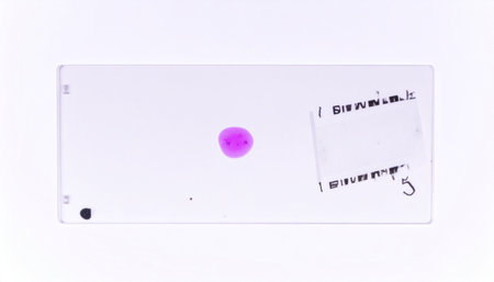
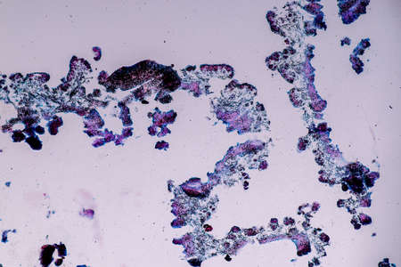
A microscope slide containing a pink stained sample for medical or scientific analysis.
Коллекция по умолчанию
Коллекция по умолчанию
Создать новую
This vibrant image showcases abstract cellular structures captured through microscopy, highlighting intricate details and patterns significant in biological research and education.
Коллекция по умолчанию
Коллекция по умолчанию
Создать новую

Stomach tissue under the microscope 100x
Коллекция по умолчанию
Коллекция по умолчанию
Создать новую
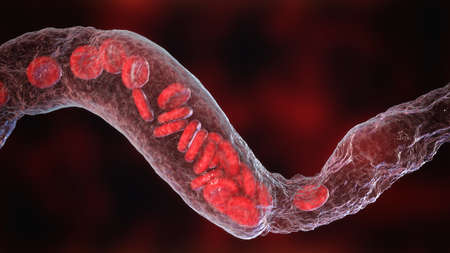
Vasoconstriction, clot formation in blood vessel. Erythrocytes flowing through narrowing blood vessel, 3D illustration
Коллекция по умолчанию
Коллекция по умолчанию
Создать новую

red Mycobacterium tuberculosis on blue background.
Коллекция по умолчанию
Коллекция по умолчанию
Создать новую
diseased ear tissue infected with Aspergillus 200x
Коллекция по умолчанию
Коллекция по умолчанию
Создать новую
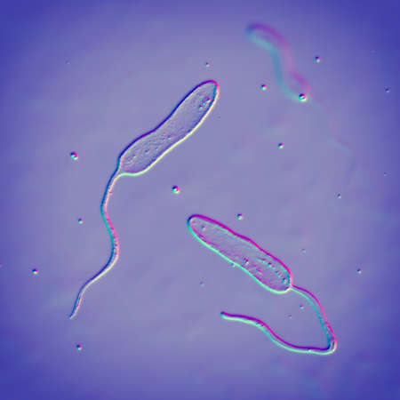
Vibrio cholerae bacteria, 3D illustration. Bacterium which causes cholera disease and is transmitted by contaminated water
Коллекция по умолчанию
Коллекция по умолчанию
Создать новую
blood films for Malaria parasite.
Коллекция по умолчанию
Коллекция по умолчанию
Создать новую
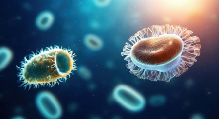
Close-up view of glowing bacteria and viruses with spiky exteriors, floating in a dark blue, luminous, and abstract background.
Коллекция по умолчанию
Коллекция по умолчанию
Создать новую
Cerebellum, Thalamus, Medulla oblongata, Spinal cord and Motor Neuron human under the microscope in Lab.
Коллекция по умолчанию
Коллекция по умолчанию
Создать новую
Characteristics of Lichen, hyphae and Symbiotic algae under the microscope for education.
Коллекция по умолчанию
Коллекция по умолчанию
Создать новую
Parasitic protozoans Toxoplasma gondii, the causative agent of toxoplasmosis in tachyzoite stage, 3D illustration
Коллекция по умолчанию
Коллекция по умолчанию
Создать новую
White blood cells of a human, photomicrograph panorama as seen under the microscope
Коллекция по умолчанию
Коллекция по умолчанию
Создать новую
Biological histological fixed colored preparation of the spleen - a secondary organ of the immune system
Коллекция по умолчанию
Коллекция по умолчанию
Создать новую
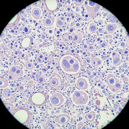
Coccidiosis, coccidia in liver, light micrograph. Micrograph shows bile duct hyperplasia and fibrosis with periductal inflammation, groups of coccidia, large violet cells
Коллекция по умолчанию
Коллекция по умолчанию
Создать новую

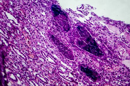
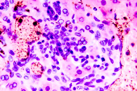
Histopathology of alcoholic hepatitis, light micrograph, photo under microscope. High magnification
Коллекция по умолчанию
Коллекция по умолчанию
Создать новую

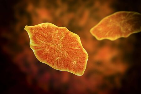
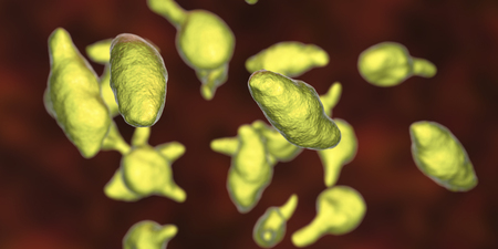
Balantidium coli protozoan, 3D illustration. Ciliated intestinal parasite that causes balantidiasis
Коллекция по умолчанию
Коллекция по умолчанию
Создать новую

brucella bacteria is a pathogen that causes brucellosis zoonosis. 3D rendering
Коллекция по умолчанию
Коллекция по умолчанию
Создать новую
Bacteria Mycoplasma genitalium, 3D illustration. The causative agent of sexually transmitted infections and infertility
Коллекция по умолчанию
Коллекция по умолчанию
Создать новую
The malaria-infected red blood cells. 3D illustration showing malaria parasite Plasmodium falciparum in schizont stage inside red blood cells, the causative agent of tropical malaria
Коллекция по умолчанию
Коллекция по умолчанию
Создать новую
cell find with microscope.
Коллекция по умолчанию
Коллекция по умолчанию
Создать новую

Histological Uterus human, Uterine tube human, Placenta human and Umbilical cord Human under the microscope for education
Коллекция по умолчанию
Коллекция по умолчанию
Создать новую
Legion-Media
Создайте свои проекты на основе качественных стоковых фотографий и видео.


Copyright © Legion-Media.