
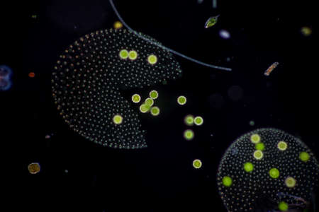
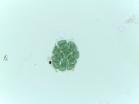
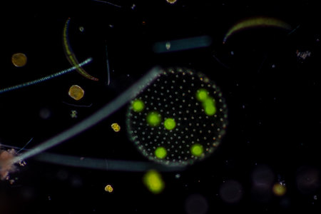
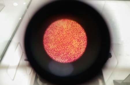
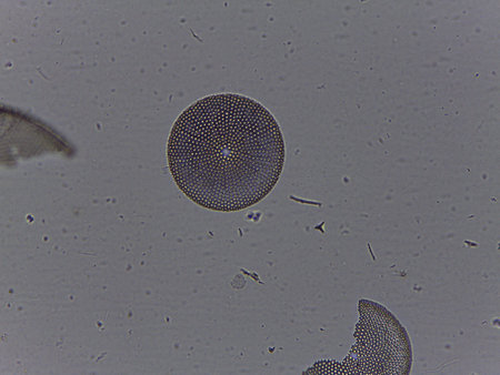
Volvox in drop of water under the microscope for classroom education.
Коллекция по умолчанию
Коллекция по умолчанию
Создать новую
Volvox in drop of water under the microscope for classroom education.
Коллекция по умолчанию
Коллекция по умолчанию
Создать новую
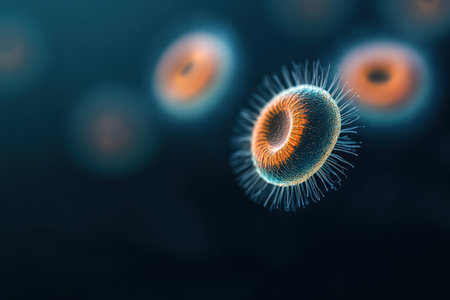
Cytomegalovirus CMV in a human cell, owl's eye inclusion in nucleus, multinucleated cell, 3D illustration. It is herpes virus, causes diseases in fetus, organ transplant patients, HIV infected people
Коллекция по умолчанию
Коллекция по умолчанию
Создать новую
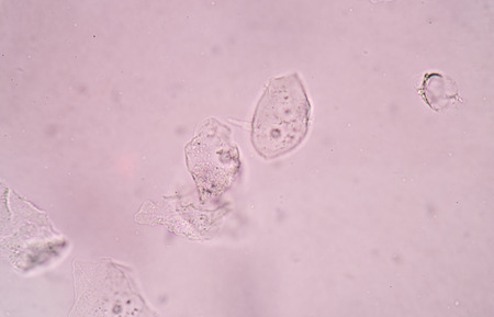
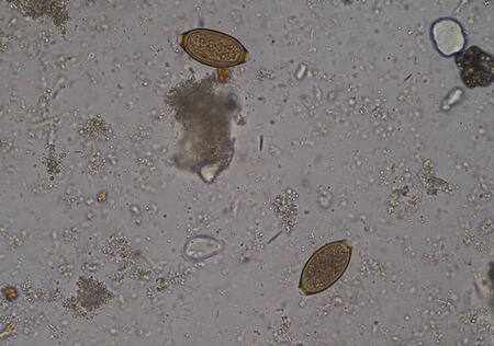
Protozoa and Green Algae in waste water under the microscope.
Коллекция по умолчанию
Коллекция по умолчанию
Создать новую
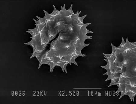
Scanning electron micrograph of one daisy pollen grain. Nottingham, UK
Коллекция по умолчанию
Коллекция по умолчанию
Создать новую

Apple pollen from a blossom in spring under the microscope
Коллекция по умолчанию
Коллекция по умолчанию
Создать новую
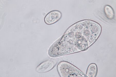
Paramecium caudatum is a genus of unicellular ciliated protozoan and Bacterium under the microscope.
Коллекция по умолчанию
Коллекция по умолчанию
Создать новую
Protozoa and Green Algae in waste water under the microscope.
Коллекция по умолчанию
Коллекция по умолчанию
Создать новую
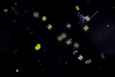
Protozoa and Green Algae in waste water under the microscope.
Коллекция по умолчанию
Коллекция по умолчанию
Создать новую

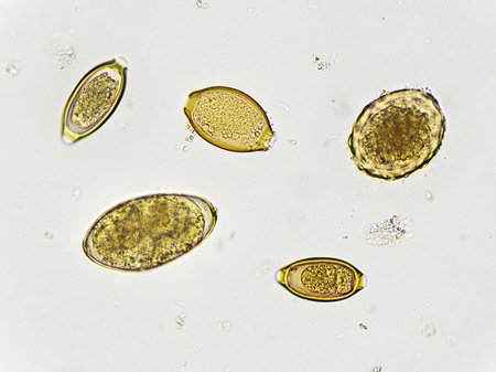
parasite eggs.
Коллекция по умолчанию
Коллекция по умолчанию
Создать новую

Protozoa and Green Algae in waste water under the microscope.
Коллекция по умолчанию
Коллекция по умолчанию
Создать новую

Euglena is a genus of single-celled flagellate Eukaryotes under microscopic view for education.
Коллекция по умолчанию
Коллекция по умолчанию
Создать новую

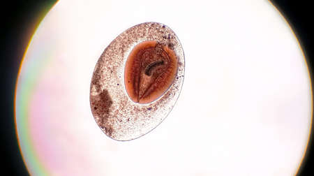
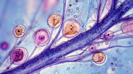
Opisthorchis viverrini, common name Southeast Asian liver fluke, is a trematode parasite.
Коллекция по умолчанию
Коллекция по умолчанию
Создать новую
bladder epithelial cells in urine.
Коллекция по умолчанию
Коллекция по умолчанию
Создать новую

Proglottid (body unit) of tapeworm Taenia saginata, 3D illustration. A flatworm parasitizing animal and human intestine. Proglottid contains uterus with 12-30 primary lateral branches filled with eggs
Коллекция по умолчанию
Коллекция по умолчанию
Создать новую
destructive mushroom in wood fabric 100x
Коллекция по умолчанию
Коллекция по умолчанию
Создать новую
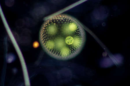
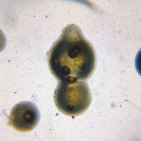
Volvox is a polyphyletic genus of chlorophyte green algae or phytoplankton.They live in a variety of freshwater and marine habitats
Коллекция по умолчанию
Коллекция по умолчанию
Создать новую

Close up Plant epidermis with stomata or Leaf Epidermis (Stomata) under microscope.
Коллекция по умолчанию
Коллекция по умолчанию
Создать новую
squamous cell
Коллекция по умолчанию
Коллекция по умолчанию
Создать новую

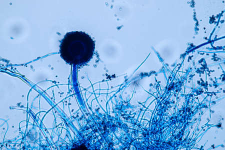
Microscope view of mold sporangia.
Коллекция по умолчанию
Коллекция по умолчанию
Создать новую
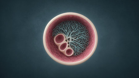
A close up of a pink and blue cell with a tree inside of it. The cell is surrounded by a pink and blue background
Коллекция по умолчанию
Коллекция по умолчанию
Создать новую
Protozoa and Green Algae in waste water under the microscope.
Коллекция по умолчанию
Коллекция по умолчанию
Создать новую
Aspergillus niger and Aspergillus oryzae (mold) under microscope for Microbiology in Lab.
Коллекция по умолчанию
Коллекция по умолчанию
Создать новую
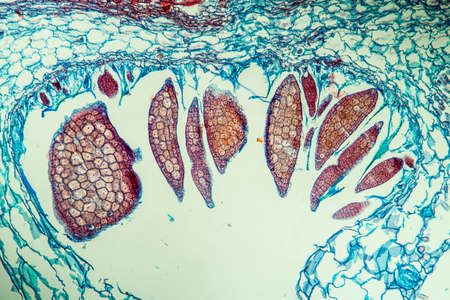
Nestwurz orchid root cross 100x
Коллекция по умолчанию
Коллекция по умолчанию
Создать новую
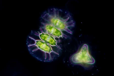
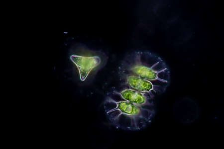
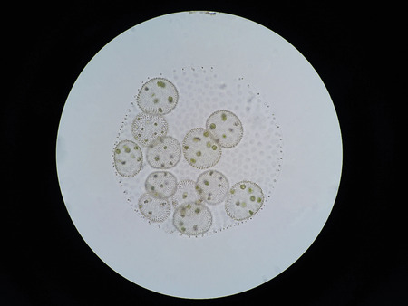
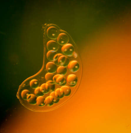
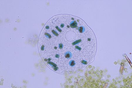
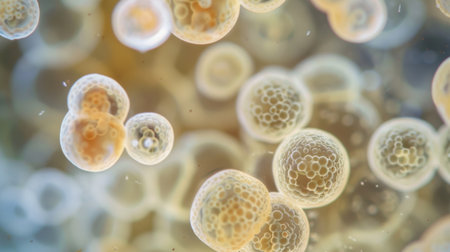
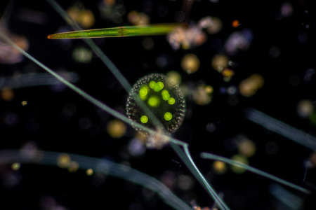
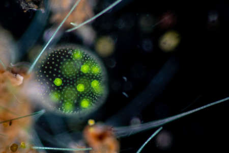
Microscopic View of the Colonial Green Alga, Pandorina
Коллекция по умолчанию
Коллекция по умолчанию
Создать новую
Moniliformis dubius in the Intestine of rat, intermediate host
Коллекция по умолчанию
Коллекция по умолчанию
Создать новую
this is a close up of a bubble snails, eggs of snails
Коллекция по умолчанию
Коллекция по умолчанию
Создать новую
Some pollen of the amaryllis under the microscope.
Коллекция по умолчанию
Коллекция по умолчанию
Создать новую
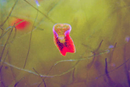
Red planaria flatworms - Convolutriloba retrogemma
Коллекция по умолчанию
Коллекция по умолчанию
Создать новую
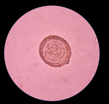
Eggs of helminth in stool
Коллекция по умолчанию
Коллекция по умолчанию
Создать новую

Characteristics of Squamous epithelial cell (Cell structure) of human under microscope view for education in laboratory.
Коллекция по умолчанию
Коллекция по умолчанию
Создать новую
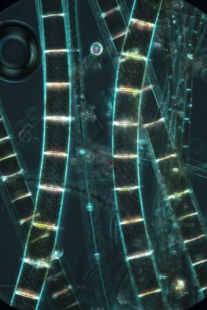
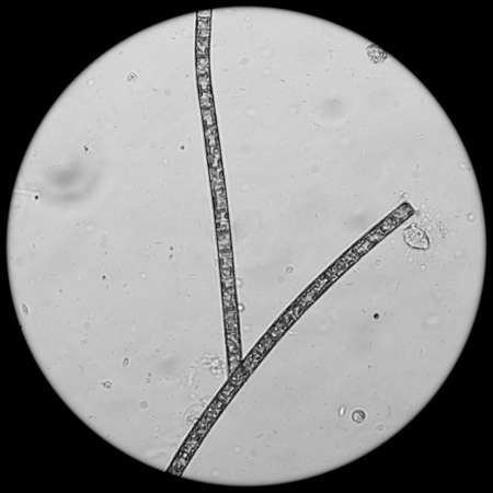
Dark, murky image of filamentous algae glowing like rungs on a ladder, in an abstract micrograph at 100x with polarization.
Коллекция по умолчанию
Коллекция по умолчанию
Создать новую
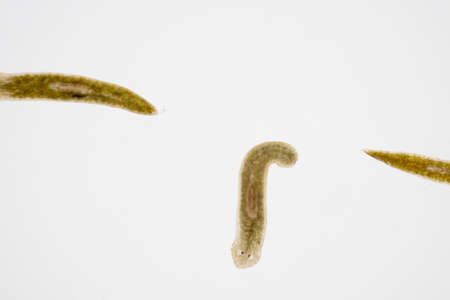
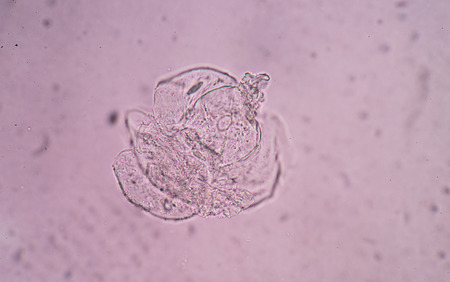
Planarian parasite (flatworm) under microscope view.
Коллекция по умолчанию
Коллекция по умолчанию
Создать новую
Volvox in drop of water under the microscope for classroom education.
Коллекция по умолчанию
Коллекция по умолчанию
Создать новую
Trumpet animal as a microscopic plankton animal in drops of water
Коллекция по умолчанию
Коллекция по умолчанию
Создать новую
cell find with microscope.
Коллекция по умолчанию
Коллекция по умолчанию
Создать новую

Aspergillus (mold) for Microbiology in Lab.
Коллекция по умолчанию
Коллекция по умолчанию
Создать новую
Cell division under microscope view. Microscopic view of cell.
Коллекция по умолчанию
Коллекция по умолчанию
Создать новую

Characteristics of Rhizopus is a genus of common saprophytic fungi on Slide under the microscope for education.
Коллекция по умолчанию
Коллекция по умолчанию
Создать новую
microscope lens, viewing Trypanosoma cruzi parasitic protozoan, causer of Chagas disease
Коллекция по умолчанию
Коллекция по умолчанию
Создать новую

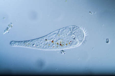
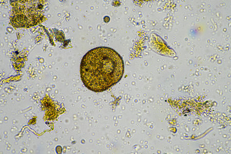
Frontonia sp. is a genus of free-living unicellular ciliate protists under the microscope.
Коллекция по умолчанию
Коллекция по умолчанию
Создать новую
Cell Gene Microscopic Series
Коллекция по умолчанию
Коллекция по умолчанию
Создать новую
Beautiful image in microscope of microorganisms in the lab
Коллекция по умолчанию
Коллекция по умолчанию
Создать новую
microscope slide with detailed view of plant stem, complete with cells and minutiae, created with generative ai
Коллекция по умолчанию
Коллекция по умолчанию
Создать новую
water algae cell macro, micrograph.
Коллекция по умолчанию
Коллекция по умолчанию
Создать новую

Eggs of Trichuris trichiura in stool
Коллекция по умолчанию
Коллекция по умолчанию
Создать новую
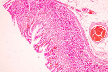
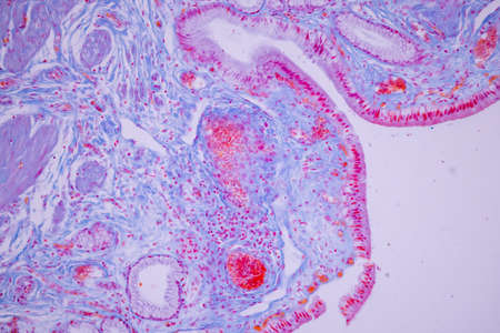
Tissue of Stomach Human under the microscope in Lab.
Коллекция по умолчанию
Коллекция по умолчанию
Создать новую
Frontonia sp. is a genus of free-living unicellular ciliate protists under the microscope.
Коллекция по умолчанию
Коллекция по умолчанию
Создать новую
Sister bacterial colonies
Коллекция по умолчанию
Коллекция по умолчанию
Создать новую
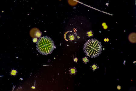
Microscopic Atlas of Diverse Biological Specimens Including Diatoms and Primitive Chordates
Коллекция по умолчанию
Коллекция по умолчанию
Создать новую
Numerous tiny white bubbles float gracefully on the surface of the water. The bubbles appear to be delicate and light, moving with the gentle current in a mesmerizing manner.
Коллекция по умолчанию
Коллекция по умолчанию
Создать новую
soil sample containing soil biology, with bacteria, fungi, amoeba, flagellate, and arcella, on a sustainable agricultural farm in australia
Коллекция по умолчанию
Коллекция по умолчанию
Создать новую
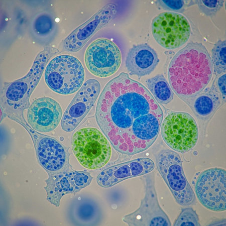
Cellular structures are magnified to reveal their complex shapes and vibrant colors, emphasizing the beauty of biology at a microscopic level.
Коллекция по умолчанию
Коллекция по умолчанию
Создать новую
Egg of parasite in stool examition testing finding with microscope.
Коллекция по умолчанию
Коллекция по умолчанию
Создать новую
Numerous small blue bubbles are seen floating on the surface of the water, creating a mesmerizing pattern in the sunlight. The bubbles gently rise and fall with the movement of the water.
Коллекция по умолчанию
Коллекция по умолчанию
Создать новую
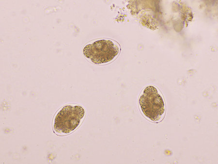
Hymenolepis diminuta parasite egg in stool exam.
Коллекция по умолчанию
Коллекция по умолчанию
Создать новую
Explore a stunning microscopic view featuring colorful cells and thin filaments against a vibrant blue background, showcasing scientific beauty in detail.
Коллекция по умолчанию
Коллекция по умолчанию
Создать новую

Stomach tissue under the microscope 100x
Коллекция по умолчанию
Коллекция по умолчанию
Создать новую
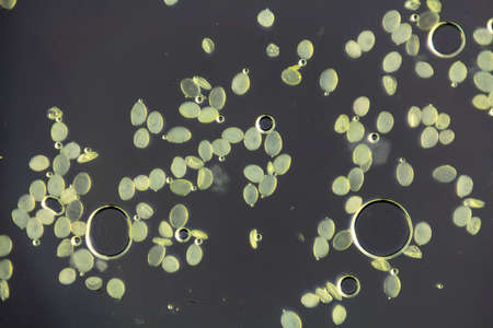
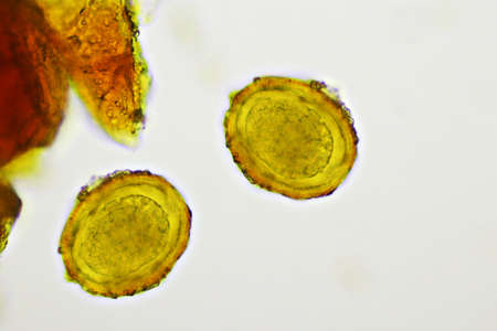
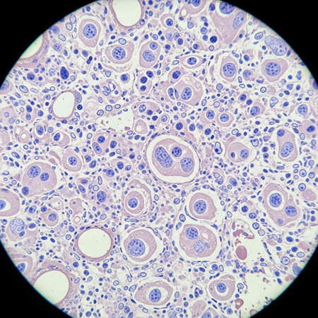
Eggs of a Taenia tapeworm. Taenia is a genus of tapeworm parasites on livestock and humans.
Коллекция по умолчанию
Коллекция по умолчанию
Создать новую
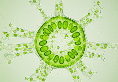
Vibrant green radial cellular structure under a microscope, displaying numerous oval chloroplasts within the central body and extending arms. ideal for biology and science education.
Коллекция по умолчанию
Коллекция по умолчанию
Создать новую
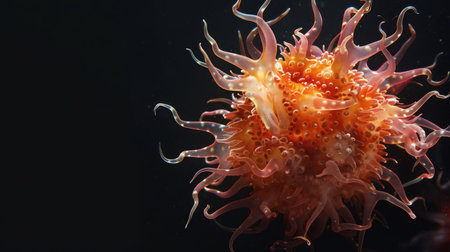
A detailed photograph of a vibrant orange sea anemone, showcasing its complex structure against a stark black backdrop.
Коллекция по умолчанию
Коллекция по умолчанию
Создать новую
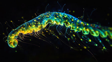
Neon glowing jellyfish isolated on black background. 3d illustration
Коллекция по умолчанию
Коллекция по умолчанию
Создать новую
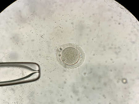
View through microscope at in vitro fertilization process
Коллекция по умолчанию
Коллекция по умолчанию
Создать новую
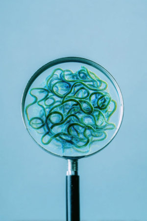
Close up of a magnifying glass revealing a complex network of intertwined green fibers, highlighting the intricate details and textures
Коллекция по умолчанию
Коллекция по умолчанию
Создать новую
microorganisms and soil biology, with nematodes and fungi under the microscope. in a soil and compost sample in australia
Коллекция по умолчанию
Коллекция по умолчанию
Создать новую
Volvox in drop of water under the microscope for classroom education.
Коллекция по умолчанию
Коллекция по умолчанию
Создать новую

Ovarian cancer, light micrograph, photo under microscope. Photograph shows a fragment of a cancerous tumor in the female ovary. Selective focus
Коллекция по умолчанию
Коллекция по умолчанию
Создать новую

Calcium oxalate in urine find with microscope.
Коллекция по умолчанию
Коллекция по умолчанию
Создать новую
Blood cells in human body under microscope view for education in laboratory.
Коллекция по умолчанию
Коллекция по умолчанию
Создать новую

Paramecium caudatum is a genus of unicellular ciliated protozoan and Bacterium under the microscope.
Коллекция по умолчанию
Коллекция по умолчанию
Создать новую

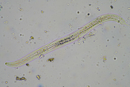
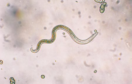
Egg of Parasitic nematode worm (roundworm) Ascaris lumbricoides which inhabits human intestine and causes disease ascariasis
Коллекция по умолчанию
Коллекция по умолчанию
Создать новую
Lab-based embryo manipulation techniques for precise experimental interventions in controlled environments
Коллекция по умолчанию
Коллекция по умолчанию
Создать новую
Microscope with metal lens at laboratory. Medical equipment.
Коллекция по умолчанию
Коллекция по умолчанию
Создать новую
Cell Gene Microscopic Series
Коллекция по умолчанию
Коллекция по умолчанию
Создать новую

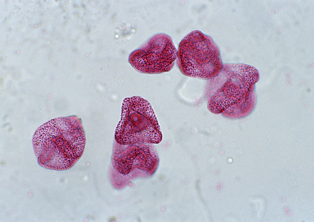
Pollen grain is a microscopic body that contains the male reproductive cell of plant.
Коллекция по умолчанию
Коллекция по умолчанию
Создать новую
Microscopic of human cells with colored cells.
Коллекция по умолчанию
Коллекция по умолчанию
Создать новую
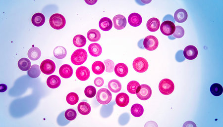
Malaria blood parasite infected red blood cells laboratory background.
Коллекция по умолчанию
Коллекция по умолчанию
Создать новую
beautiful electronic microscopy of bacteria fungi fantasy microbiology in blue tones microscopic life generative ai
Коллекция по умолчанию
Коллекция по умолчанию
Создать новую
Gloved hand holds a translucent cell model under focused laboratory light with visible nucleus and organelle structures, suggesting scientific research and educational demonstration, with empty background space available for text
Коллекция по умолчанию
Коллекция по умолчанию
Создать новую
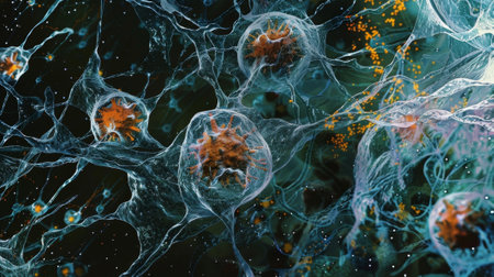
Vibrant microorganisms are suspended in a dark backdrop, showing detailed structures.
Коллекция по умолчанию
Коллекция по умолчанию
Создать новую

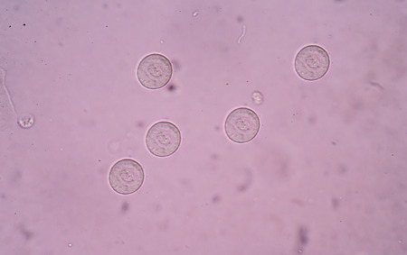
Egg of Ascaris lumbricoides (roundworm) in human stool, analyze by microscope, 400x
Коллекция по умолчанию
Коллекция по умолчанию
Создать новую
Volvox in drop of water under the microscope for classroom education.
Коллекция по умолчанию
Коллекция по умолчанию
Создать новую
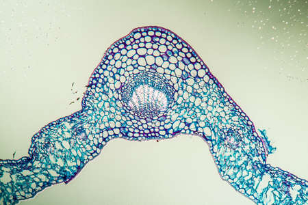
Linguster leaf cross section under the microscope 100x
Коллекция по умолчанию
Коллекция по умолчанию
Создать новую
Transparent gel with bubbles close-up. Smear of face gel cream. The texture of gel cream. A sample of a cosmetic product. Antibacterial gel.
Коллекция по умолчанию
Коллекция по умолчанию
Создать новую
Rare image of Ghost flatworm - Maricola (Planarian) triclad flatworms in reef aquarium glass
Коллекция по умолчанию
Коллекция по умолчанию
Создать новую
Egg of Ascaris lumbricoides (roundworm) in human stool, analyze by microscope, 400x
Коллекция по умолчанию
Коллекция по умолчанию
Создать новую
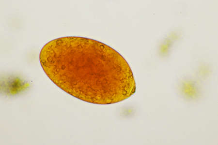
Egg of intestinal fluke in human stool, analyze by microscope, original magnification 400x
Коллекция по умолчанию
Коллекция по умолчанию
Создать новую
Egg of Hookworm in human stool, analyze by microscope
Коллекция по умолчанию
Коллекция по умолчанию
Создать новую
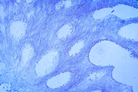
Columnar epithelium of human gall bladder under the microscope in Lab.
Коллекция по умолчанию
Коллекция по умолчанию
Создать новую
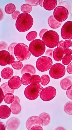
Red blood cells are visibly floating and interacting under a microscope showcasing their circular shape and vibrant red color in detail.
Коллекция по умолчанию
Коллекция по умолчанию
Создать новую
The activation of TLRs leading to the production of proinflammatory cytokines as seen in a micrograph of immune cells
Коллекция по умолчанию
Коллекция по умолчанию
Создать новую
Bacillary dysentery, light micrograph, photo under microscope showing presence of bacteria and accumulation of inflammatory cells in intestinal epithelium
Коллекция по умолчанию
Коллекция по умолчанию
Создать новую
In this close-up shot, a translucent jelly-like substance is shown in detail, with its squishy texture and semi-transparent appearance clearly visible. The substance sits in a pool of liquid, glistening under the light.
Коллекция по умолчанию
Коллекция по умолчанию
Создать новую
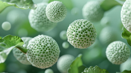
close-up photograph of small, green spheres with a textured surface, floating gracefully against a blurred background of green leaves.
Коллекция по умолчанию
Коллекция по умолчанию
Создать новую
Toxocara canis second stage larvae hatch from eggs
Коллекция по умолчанию
Коллекция по умолчанию
Создать новую
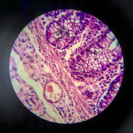
Histopathology of human liver under microscope view for medical education.
Коллекция по умолчанию
Коллекция по умолчанию
Создать новую

Human hyaline cartilage bone under microscope view for education pathology. Human tissue.
Коллекция по умолчанию
Коллекция по умолчанию
Создать новую
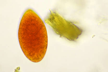
Egg of intestinal fluke in human stool, analyze by microscope, original magnification 400x
Коллекция по умолчанию
Коллекция по умолчанию
Создать новую
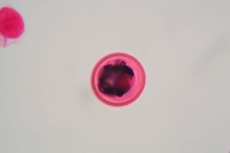
Paraffin Embedded Tissue Blocks of Cancer on iSolated White Background.
Коллекция по умолчанию
Коллекция по умолчанию
Создать новую
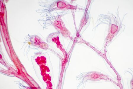
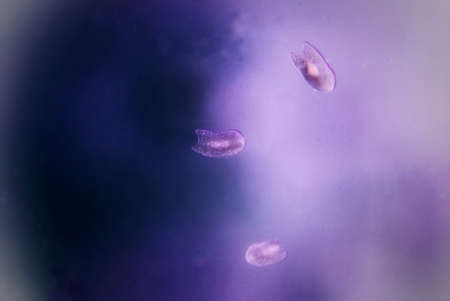
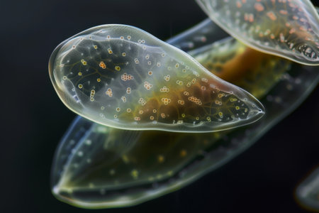
Microscope view of Obelia hydroid, a marine and sometimes freshwater animal.
Коллекция по умолчанию
Коллекция по умолчанию
Создать новую
Legion-Media
Создайте свои проекты на основе качественных стоковых фотографий и видео.


Copyright © Legion-Media.