

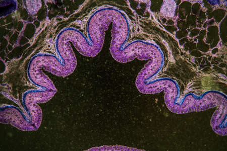
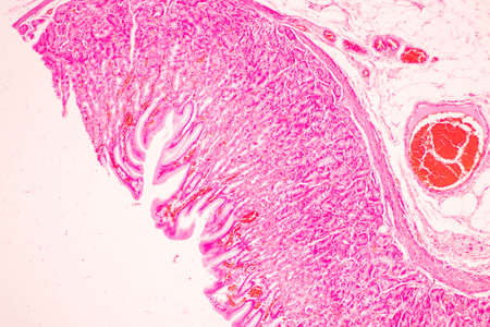
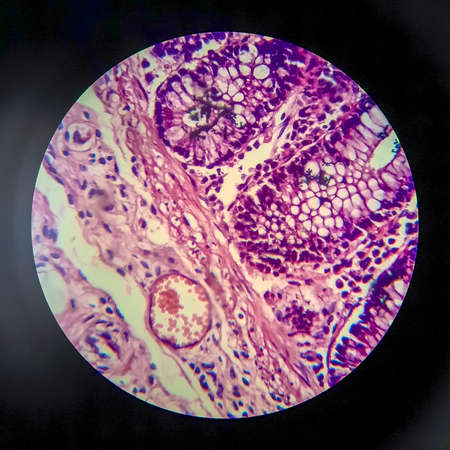
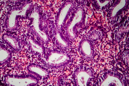
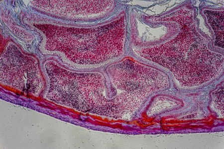
Stomach tissue under the microscope 100x
Коллекция по умолчанию
Коллекция по умолчанию
Создать новую

Ovarian cancer, light micrograph, photo under microscope. Photograph shows a fragment of a cancerous tumor in the female ovary. Selective focus
Коллекция по умолчанию
Коллекция по умолчанию
Создать новую
Tongue Tissue with taste buds across 200x
Коллекция по умолчанию
Коллекция по умолчанию
Создать новую
Fibrin deposits in the kidney, microscopy 100x
Коллекция по умолчанию
Коллекция по умолчанию
Создать новую
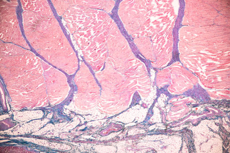
Education anatomy and Histological sample of Human under the microscope.
Коллекция по умолчанию
Коллекция по умолчанию
Создать новую
destructive mushroom in wood fabric 100x
Коллекция по умолчанию
Коллекция по умолчанию
Создать новую

Characteristics of Lichen, hyphae and Symbiotic algae under the microscope for education.
Коллекция по умолчанию
Коллекция по умолчанию
Создать новую
Characteristics of Lichen, hyphae and Symbiotic algae under the microscope for education.
Коллекция по умолчанию
Коллекция по умолчанию
Создать новую

Thyroid follicular carcinoma, light micrograph, photo under microscope
Коллекция по умолчанию
Коллекция по умолчанию
Создать новую
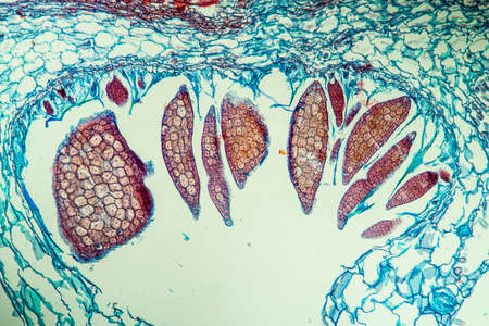
Earthworm histology cross section 10th segment 100x
Коллекция по умолчанию
Коллекция по умолчанию
Создать новую

Histopathology of human under microscope view for education in laboratory.
Коллекция по умолчанию
Коллекция по умолчанию
Создать новую
Abstract ink design template mixed texture background. Blue abstract texture. Multicolored liquid marble pattern. Fluid Art
Коллекция по умолчанию
Коллекция по умолчанию
Создать новую
Columnar epithelium of human gall bladder under the microscope in Lab.
Коллекция по умолчанию
Коллекция по умолчанию
Создать новую
Bowel with goblet cells in the dark field 100x
Коллекция по умолчанию
Коллекция по умолчанию
Создать новую

Tongue with taste buds Papilla across 100x
Коллекция по умолчанию
Коллекция по умолчанию
Создать новую
A highresolution image of tight junctions in epithelial cells displaying the various types and arrangements of proteins that make up these junctions
Коллекция по умолчанию
Коллекция по умолчанию
Создать новую

Scalp and hair follicles of human under the microscope in Lab.
Коллекция по умолчанию
Коллекция по умолчанию
Создать новую
Characteristics of Lichen, hyphae and Symbiotic algae under the microscope for education.
Коллекция по умолчанию
Коллекция по умолчанию
Создать новую

Bladder cancer, light micrograph, photo under microscope
Коллекция по умолчанию
Коллекция по умолчанию
Создать новую
Lung adenocarcinoma, light micrograph, photo under microscope
Коллекция по умолчанию
Коллекция по умолчанию
Создать новую
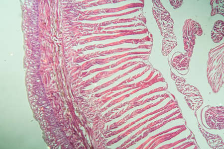
Tissue of Stomach Human under the microscope in Lab.
Коллекция по умолчанию
Коллекция по умолчанию
Создать новую
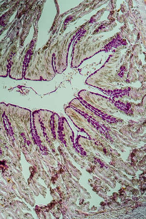
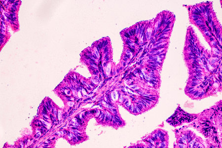
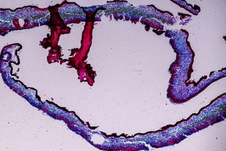
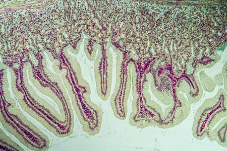
Cross-section through the intestine with glands 200x
Коллекция по умолчанию
Коллекция по умолчанию
Создать новую
Characteristics of Lichen, hyphae and Symbiotic algae under the microscope for education.
Коллекция по умолчанию
Коллекция по умолчанию
Создать новую
Pancreas cancer cell under microscope view for medical education.
Коллекция по умолчанию
Коллекция по умолчанию
Создать новую
Nestwurz orchid root cross 100x
Коллекция по умолчанию
Коллекция по умолчанию
Создать новую
Characteristics of Lichen, hyphae and Symbiotic algae under the microscope for education.
Коллекция по умолчанию
Коллекция по умолчанию
Создать новую
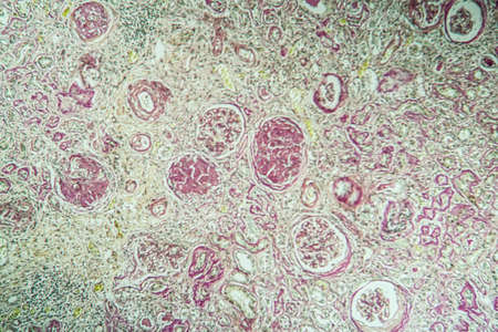
Diffuse proliferative glomerulonephritis, light micrograph, photo under microscope. High magnification
Коллекция по умолчанию
Коллекция по умолчанию
Создать новую
Microscopic fungi Cunninghamella, scientific 3D illustration. Pathogenic fungi from the order Mucorales, cause sinopulmonary and disseminated infections, one of the causative agents of mucormycosis
Коллекция по умолчанию
Коллекция по умолчанию
Создать новую
Lungworm under the microscope 100x
Коллекция по умолчанию
Коллекция по умолчанию
Создать новую
Detailed image of rod-shaped bacteria under a high-powered microscope, showing cell structure
Коллекция по умолчанию
Коллекция по умолчанию
Создать новую
Histopathology of acute nephritis, light micrograph, photo under microscope
Коллекция по умолчанию
Коллекция по умолчанию
Создать новую
Uterine cancer, light micrograph, photo under microscope
Коллекция по умолчанию
Коллекция по умолчанию
Создать новую
Squamous cell carcinoma of the uterus, light micrograph, photo under microscope
Коллекция по умолчанию
Коллекция по умолчанию
Создать новую

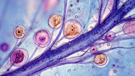
Apple pollen from a blossom in spring under the microscope
Коллекция по умолчанию
Коллекция по умолчанию
Создать новую
Breast fibroadenosis, light micrograph, photo under microscope. Common benign hyperplastic process involving breast glands
Коллекция по умолчанию
Коллекция по умолчанию
Создать новую
A close up of a pink and blue cell with a tree inside of it. The cell is surrounded by a pink and blue background
Коллекция по умолчанию
Коллекция по умолчанию
Создать новую
This detailed microscopic image showcases various cellular structures, highlighted in striking purple tones. The intricate patterns and textures reveal the complexity of biological tissues, making it a valuable resource for educational and scientific purposes
Коллекция по умолчанию
Коллекция по умолчанию
Создать новую
Colon tissue with diverticulum 100x
Коллекция по умолчанию
Коллекция по умолчанию
Создать новую

Condyloma acuminatum, also known as genital warts. Light micrograph, photo under microscope
Коллекция по умолчанию
Коллекция по умолчанию
Создать новую
Characteristics of Lichen, hyphae and Symbiotic algae under the microscope for education.
Коллекция по умолчанию
Коллекция по умолчанию
Создать новую

Protozoa and Green Algae in waste water under the microscope.
Коллекция по умолчанию
Коллекция по умолчанию
Создать новую
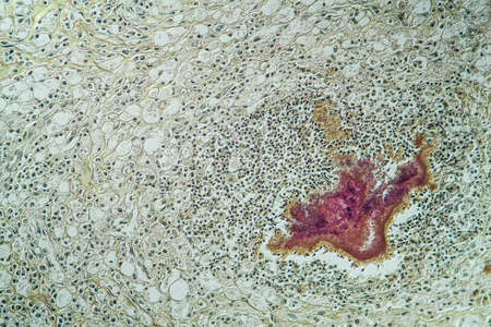
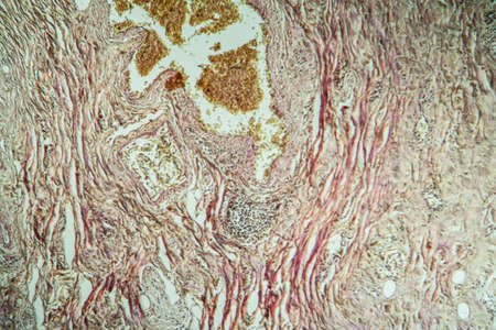
Actinomyces in the jaw diseased tissue 200x
Коллекция по умолчанию
Коллекция по умолчанию
Создать новую
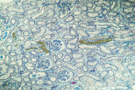
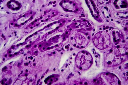
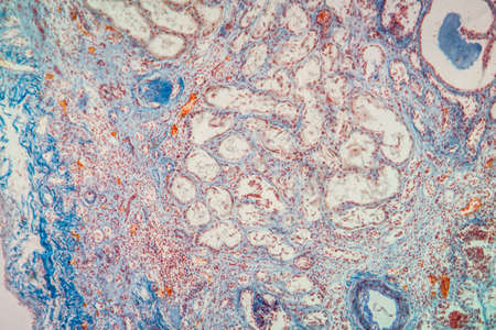
science medical anthropotomy physiology microscopic section of human kidney tissue background
Коллекция по умолчанию
Коллекция по умолчанию
Создать новую
Bacillary dysentery, light micrograph, photo under microscope showing presence of bacteria and accumulation of inflammatory cells in intestinal epithelium
Коллекция по умолчанию
Коллекция по умолчанию
Создать новую
water flowing over boulders, rocks, and stones
Коллекция по умолчанию
Коллекция по умолчанию
Создать новую
Histopathology of human under microscope view for education in laboratory.
Коллекция по умолчанию
Коллекция по умолчанию
Создать новую
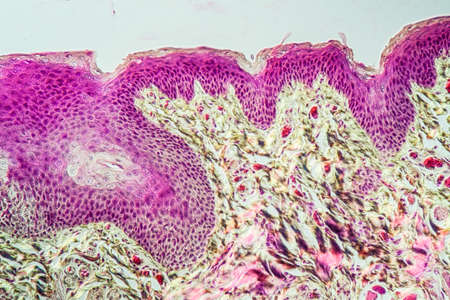
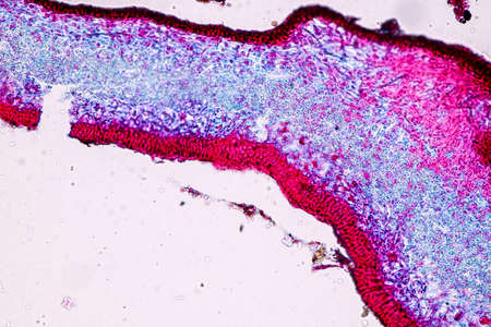
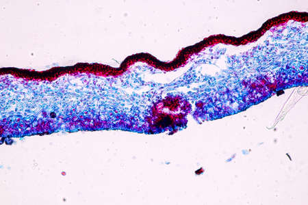
Atypical squamous epithelial hyperplasia, photomicrograph showing thickened stratified epithelium with nuclear atypia and basal layer crowding.
Коллекция по умолчанию
Коллекция по умолчанию
Создать новую
Shrinked kidney diseased tissue under the microscope 100x
Коллекция по умолчанию
Коллекция по умолчанию
Создать новую

Uterine cancer, light micrograph, photo under microscope
Коллекция по умолчанию
Коллекция по умолчанию
Создать новую

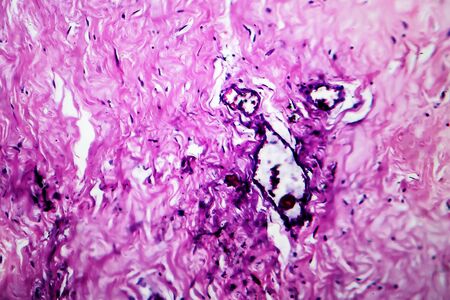
Human hyaline cartilage bone under microscope view for education pathology. Human tissue.
Коллекция по умолчанию
Коллекция по умолчанию
Создать новую
beautiful electronic microscopy of bacteria fungi fantasy microbiology in blue tones microscopic life generative ai
Коллекция по умолчанию
Коллекция по умолчанию
Создать новую
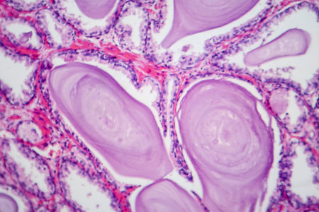
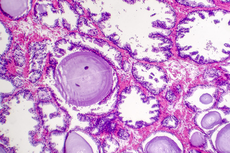
Photomicrograph showing histological features of benign prostatic hyperplasia. Enlarged prostate gland with nodular proliferation of glandular and stromal components.
Коллекция по умолчанию
Коллекция по умолчанию
Создать новую
Characteristics of Lichen, hyphae and Symbiotic algae under the microscope for education.
Коллекция по умолчанию
Коллекция по умолчанию
Создать новую
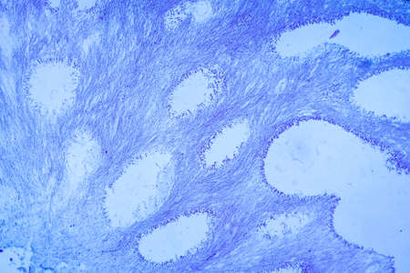
Large intestine with diverticular tissue under the microscope 100x
Коллекция по умолчанию
Коллекция по умолчанию
Создать новую
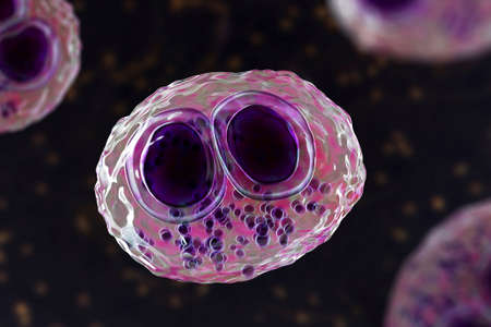
Cytomegalovirus CMV in a human cell, owl's eye inclusion in nucleus, multinucleated cell, 3D illustration. It is herpes virus, causes diseases in fetus, organ transplant patients, HIV infected people
Коллекция по умолчанию
Коллекция по умолчанию
Создать новую
close up of red cabbage leaf with water droplets, abstract background
Коллекция по умолчанию
Коллекция по умолчанию
Создать новую
Embryo with endosperm across 100x
Коллекция по умолчанию
Коллекция по умолчанию
Создать новую
Diffuse proliferative glomerulonephritis, DPGN, light micrograph, photo under microscope. High magnification
Коллекция по умолчанию
Коллекция по умолчанию
Создать новую
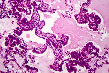
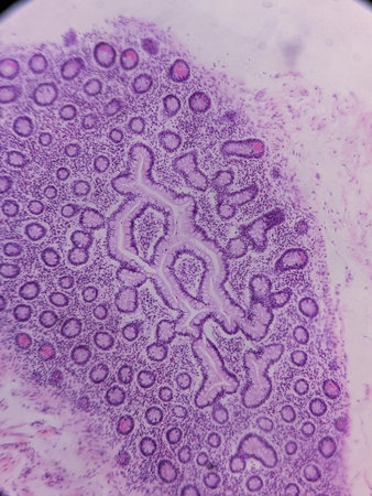
serous gland tissue under the microscope 100x
Коллекция по умолчанию
Коллекция по умолчанию
Создать новую

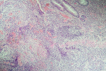
picture of histology human tissue with microscope from laboratory (not Illustration Designation)
Коллекция по умолчанию
Коллекция по умолчанию
Создать новую
A closeup examination of a cultured cell on a nanoengineered substrate with bright staining revealing cellular morphology and interaction with the surface at the nanoscale
Коллекция по умолчанию
Коллекция по умолчанию
Создать новую
Chaos ink texture background, ink in water pattern frost. Crystal winter design
Коллекция по умолчанию
Коллекция по умолчанию
Создать новую
Endometriosis, a disorder in which cells similar to those in the endometrium grow outside the uterus. Light micrograph, photo under microscope
Коллекция по умолчанию
Коллекция по умолчанию
Создать новую
Abstract view of semiprecious resin surface with vivid grape-like purple clusters scattered across cloudy blue and white texture.
Коллекция по умолчанию
Коллекция по умолчанию
Создать новую
Characteristics of fungi living in wood as a group, are polyphyletic under the microscope for education.
Коллекция по умолчанию
Коллекция по умолчанию
Создать новую
Viscous thick oil texture with industrial dark background showcasing vibrant patterns
Коллекция по умолчанию
Коллекция по умолчанию
Создать новую
An illustration of the rie assembly process with rRNA molecules and rial proteins coming together to form functional ries
Коллекция по умолчанию
Коллекция по умолчанию
Создать новую
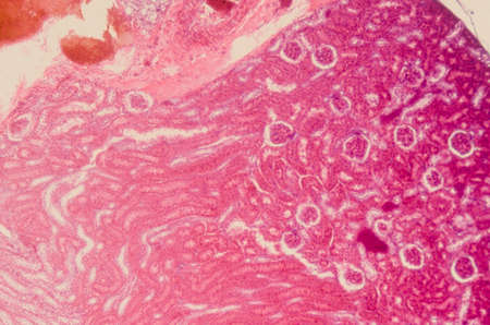
Chronic pyelonephritis, light micrograph, photo under microscope. High magnification
Коллекция по умолчанию
Коллекция по умолчанию
Создать новую

microscopy micrograph plant tissue, corn embryo
Коллекция по умолчанию
Коллекция по умолчанию
Создать новую
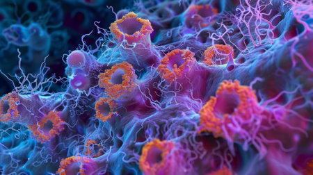
A colorful image of a cell with a purple and blue blob in the center. The image is abstract and has a mood of curiosity and wonder
Коллекция по умолчанию
Коллекция по умолчанию
Создать новую
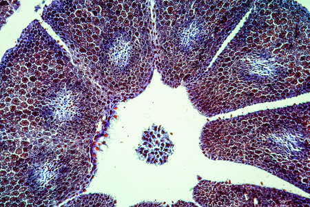
Lymph node tissue under the microscope 100x
Коллекция по умолчанию
Коллекция по умолчанию
Создать новую

Itch mites under the skin cross-section 100x
Коллекция по умолчанию
Коллекция по умолчанию
Создать новую
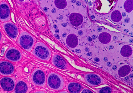
The study of tissue samples of Trachea of Cat, Epididymis, Prostate, Uterus with embryo of rat and Mammary gland cow under the microscope in Lab.
Коллекция по умолчанию
Коллекция по умолчанию
Создать новую
Colon inflammation in Crohn's disease 100x
Коллекция по умолчанию
Коллекция по умолчанию
Создать новую
Benign prostatic hyperplasia. Micrograph shows dilated glands, papillary projections inside the lumen of the glands, cystic dilatation with accumulation of secretory material. Photo under microscope
Коллекция по умолчанию
Коллекция по умолчанию
Создать новую
Creative colorful sand flows on blue background. Place for text or product promotion. Copy space. High quality photo
Коллекция по умолчанию
Коллекция по умолчанию
Создать новую
Close up of a pink coral with many small pink points. The coral is very detailed and has a lot of texture
Коллекция по умолчанию
Коллекция по умолчанию
Создать новую

Papillary thyroid carcinoma, light micrograph, photo under microscope. The most common type of thyroid cancer
Коллекция по умолчанию
Коллекция по умолчанию
Создать новую

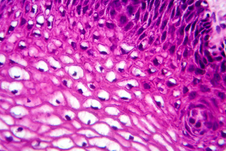
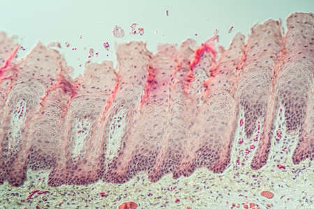
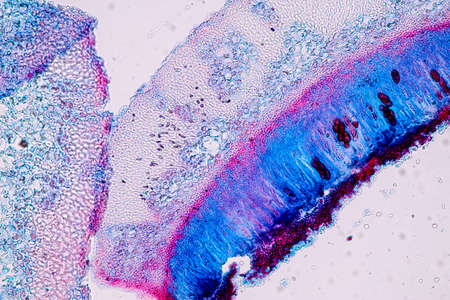
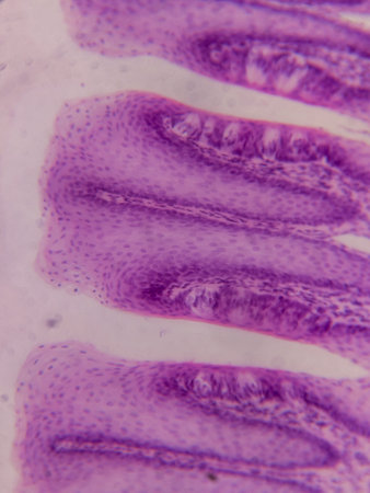
Human tongue section showing papillae and stratified epithelium
Коллекция по умолчанию
Коллекция по умолчанию
Создать новую
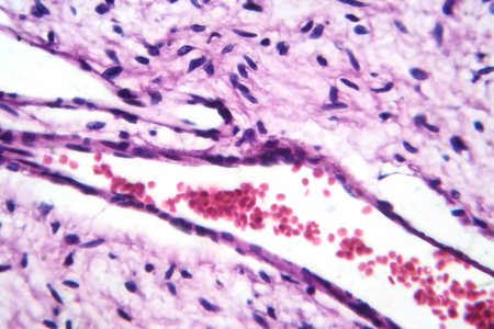
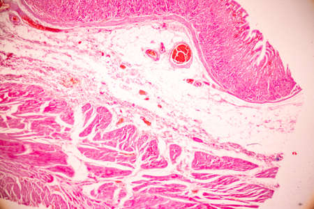
A small blood vessel with red blood cells in neurofibroma tissue sample, light photomicrograph.
Коллекция по умолчанию
Коллекция по умолчанию
Создать новую
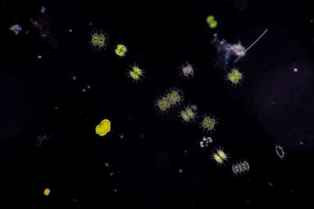
Protozoa and Green Algae in waste water under the microscope.
Коллекция по умолчанию
Коллекция по умолчанию
Создать новую
Hodgkin's lymphoma, light micrograph, photo under microscope. High magnification
Коллекция по умолчанию
Коллекция по умолчанию
Создать новую
Thermal photography of an abstract, colorful pattern with heat waves in the style of Matisse. The color palette features neon pink and blue, with vivid colors. Captured using a macro lens, the image has an x-ray effect and a dynamic composition. Created using a redshift renderer, the result is hyper-realistic, with detailed textures and a cinematic, high-resolution appearance. --ar 3:2 --v 6.1 Job ID: 06163584-6a82-45d4-b422-f688ac3881ef
Коллекция по умолчанию
Коллекция по умолчанию
Создать новую

Cells in reproductive female cytology and histology education concept.
Коллекция по умолчанию
Коллекция по умолчанию
Создать новую
Tongue Tissue with taste buds across 200x
Коллекция по умолчанию
Коллекция по умолчанию
Создать новую
Wilms tumor, or nephroblastoma, light micrograph, photo under microscope. High magnification
Коллекция по умолчанию
Коллекция по умолчанию
Создать новую
Tissue of Stomach Human under the microscope in Lab.
Коллекция по умолчанию
Коллекция по умолчанию
Создать новую
Detailed macro image reveals microorganisms with thin cell walls in linear arrangement, Ai Generated.
Коллекция по умолчанию
Коллекция по умолчанию
Создать новую
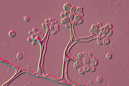
Aspergillus niger and Aspergillus oryzae (mold) under microscope for Microbiology in Lab.
Коллекция по умолчанию
Коллекция по умолчанию
Создать новую
Chronic glomerulonephritis, light micrograph, photo under microscope. High magnification
Коллекция по умолчанию
Коллекция по умолчанию
Создать новую
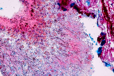
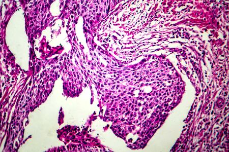
Ovarian cancer, light micrograph, photo under microscope. Photograph shows a fragment of a cancerous tumor in the female ovary. Selective focus
Коллекция по умолчанию
Коллекция по умолчанию
Создать новую
Anatomy and Histological Bone, Elastic cartilage human and Joint of human foetus under the microscope for education.
Коллекция по умолчанию
Коллекция по умолчанию
Создать новую
Characteristics of Lichen, hyphae and Symbiotic algae under the microscope for education.
Коллекция по умолчанию
Коллекция по умолчанию
Создать новую
Earthworm histology cross section 10th segment 100x
Коллекция по умолчанию
Коллекция по умолчанию
Создать новую
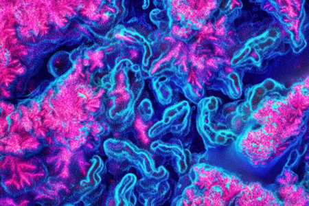
Explore a stunning microscopic view featuring colorful cells and thin filaments against a vibrant blue background, showcasing scientific beauty in detail.
Коллекция по умолчанию
Коллекция по умолчанию
Создать новую
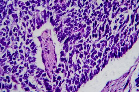
Microscopic view of tissue sections showing purple-stained cells, possibly from a biological sample
Коллекция по умолчанию
Коллекция по умолчанию
Создать новую
An electron microscope image of a rie attached to the endoplasmic with newly synthesized proteins being transported through the cell
Коллекция по умолчанию
Коллекция по умолчанию
Создать новую
Atrophy kidney tissue under the microscope 100x
Коллекция по умолчанию
Коллекция по умолчанию
Создать новую

science microscopy micrograph earthworm crosscutting
Коллекция по умолчанию
Коллекция по умолчанию
Создать новую
Microscope with metal lens at laboratory. Medical equipment.
Коллекция по умолчанию
Коллекция по умолчанию
Создать новую
Legion-Media
Создайте свои проекты на основе качественных стоковых фотографий и видео.


Copyright © Legion-Media.