

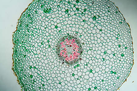
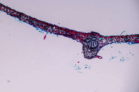
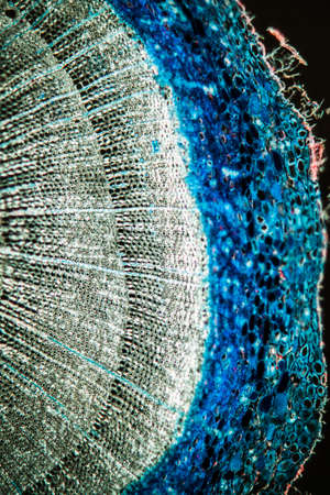
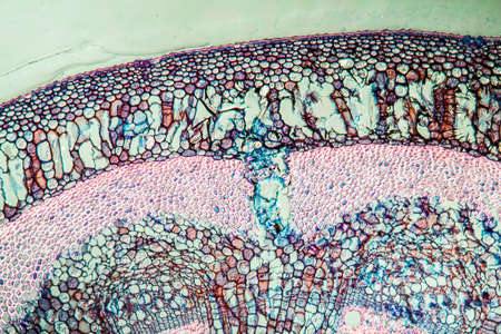
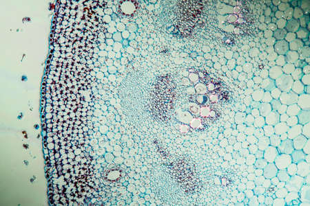
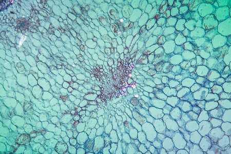
Cross-section leaf Plant of under the microscope for classroom education.
Коллекция по умолчанию
Коллекция по умолчанию
Создать новую
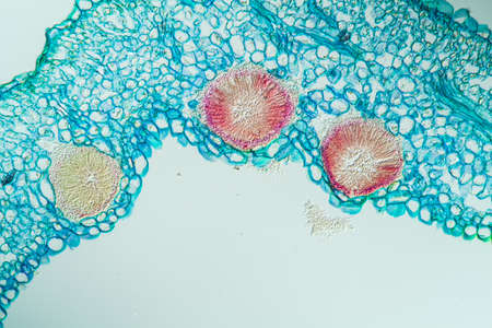
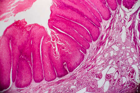
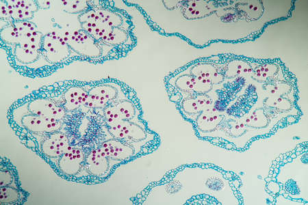
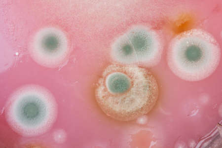
Rust fungus on milkweed plant, 200x
Коллекция по умолчанию
Коллекция по умолчанию
Создать новую
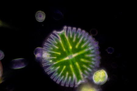
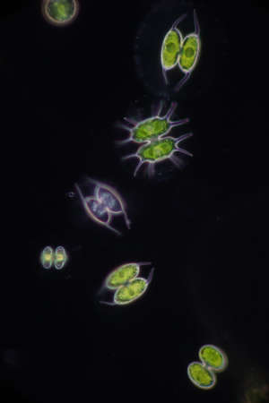
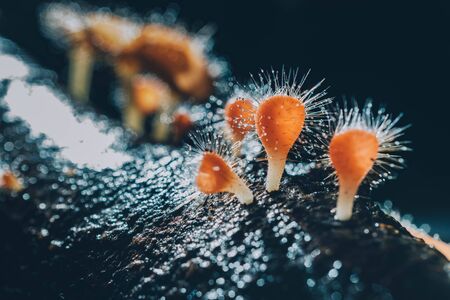
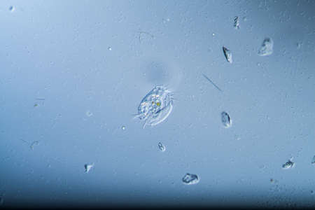
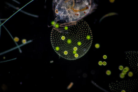
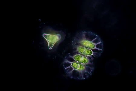
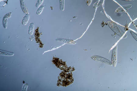
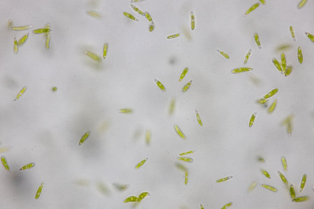
Protozoa and Green Algae in waste water under the microscope.
Коллекция по умолчанию
Коллекция по умолчанию
Создать новую
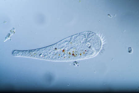
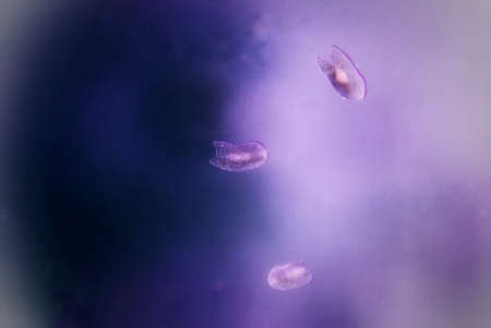
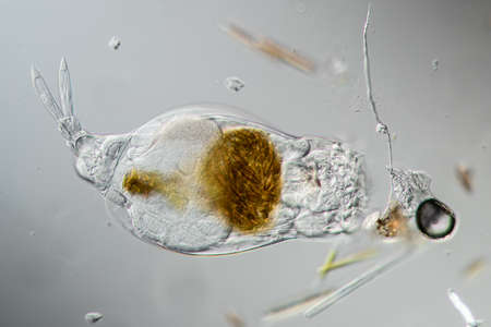
Trumpet animal as a microscopic plankton animal in drops of water
Коллекция по умолчанию
Коллекция по умолчанию
Создать новую
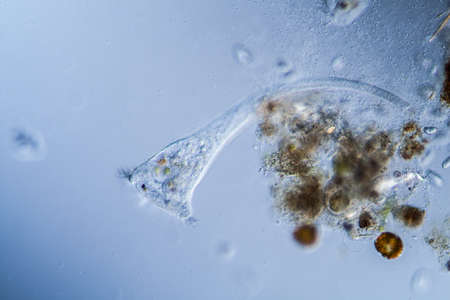
Trumpet animal as a microscopic plankton animal in drops of water
Коллекция по умолчанию
Коллекция по умолчанию
Создать новую

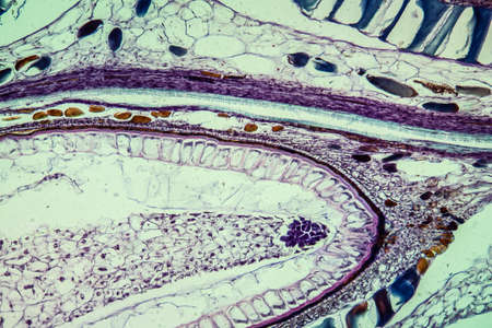
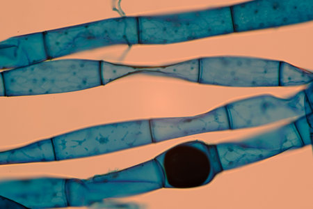
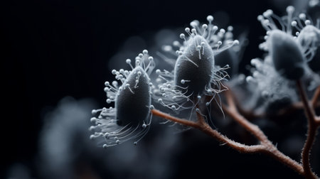
Host cells with spores (mold) are inside wood under the microscope for education.
Коллекция по умолчанию
Коллекция по умолчанию
Создать новую
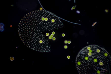
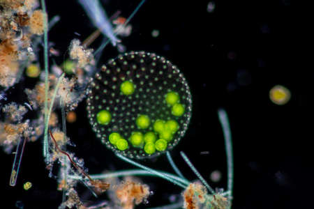
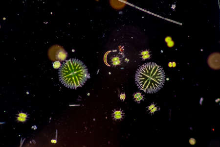
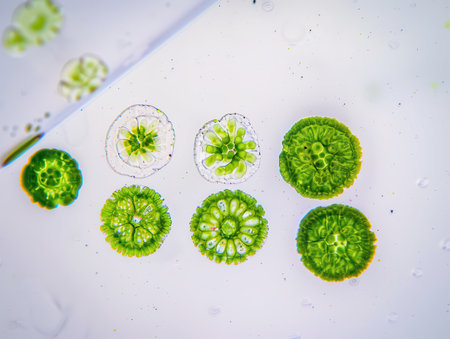
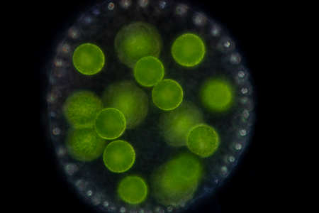
Volvox in drop of water under the microscope for classroom education.
Коллекция по умолчанию
Коллекция по умолчанию
Создать новую
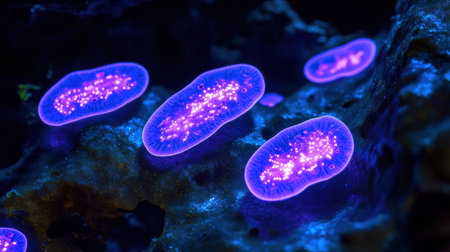
Rare image of Ghost flatworm - Maricola (Planarian) triclad flatworms in reef aquarium glass
Коллекция по умолчанию
Коллекция по умолчанию
Создать новую
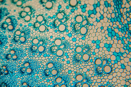
Johannes berry fruit cross 100x
Коллекция по умолчанию
Коллекция по умолчанию
Создать новую

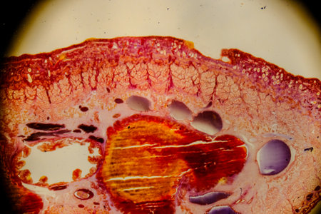
Field buttercup fruit cross 100x
Коллекция по умолчанию
Коллекция по умолчанию
Создать новую
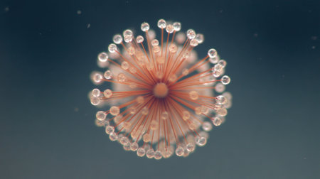
A close-up, detailed view of a single cell exhibiting a complex structure with numerous small tentacles or appendages.
Коллекция по умолчанию
Коллекция по умолчанию
Создать новую

Host cells with spores (mold) are inside wood under the microscope for education.
Коллекция по умолчанию
Коллекция по умолчанию
Создать новую
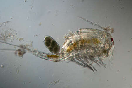
Copepod crab swims through the water
Коллекция по умолчанию
Коллекция по умолчанию
Создать новую
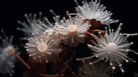
This is a close-up image of a green, spiky flower under a microscope
Коллекция по умолчанию
Коллекция по умолчанию
Создать новую
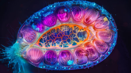
Vibrant cross-section of a developing seed under UV light, highlighting the embryo and endosperm in bright colors
Коллекция по умолчанию
Коллекция по умолчанию
Создать новую
Protozoa and Green Algae in waste water under the microscope.
Коллекция по умолчанию
Коллекция по умолчанию
Создать новую
Rotifer foraging in the stream 200x
Коллекция по умолчанию
Коллекция по умолчанию
Создать новую
Cowslip stem in cross section 100x
Коллекция по умолчанию
Коллекция по умолчанию
Создать новую

Rhizopus (bread mold) is a genus of common saprophytic fungi,Rhizopus (bread mold) under the microscope.
Коллекция по умолчанию
Коллекция по умолчанию
Создать новую
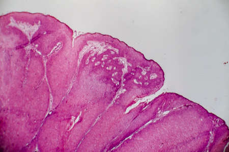
Condyloma acuminatum, also known as genital warts. Light micrograph, photo under microscope
Коллекция по умолчанию
Коллекция по умолчанию
Создать новую
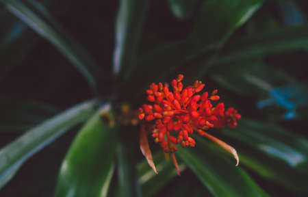
Orange mushroom ,Champagne mushroom or eyelash cup mushroom with sparkling droplets in the forest. Ecosystem or biological diversity concept.
Коллекция по умолчанию
Коллекция по умолчанию
Создать новую

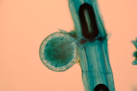
Cross sections of plant root under microscope view
Коллекция по умолчанию
Коллекция по умолчанию
Создать новую
Microscopic view of blue-stained fungal hyphae with spores.
Коллекция по умолчанию
Коллекция по умолчанию
Создать новую
Ciliates as single-cell plankton animals in drops of water
Коллекция по умолчанию
Коллекция по умолчанию
Создать новую
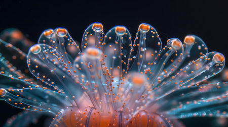
A closeup of the transparent jellyfish tentacles, each petal with tiny beads that glow in shades of orange and blue against an isolated black background.
Коллекция по умолчанию
Коллекция по умолчанию
Создать новую
Host cells with spores (mold) are inside wood under the microscope for education.
Коллекция по умолчанию
Коллекция по умолчанию
Создать новую
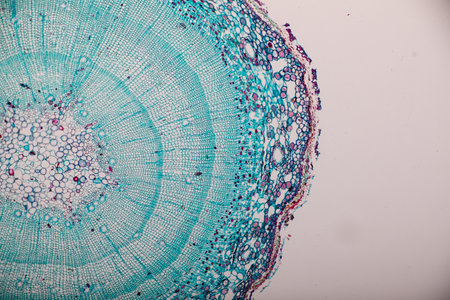
Pine tree trunk across 100x
Коллекция по умолчанию
Коллекция по умолчанию
Создать новую

Protozoa and Green Algae in waste water under the microscope.
Коллекция по умолчанию
Коллекция по умолчанию
Создать новую

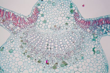
micrograph plant tissue, stem of pumpkin
Коллекция по умолчанию
Коллекция по умолчанию
Создать новую
Fern stems in cross section 100x
Коллекция по умолчанию
Коллекция по умолчанию
Создать новую

Spherical colony of freshwater green algae (Volvox). Microscopic view, Rheinberg illumination.
Коллекция по умолчанию
Коллекция по умолчанию
Создать новую
Condyloma acuminatum, also known as genital warts. Light micrograph, photo under microscope
Коллекция по умолчанию
Коллекция по умолчанию
Создать новую
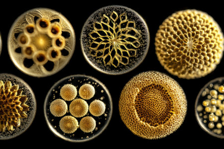
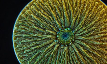
Intricate golden-hued circular patterns resembling microscopic organisms, each with unique designs, highlighted against a contrasting background, showing delicate textures and symmetry
Коллекция по умолчанию
Коллекция по умолчанию
Создать новую

Protozoa and Green Algae in waste water under the microscope.
Коллекция по умолчанию
Коллекция по умолчанию
Создать новую
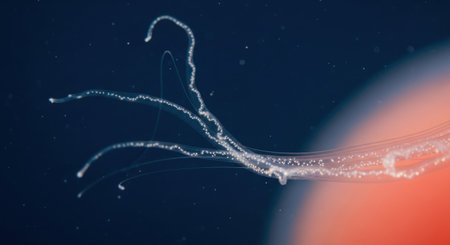
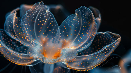
A mesmerizing view of a marine organism with luminous, branching appendages drifting in the abyss.
Коллекция по умолчанию
Коллекция по умолчанию
Создать новую
a close-up portrait photograph showcasing the intricate details of unknown escherichia coli biological virus alien flowers. the image captures the brittle yet beautiful essence of these flowers, with dry and elegant petals. the rich and vivid contrast, along with the depth of field and black tones, creates a crisp and realistic depiction. shot on a 100mm lens with an aperture of f/2.0, the natural lighting
Коллекция по умолчанию
Коллекция по умолчанию
Создать новую
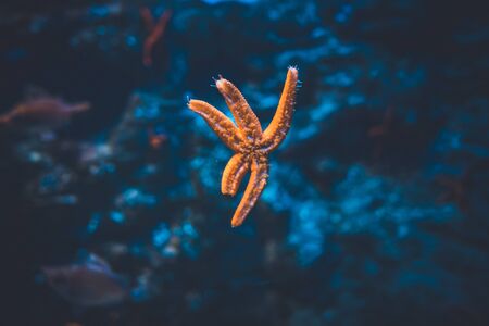
Starfish attached to the glass wall of the aquarium of San Sebastian, Spain
Коллекция по умолчанию
Коллекция по умолчанию
Создать новую
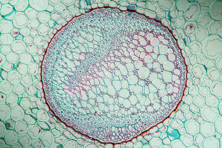
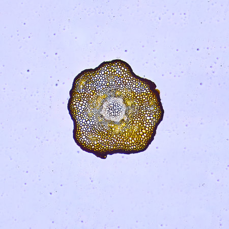
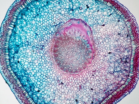
This visual representation reveals a cross-section of a plant stem, displaying the intricate structures essential for plant physiology.
Коллекция по умолчанию
Коллекция по умолчанию
Создать новую
Volvox in drop of water under the microscope for classroom education.
Коллекция по умолчанию
Коллекция по умолчанию
Создать новую

natural snowflakes
Коллекция по умолчанию
Коллекция по умолчанию
Создать новую
Host cells with spores (mold) are inside wood under the microscope for education.
Коллекция по умолчанию
Коллекция по умолчанию
Создать новую
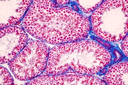
Anatomy and Histological Epididymis and Testis human cells under microscope.
Коллекция по умолчанию
Коллекция по умолчанию
Создать новую
Volvox in drop of water under the microscope for classroom education.
Коллекция по умолчанию
Коллекция по умолчанию
Создать новую
close-up shot of beautiful fern leaves on light natural background
Коллекция по умолчанию
Коллекция по умолчанию
Создать новую
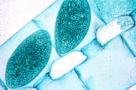
Microscopic view of plant cells with a spore and vascular tissue.
Коллекция по умолчанию
Коллекция по умолчанию
Создать новую
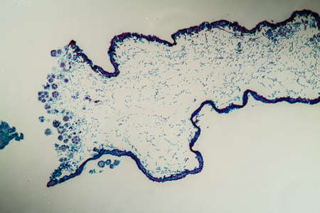
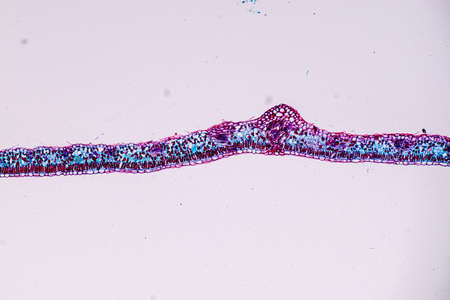
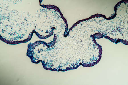
Cross-section through the lichen symbiote body 100x
Коллекция по умолчанию
Коллекция по умолчанию
Создать новую
Protozoa and Green Algae in waste water under the microscope.
Коллекция по умолчанию
Коллекция по умолчанию
Создать новую
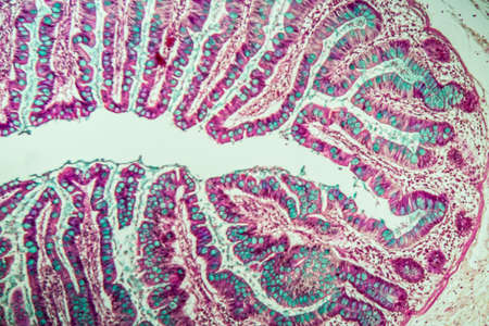
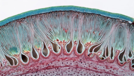
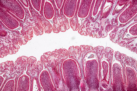
Small intestine with villi under the microscope 100x
Коллекция по умолчанию
Коллекция по умолчанию
Создать новую
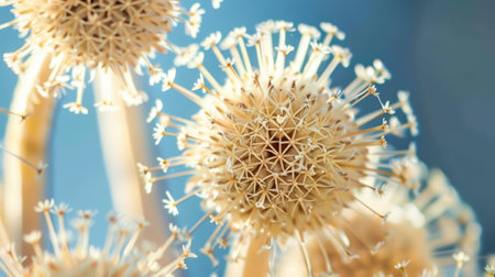
A close-up view of a dried flower head with white seeds. The seeds are arranged in a circular pattern around the center of the flower. The flower is surrounded by a blue background, creating a beautiful contrast between the delicate white seeds and the bright blue sky. The texture of the flower head and the intricate details of the seeds are visible.
Коллекция по умолчанию
Коллекция по умолчанию
Создать новую
A highly detailed 3D illustration showing various bacteria and microbes in different colors, providing a close-up microscopic view of microorganisms
Коллекция по умолчанию
Коллекция по умолчанию
Создать новую
Study of protozoa and plant cells under the microscope for education.
Коллекция по умолчанию
Коллекция по умолчанию
Создать новую
Microscope view of green plant cells on white background. AI-generated.
Коллекция по умолчанию
Коллекция по умолчанию
Создать новую

Ovarian cancer, light micrograph, photo under microscope. Photograph shows a fragment of a cancerous tumor in the female ovary. Selective focus
Коллекция по умолчанию
Коллекция по умолчанию
Создать новую
Larch with trunk 100x in cross section
Коллекция по умолчанию
Коллекция по умолчанию
Создать новую
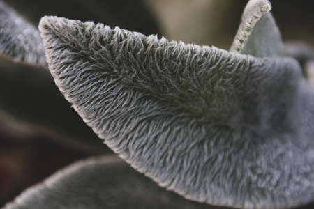
Photo leaves a plant with texture of hair on the plant
Коллекция по умолчанию
Коллекция по умолчанию
Создать новую
Tansy inflorescence in cross section 100x
Коллекция по умолчанию
Коллекция по умолчанию
Создать новую
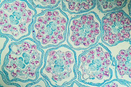
Tansy inflorescence in cross section 100x
Коллекция по умолчанию
Коллекция по умолчанию
Создать новую
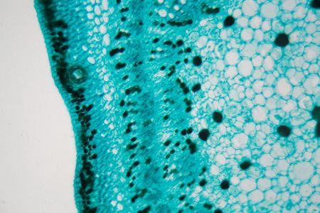
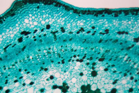
Cross section of a cotton leaf under the microscope.
Коллекция по умолчанию
Коллекция по умолчанию
Создать новую
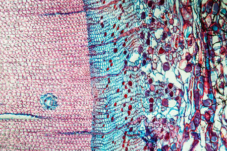
Cross-section Dicot, Monocot and Root of Plant Stem under the microscope for classroom education.
Коллекция по умолчанию
Коллекция по умолчанию
Создать новую
Microscope photo of a cross section of a cotton stem.
Коллекция по умолчанию
Коллекция по умолчанию
Создать новую
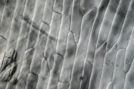
Onion skin with plant cells under the microscope
Коллекция по умолчанию
Коллекция по умолчанию
Создать новую
Protozoa and Green Algae in waste water under the microscope.
Коллекция по умолчанию
Коллекция по умолчанию
Создать новую
Pipe bush with trunk 100x across
Коллекция по умолчанию
Коллекция по умолчанию
Создать новую
Bacteria emit a brilliant glow under UV light in a dark setting, showing vibrant colors and intricate patterns.
Коллекция по умолчанию
Коллекция по умолчанию
Создать новую
Sunflower with stem across 100x
Коллекция по умолчанию
Коллекция по умолчанию
Создать новую
Cross-section through the lichen symbiote body 100x
Коллекция по умолчанию
Коллекция по умолчанию
Создать новую
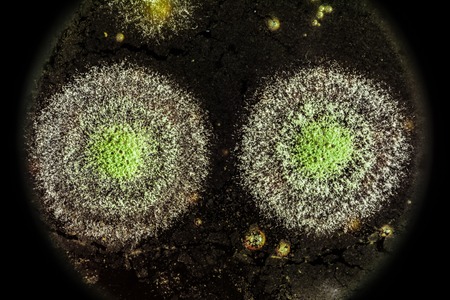
view of the microscope development of green mold on organic basis, macro abstract background
Коллекция по умолчанию
Коллекция по умолчанию
Создать новую
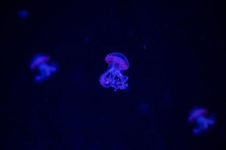
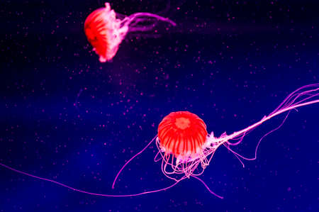
Beautiful jellyfish or medusa in the neon light in aquarium in new opened Prague medusarium in Czech Republic
Коллекция по умолчанию
Коллекция по умолчанию
Создать новую

thai style decoration made from flower
Коллекция по умолчанию
Коллекция по умолчанию
Создать новую
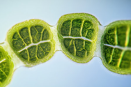
Microscopic View of Filamentous Algae (Oedogonium) with Potential Dwarf Male Filaments
Коллекция по умолчанию
Коллекция по умолчанию
Создать новую
sour and moldy food, top view close-up
Коллекция по умолчанию
Коллекция по умолчанию
Создать новую
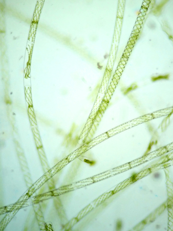
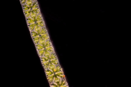
Filamentous algae are single algae cells that form long visible chains, threads, or filaments.
Коллекция по умолчанию
Коллекция по умолчанию
Создать новую

Cross-section through the lichen symbiote body 100x
Коллекция по умолчанию
Коллекция по умолчанию
Создать новую
Plankton with microscopic ciliates
Коллекция по умолчанию
Коллекция по умолчанию
Создать новую

Fungus on old coffee ground for the background
Коллекция по умолчанию
Коллекция по умолчанию
Создать новую
Volvox in drop of water under the microscope for classroom education.
Коллекция по умолчанию
Коллекция по умолчанию
Создать новую
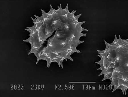
Scanning electron micrograph of one daisy pollen grain. Nottingham, UK
Коллекция по умолчанию
Коллекция по умолчанию
Создать новую

Lesser celandine with roots 100x across
Коллекция по умолчанию
Коллекция по умолчанию
Создать новую
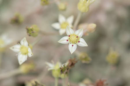
All kinds of small and lovely succulent plants
Коллекция по умолчанию
Коллекция по умолчанию
Создать новую
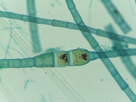
Green algae or spirigyra conjugation, sexual reproduction under light microscope
Коллекция по умолчанию
Коллекция по умолчанию
Создать новую
Apple fruit in cross section 100x
Коллекция по умолчанию
Коллекция по умолчанию
Создать новую
A closeup of the transparent jellyfish tentacles, each petal with tiny beads that glow in shades of orange and blue against an isolated black background.
Коллекция по умолчанию
Коллекция по умолчанию
Создать новую

Histological Uterus human, Uterine tube human, Placenta human and Umbilical cord Human under the microscope for education.
Коллекция по умолчанию
Коллекция по умолчанию
Создать новую
Beautiful jellyfish close up. Abstract background .
Коллекция по умолчанию
Коллекция по умолчанию
Создать новую
Microscope photo of a cross section of a cotton stem.
Коллекция по умолчанию
Коллекция по умолчанию
Создать новую
Euglena is a genus of single cell flagellate eukaryotes under microscopic view for study.
Коллекция по умолчанию
Коллекция по умолчанию
Создать новую

Host cells with spores (mold) are inside wood under the microscope for education.
Коллекция по умолчанию
Коллекция по умолчанию
Создать новую
a close-up portrait photograph showcasing the intricate details of unknown helicobacter pylori biological virus alien flowers. the image captures the brittle yet beautiful and dry elegance of the flowers, with rich and vivid contrast. shot on a 100mm lens at f/2.0, the natural lighting enhances the realistic and impressive qualities of the photo. the black tones and crispness add depth of field, resulting in an
Коллекция по умолчанию
Коллекция по умолчанию
Создать новую
Close-up view of colorful cellular structures, highlighting unique patterns and textures, illustrating the complexity of microscopic life and its natural beauty
Коллекция по умолчанию
Коллекция по умолчанию
Создать новую
Leech cross section showing internal anatomical structures stained
Коллекция по умолчанию
Коллекция по умолчанию
Создать новую
amazing inhabitants of the microworld under a microscope
Коллекция по умолчанию
Коллекция по умолчанию
Создать новую
Columnar epithelium of human gall bladder under the microscope in Lab.
Коллекция по умолчанию
Коллекция по умолчанию
Создать новую
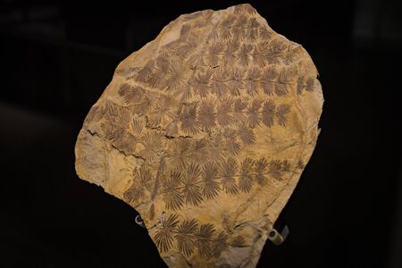
7 JUNE 2018, MILAN, ITALY: Expositions of prehistoric dinosaurs in the Museum of Natural History in Milan.
Коллекция по умолчанию
Коллекция по умолчанию
Создать новую
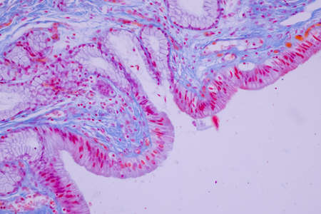
Pathology and Histology Tissue of Mammals under microscope.
Коллекция по умолчанию
Коллекция по умолчанию
Создать новую
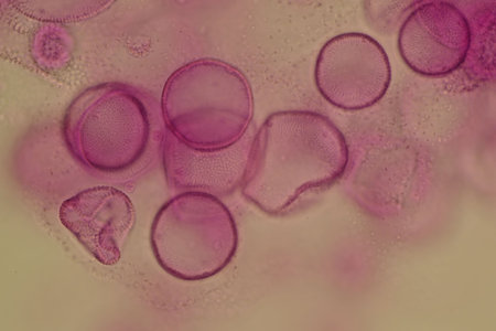
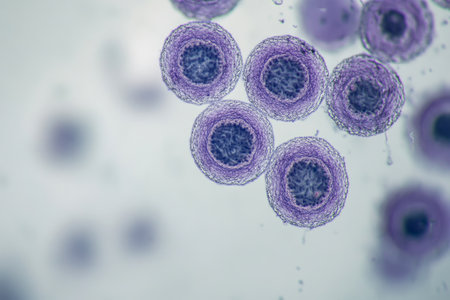
Close-up of pink circular cells under a microscope
Коллекция по умолчанию
Коллекция по умолчанию
Создать новую
Cross sections of plant root under microscope view
Коллекция по умолчанию
Коллекция по умолчанию
Создать новую

Paramecium caudatum is a genus of unicellular ciliated protozoan and Bacterium under the microscope
Коллекция по умолчанию
Коллекция по умолчанию
Создать новую
Microscopic view of leaf stoma showing gas exchange involved in photosynthesis, emphasizing plant biology and cellular structure.
Коллекция по умолчанию
Коллекция по умолчанию
Создать новую
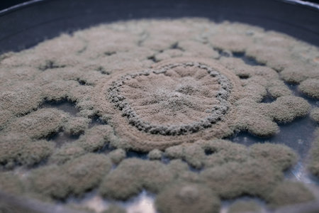
A close-up view of a circular pattern formed by mold or fungus on a surface, showcasing intricate textures and shapes
Коллекция по умолчанию
Коллекция по умолчанию
Создать новую
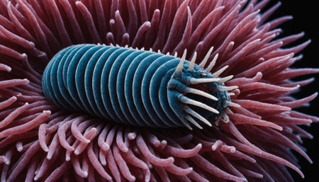
A blue segmented worm with white bristles is nestled inside a bed of pink tentacles
Коллекция по умолчанию
Коллекция по умолчанию
Создать новую
Legion-Media
Создайте свои проекты на основе качественных стоковых фотографий и видео.


Copyright © Legion-Media.