
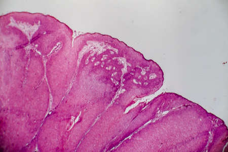
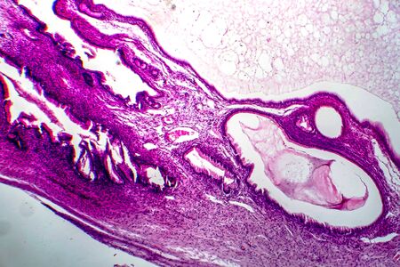
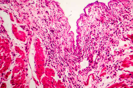
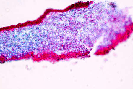
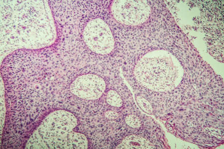
Condyloma acuminatum, also known as genital warts. Light micrograph, photo under microscope
Коллекция по умолчанию
Коллекция по умолчанию
Создать новую
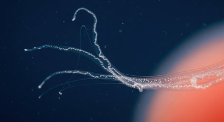
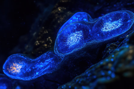
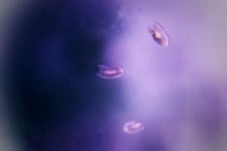
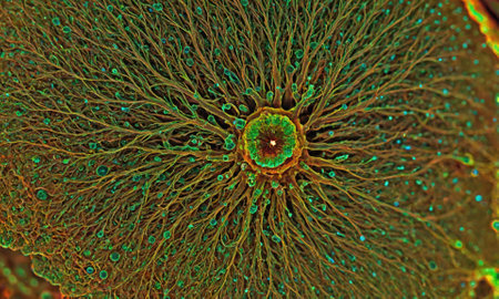
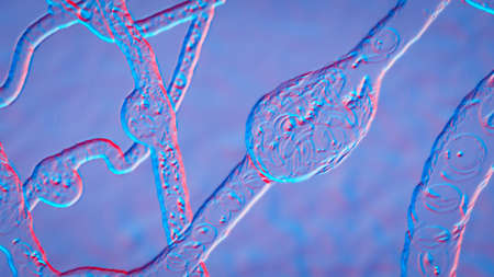
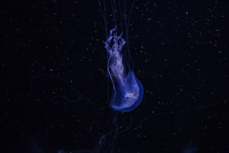
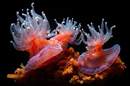
A mesmerizing view of a marine organism with luminous, branching appendages drifting in the abyss.
Коллекция по умолчанию
Коллекция по умолчанию
Создать новую
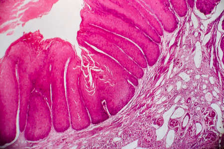
Condyloma acuminatum, also known as genital warts. Light micrograph, photo under microscope
Коллекция по умолчанию
Коллекция по умолчанию
Создать новую
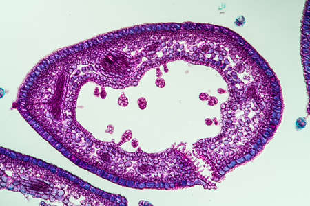
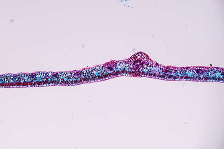
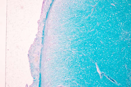
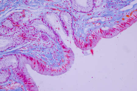
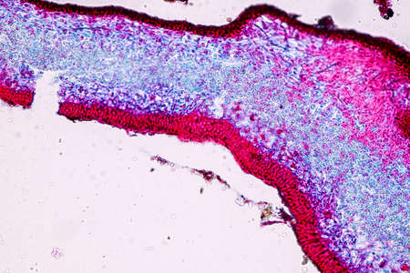
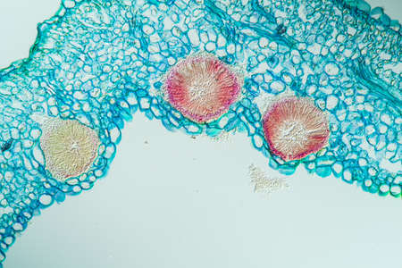
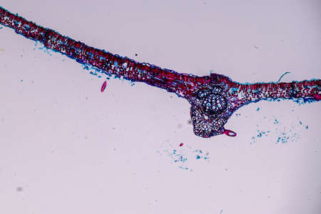
Heather leaf cross section under the microscope, 200x
Коллекция по умолчанию
Коллекция по умолчанию
Создать новую
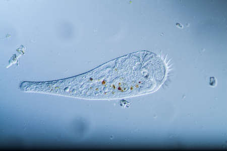
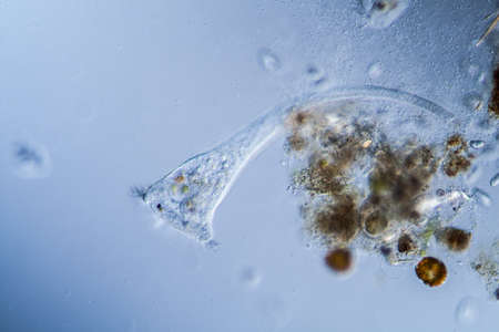
Trumpet animal as a microscopic plankton animal in drops of water
Коллекция по умолчанию
Коллекция по умолчанию
Создать новую
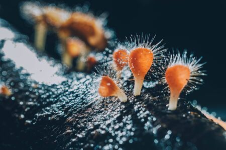
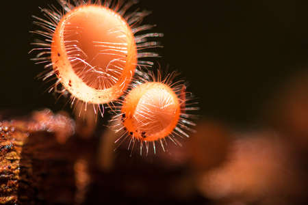
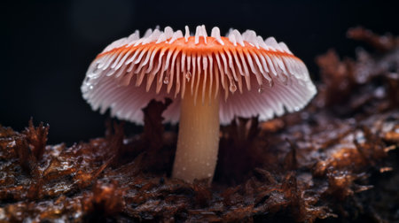
Orange mushroom ,Champagne mushroom or eyelash cup mushroom with sparkling droplets in the forest. Ecosystem or biological diversity concept.
Коллекция по умолчанию
Коллекция по умолчанию
Создать новую
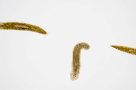
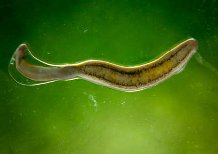
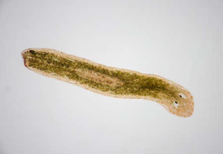
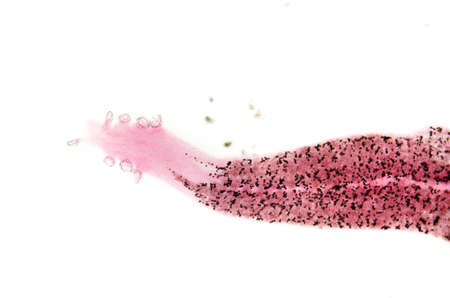
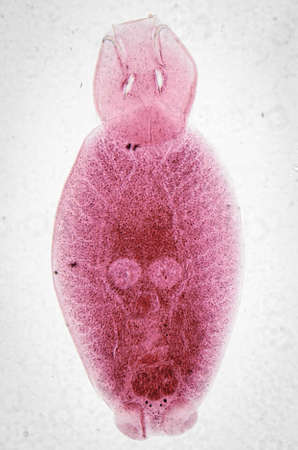
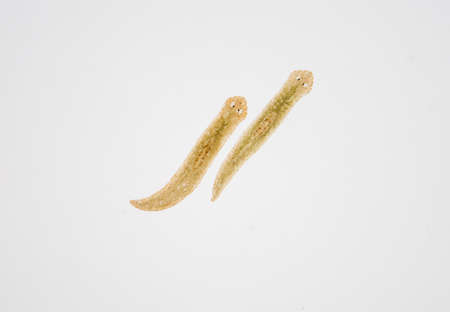
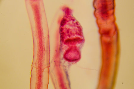
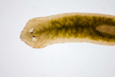
Planarian parasite (flatworm) under microscope view.
Коллекция по умолчанию
Коллекция по умолчанию
Создать новую
Johannes berry fruit cross 100x
Коллекция по умолчанию
Коллекция по умолчанию
Создать новую
Under ultraviolet light, bacteria emit a vibrant glow, revealing their bioluminescent properties in a dark, captivating setting.
Коллекция по умолчанию
Коллекция по умолчанию
Создать новую

Planarian parasite (flatworm) under microscope view.
Коллекция по умолчанию
Коллекция по умолчанию
Создать новую
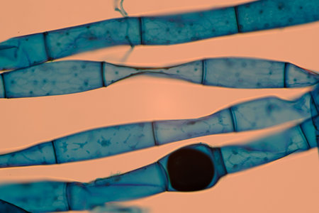
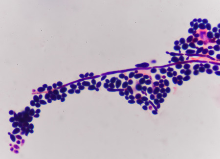
Microscopic view of blue-stained fungal hyphae with spores.
Коллекция по умолчанию
Коллекция по умолчанию
Создать новую

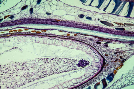
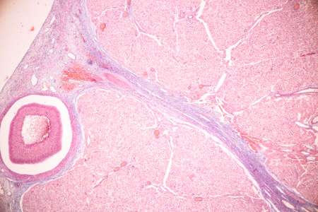
Histological Uterus human, Uterine tube human, Placenta human and Umbilical cord Human under the microscope for education.
Коллекция по умолчанию
Коллекция по умолчанию
Создать новую
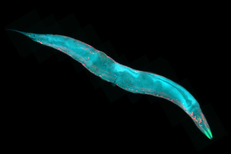
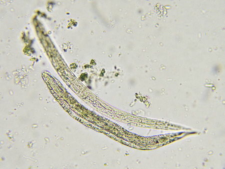
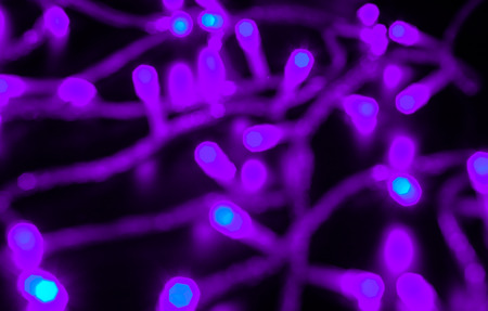
Caenorhabditis elegans, a free-living transparent nematode roundworm, about 1 mm in length. Fluorescence micrograph.
Коллекция по умолчанию
Коллекция по умолчанию
Создать новую

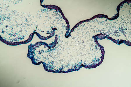
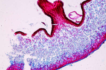
Cross-section through the lichen symbiote body 100x
Коллекция по умолчанию
Коллекция по умолчанию
Создать новую
Pathology and Histology Tissue of Mammals under microscope.
Коллекция по умолчанию
Коллекция по умолчанию
Создать новую
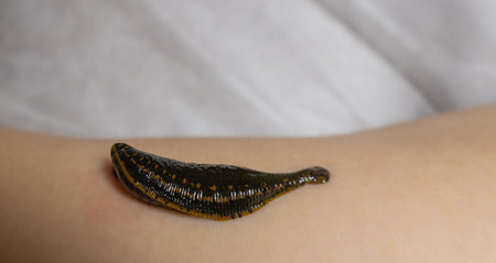
An extreme macro of an Asian leech
Коллекция по умолчанию
Коллекция по умолчанию
Создать новую

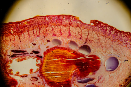
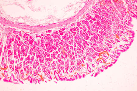
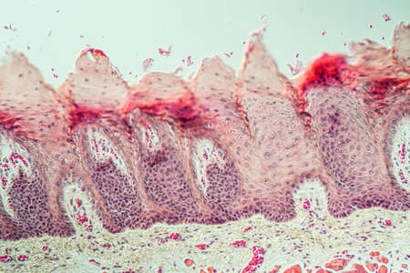
Stomach tissue under the microscope 100x
Коллекция по умолчанию
Коллекция по умолчанию
Создать новую

The study of Acanthocephala is a phylum of parasitic worms known as acanthocephalans, thorny-headed worms or spiny-headed worms in laboratory.
Коллекция по умолчанию
Коллекция по умолчанию
Создать новую
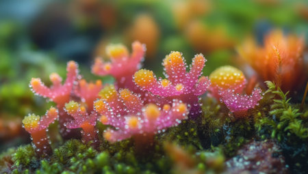
Close-up of coral-like flowers growing on the ground, with numerous corals in various shapes and colors. The petals have small dots that resemble dewdrops. This is an award-winning photo taken with a Sony A7R IV camera. It was shot from low angles using macro lenses, creating a dreamy atmosphere. In the background, there were various mosses, as well as other unique plants. --ar 53:30 --v 6.1 Job ID: 516d3a1a-9c37-4dc3-921f-11d8f4dc1826
Коллекция по умолчанию
Коллекция по умолчанию
Создать новую
A snake star with its six tentacles on the disk of a saltwater aquarium.
Коллекция по умолчанию
Коллекция по умолчанию
Создать новую

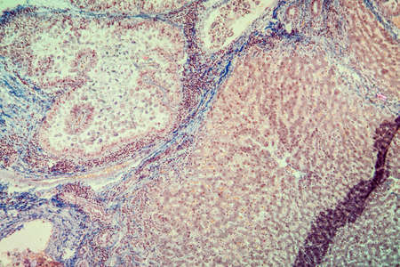
Ovarian cancer, light micrograph, photo under microscope. Photograph shows a fragment of a cancerous tumor in the female ovary. Selective focus
Коллекция по умолчанию
Коллекция по умолчанию
Создать новую

A large number of jellyfish are seen gracefully swimming together in the ocean, A close-up perspective of microscopic life forms like plankton, during a feeding frenzy, AI Generated
Коллекция по умолчанию
Коллекция по умолчанию
Создать новую
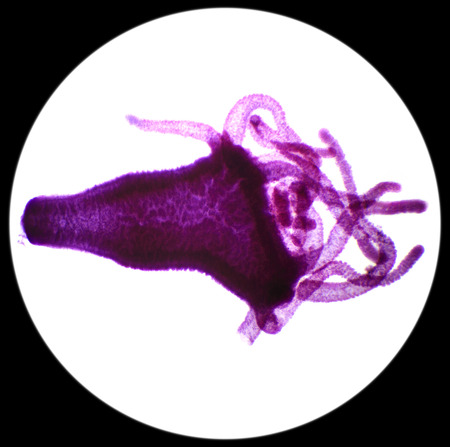
Hydra under the microscope (Hydra W.M.), 40x
Коллекция по умолчанию
Коллекция по умолчанию
Создать новую
budding yeast cell structure fine with microscope in laboratory.
Коллекция по умолчанию
Коллекция по умолчанию
Создать новую
Stomach tissue under the microscope 100x
Коллекция по умолчанию
Коллекция по умолчанию
Создать новую
Leech cross section showing internal anatomical structures stained
Коллекция по умолчанию
Коллекция по умолчанию
Создать новую
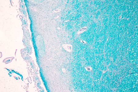
Cerebellum, Thalamus, Medulla oblongata, Spinal cord and Motor Neuron human under the microscope in Lab.
Коллекция по умолчанию
Коллекция по умолчанию
Создать новую
Planarian parasite (flatworm) under microscope view.
Коллекция по умолчанию
Коллекция по умолчанию
Создать новую
The Fungi Cup thrives on a dead log or tree. In the phylum Ascomycota (phylum Ascomycota) is a mushroom with a cone-shaped cap. Have pink, orange or red
Коллекция по умолчанию
Коллекция по умолчанию
Создать новую
Rare image of Ghost flatworm - Maricola (Planarian) triclad flatworms in reef aquarium glass
Коллекция по умолчанию
Коллекция по умолчанию
Создать новую
Trumpet animal as a microscopic plankton animal in drops of water
Коллекция по умолчанию
Коллекция по умолчанию
Создать новую
Tissue of Stomach Human under the microscope in Lab.
Коллекция по умолчанию
Коллекция по умолчанию
Создать новую
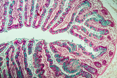
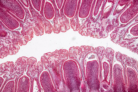
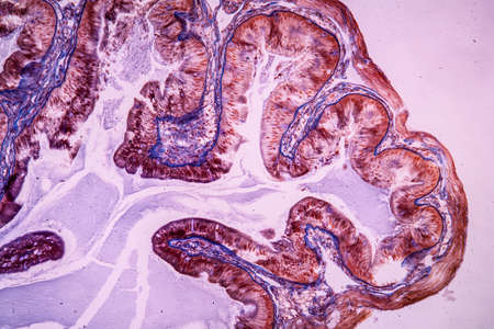
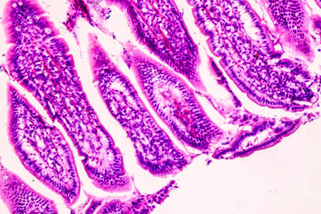
Small intestine with villi under the microscope 100x
Коллекция по умолчанию
Коллекция по умолчанию
Создать новую

Host cells with spores (mold) are inside wood under the microscope for education.
Коллекция по умолчанию
Коллекция по умолчанию
Создать новую

A deep stunning-sea siphonophore drifting gracefully in the ocean depths
Коллекция по умолчанию
Коллекция по умолчанию
Создать новую
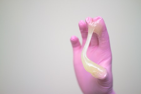
depilation and beauty concept - sugar paste or wax honey for hair removing with pink gloves hands of cosmetologist in spa salon
Коллекция по умолчанию
Коллекция по умолчанию
Создать новую

Cell- science background. Esophagus of the dog- cross section
Коллекция по умолчанию
Коллекция по умолчанию
Создать новую

Colon polyp, one of the largest polyps
Коллекция по умолчанию
Коллекция по умолчанию
Создать новую
science aquaculture fish parasite hook clip worm micrograph
Коллекция по умолчанию
Коллекция по умолчанию
Создать новую
science aquaculture fish parasite Benedenia seriolae worm micrograph
Коллекция по умолчанию
Коллекция по умолчанию
Создать новую
Host cells with spores (mold) are inside wood under the microscope for education.
Коллекция по умолчанию
Коллекция по умолчанию
Создать новую
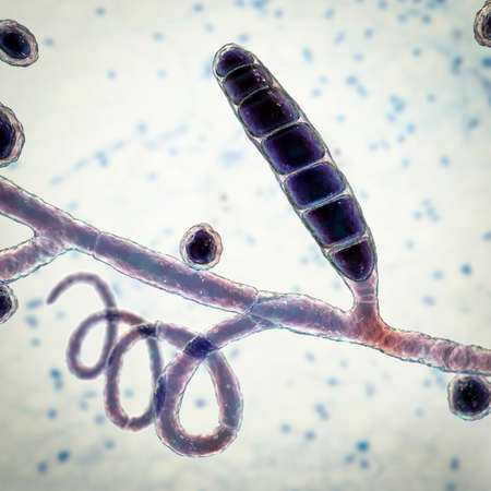
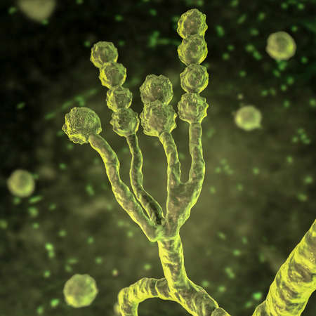
Fungi Trichophyton mentagrophytes, 3D illustration showing macroconidium, septate and spiral hyphae. Causes skin infection (ringworm, tinea capitis, tinea corporis and other), hair and nail infections
Коллекция по умолчанию
Коллекция по умолчанию
Создать новую

Cross-section leaf Plant of under the microscope for classroom education.
Коллекция по умолчанию
Коллекция по умолчанию
Создать новую
Tongue Tissue with taste buds across 200x
Коллекция по умолчанию
Коллекция по умолчанию
Создать новую

Feeler snail's eye tissue under the microscope 200x
Коллекция по умолчанию
Коллекция по умолчанию
Создать новую

Host cells with spores (mold) are inside wood under the microscope for education.
Коллекция по умолчанию
Коллекция по умолчанию
Создать новую

medical microscopy animal parasiteras schistosome blood flukes
Коллекция по умолчанию
Коллекция по умолчанию
Создать новую
Cerebellum, Thalamus, Medulla oblongata, Spinal cord and Motor Neuron human under the microscope in Lab.
Коллекция по умолчанию
Коллекция по умолчанию
Создать новую
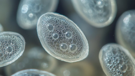
A close up of many small, round, clear objects with a lot of holes in them. The objects are all different sizes and are scattered throughout the image. Scene is one of curiosity and wonder
Коллекция по умолчанию
Коллекция по умолчанию
Создать новую
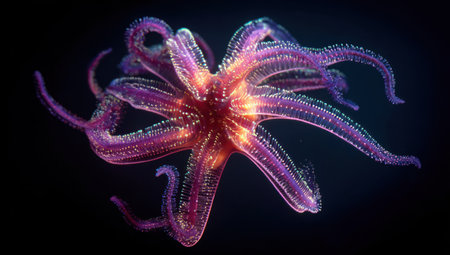
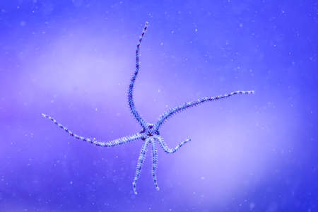
A close-up image showcases a star-shaped marine invertebrate, radiating with shades of purple and red. The translucent body reveals internal structures, highlighted by overhead lighting against a dark backdrop. Suitable for educational materials, scientific publications, and various commercial projects, the image captures the beauty of underwater life.
Коллекция по умолчанию
Коллекция по умолчанию
Создать новую
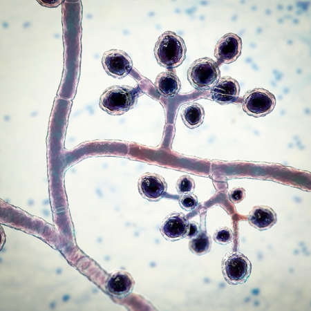
Fungi Trichophyton mentagrophytes, 3D illustration showing branched conidiophores bearing spherical microconidia. Causes skin infection (ringworm), hair and nail infections
Коллекция по умолчанию
Коллекция по умолчанию
Создать новую

Fusobacterium, 3D illustration. An oral bacterium, causes periodontal diseases, periodontal plague formation, sore throat, Lemmieres syndrome. It is also associated with preterm birth and colon cancer
Коллекция по умолчанию
Коллекция по умолчанию
Создать новую
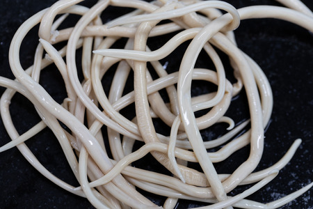
Ascariasis is a disease caused by the parasitic roundworm Ascaris lumbricoides for education in laboratories.
Коллекция по умолчанию
Коллекция по умолчанию
Создать новую
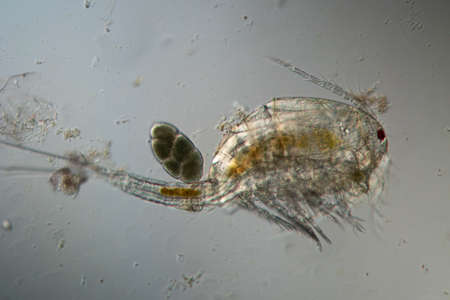
Copepod crab swims through the water
Коллекция по умолчанию
Коллекция по умолчанию
Создать новую

Cross section of human skin under microscope view for education in laboratory.
Коллекция по умолчанию
Коллекция по умолчанию
Создать новую
Coccidiosis of liver tissue under the microscope 100x
Коллекция по умолчанию
Коллекция по умолчанию
Создать новую
Planarian parasite (flatworm) under microscope view.
Коллекция по умолчанию
Коллекция по умолчанию
Создать новую
Ovarian mucinous cystadenoma, a benign tumor of ovary, light micrograph, photo under microscope
Коллекция по умолчанию
Коллекция по умолчанию
Создать новую
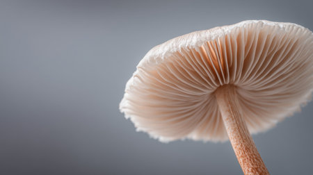
This striking close-up image showcases the intricate details of a mushroom cap, highlighting its delicate gills and soft texture against a muted gray backdrop.
Коллекция по умолчанию
Коллекция по умолчанию
Создать новую
Eudendrium female colony with developing gonophores observed
Коллекция по умолчанию
Коллекция по умолчанию
Создать новую
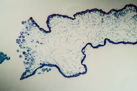
Cross-section through the lichen symbiote body 100x
Коллекция по умолчанию
Коллекция по умолчанию
Создать новую
Signet ring cell carcinoma of the stomach, light micrograph, photo under microscope
Коллекция по умолчанию
Коллекция по умолчанию
Создать новую
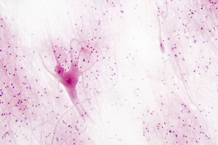
A microscopic view of a neuron with its intricate network of dendrites
Коллекция по умолчанию
Коллекция по умолчанию
Создать новую
Stunning portrayal capturing the allure of fluorite crystal
Коллекция по умолчанию
Коллекция по умолчанию
Создать новую
Columnar epithelium of human gall bladder under the microscope in Lab.
Коллекция по умолчанию
Коллекция по умолчанию
Создать новую
Motor nerves in spinal tissue.
Коллекция по умолчанию
Коллекция по умолчанию
Создать новую
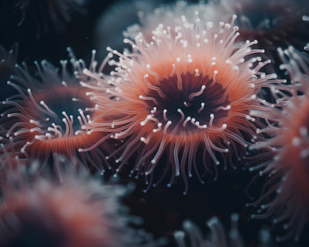
Clown anemone on a dark background. 3d illustration
Коллекция по умолчанию
Коллекция по умолчанию
Создать новую
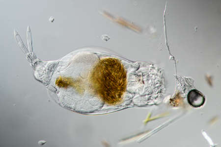
Rotifer foraging in the stream 200x
Коллекция по умолчанию
Коллекция по умолчанию
Создать новую
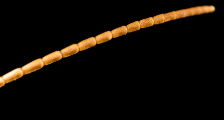
Antenna on the head of a grasshopper. macro
Коллекция по умолчанию
Коллекция по умолчанию
Создать новую

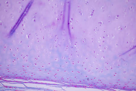
Anatomy and Histological Bone, Elastic cartilage human and Joint of human foetus under the microscope for education.
Коллекция по умолчанию
Коллекция по умолчанию
Создать новую
Characteristics of Lichen, hyphae and Symbiotic algae under the microscope for education.
Коллекция по умолчанию
Коллекция по умолчанию
Создать новую
Microaneurysms, microscopic buldges in the artery walls filled with blood, 3D illustration. Found in the eye retina in diabetic retinopathy, and also in brain (Charcot-Bouchard aneurysms)
Коллекция по умолчанию
Коллекция по умолчанию
Создать новую
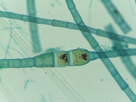
Microscopic View of Filamentous Algae (Oedogonium) with Potential Dwarf Male Filaments
Коллекция по умолчанию
Коллекция по умолчанию
Создать новую
Characteristics of Lichen, hyphae and Symbiotic algae under the microscope for education.
Коллекция по умолчанию
Коллекция по умолчанию
Создать новую
Anatomy and Histological Ovary, Testis and Sperm human cells under microscope.
Коллекция по умолчанию
Коллекция по умолчанию
Создать новую
Characteristics of Lichen, hyphae and Symbiotic algae under the microscope for education.
Коллекция по умолчанию
Коллекция по умолчанию
Создать новую

a white, green hydra in the aquarium
Коллекция по умолчанию
Коллекция по умолчанию
Создать новую
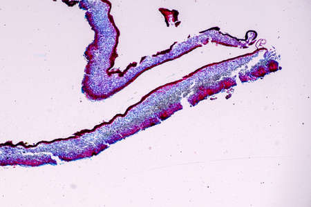
Cross-section through the lichen symbiote body 100x
Коллекция по умолчанию
Коллекция по умолчанию
Создать новую
Characteristics of Lichen, hyphae and Symbiotic algae under the microscope for education.
Коллекция по умолчанию
Коллекция по умолчанию
Создать новую
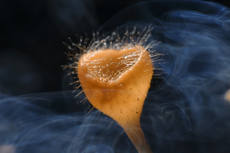
Fungi cup red Mushroom Champagne Cup with smoke in the morning
Коллекция по умолчанию
Коллекция по умолчанию
Создать новую
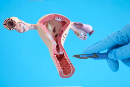
Female reproductive system anatomy uterus and scalpel closeup
Коллекция по умолчанию
Коллекция по умолчанию
Создать новую
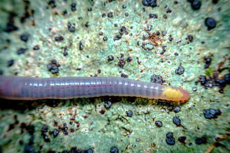
Closeup macro pink earthworm crawling through green moss on forest floor
Коллекция по умолчанию
Коллекция по умолчанию
Создать новую
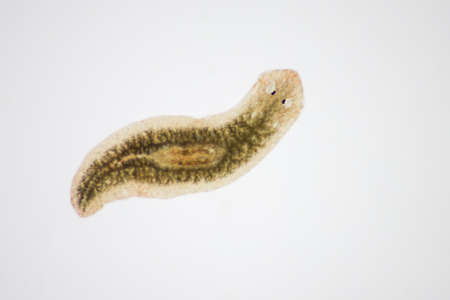
Planarian parasite (flatworm) under microscope view.
Коллекция по умолчанию
Коллекция по умолчанию
Создать новую
Planarian parasite (flatworm) under microscope view.
Коллекция по умолчанию
Коллекция по умолчанию
Создать новую
Rust fungus on milkweed plant, 200x
Коллекция по умолчанию
Коллекция по умолчанию
Создать новую

Vibrio cholerae bacteria, 3D illustration. Bacterium which causes cholera disease and is transmitted by contaminated water
Коллекция по умолчанию
Коллекция по умолчанию
Создать новую
Bowen's Disease Tumor under the microscope 100x
Коллекция по умолчанию
Коллекция по умолчанию
Создать новую

Bowen's Disease Tumor under the microscope 100x
Коллекция по умолчанию
Коллекция по умолчанию
Создать новую
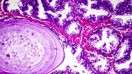
Histopathology of prostate gland hyperplasia, light micrograph, photo under microscope
Коллекция по умолчанию
Коллекция по умолчанию
Создать новую
Microscopic fungi Scopulariopsis brevicaulis, 3D illustration. Fungus that infects nails, causes subcutaneous and invasive infections, endocarditis, sinusitis, disseminated infection
Коллекция по умолчанию
Коллекция по умолчанию
Создать новую
Strongyloides stercoralis in stool
Коллекция по умолчанию
Коллекция по умолчанию
Создать новую
Host cells with spores (mold) are inside wood under the microscope for education.
Коллекция по умолчанию
Коллекция по умолчанию
Создать новую
Cliated epithelium of human under the microscope in Lab.
Коллекция по умолчанию
Коллекция по умолчанию
Создать новую
jellyfish underwater, dark background, blue color light, wildlife sea animal, beautiful scene of swimming.
Коллекция по умолчанию
Коллекция по умолчанию
Создать новую
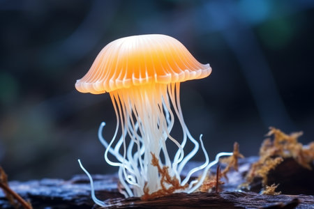
Translucent mushroom resembling a jellyfish, diverse mushroom species in natural habitat
Коллекция по умолчанию
Коллекция по умолчанию
Создать новую
Education anatomy and Histological sample of Human under the microscope.
Коллекция по умолчанию
Коллекция по умолчанию
Создать новую
Traditional alternative medicine - doctor doing procedure of hirudotherapy for young woman.
Коллекция по умолчанию
Коллекция по умолчанию
Создать новую
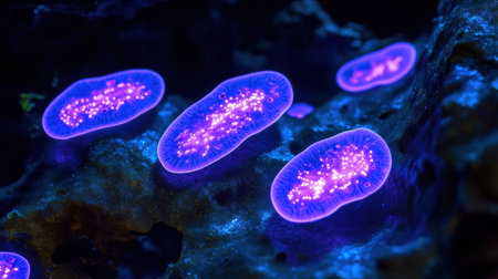
Bacteria emit a brilliant glow under UV light in a dark setting, showing vibrant colors and intricate patterns.
Коллекция по умолчанию
Коллекция по умолчанию
Создать новую
The Enchanting Encounter: Opalescent Nudibranchs Explore the Red Invasive Bryozoan at Monterey Bay National Marine Sanctuary
Коллекция по умолчанию
Коллекция по умолчанию
Создать новую
abstract small Bokeh lights. Beautiful background pattern
Коллекция по умолчанию
Коллекция по умолчанию
Создать новую
Legion-Media
Создайте свои проекты на основе качественных стоковых фотографий и видео.


Copyright © Legion-Media.