
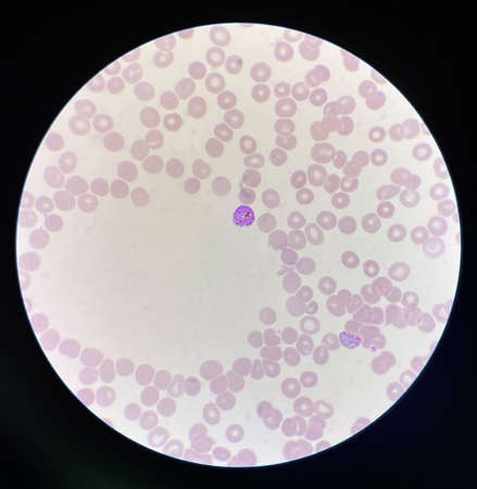
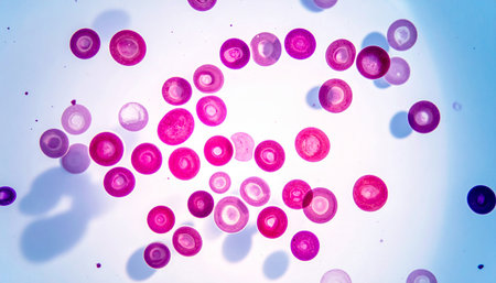
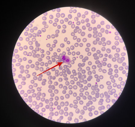
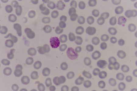
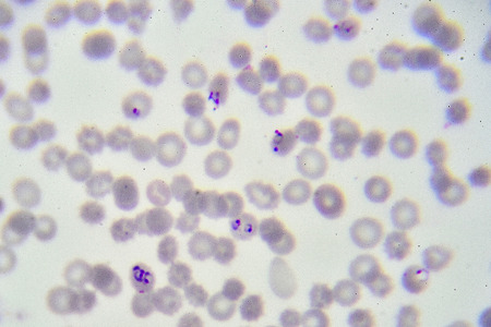
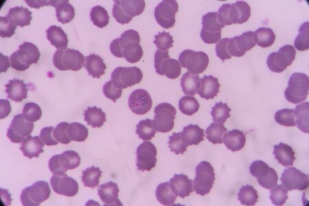
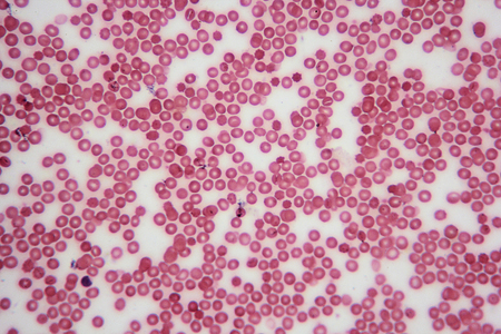
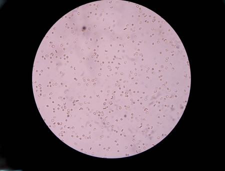
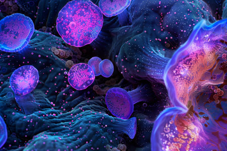
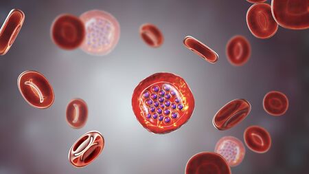
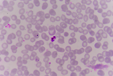
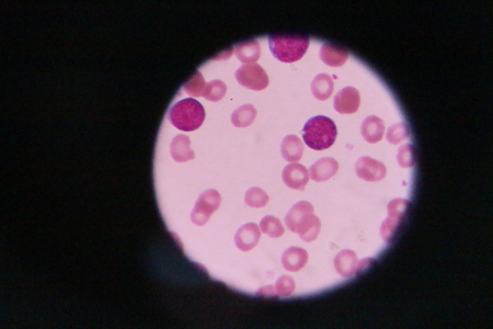
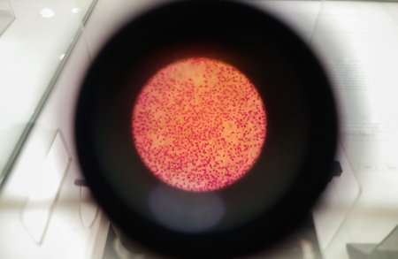
Malaria blood parasite infected red blood cells laboratory background.
Коллекция по умолчанию
Коллекция по умолчанию
Создать новую
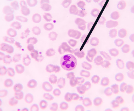
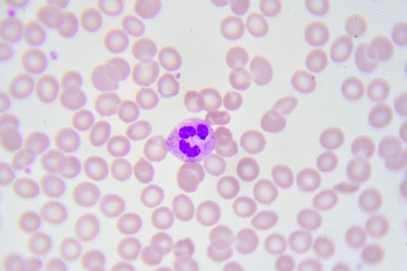
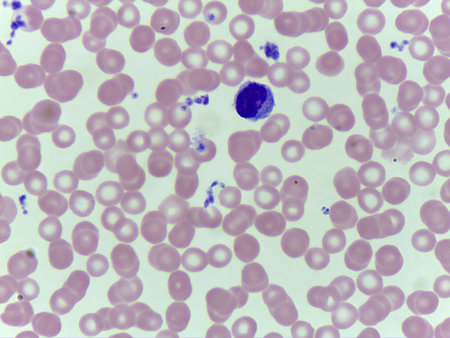
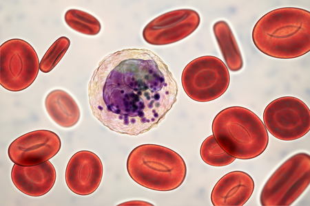
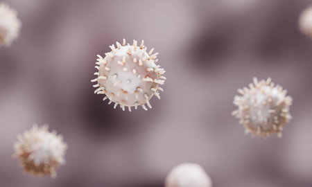
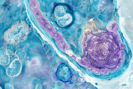
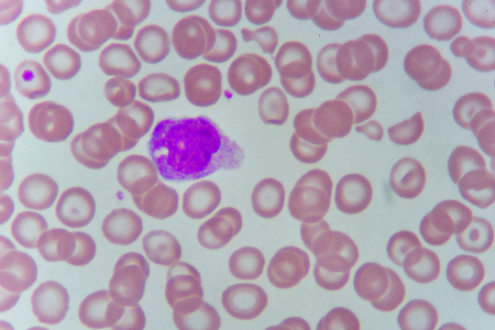
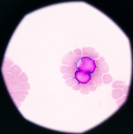
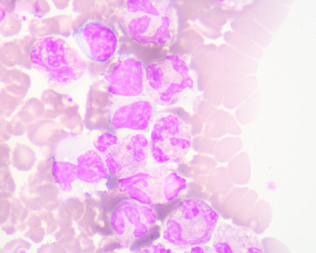
Neutrophil show in blood smear CBC test find with microscope.
Коллекция по умолчанию
Коллекция по умолчанию
Создать новую
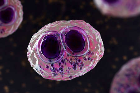
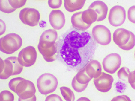
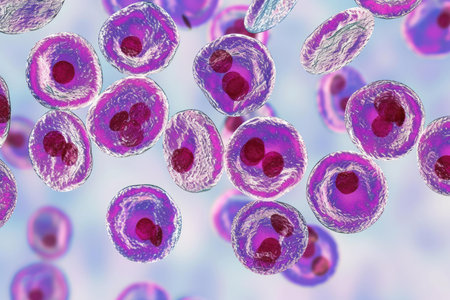
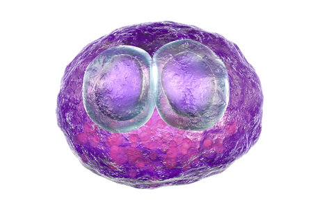
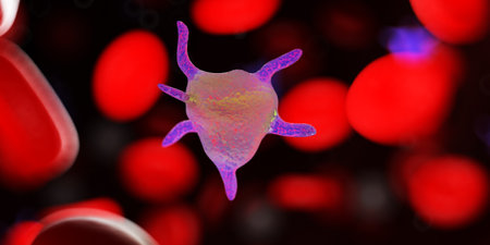
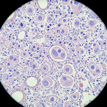
Cytomegalovirus CMV in a human cell, owl's eye inclusion in nucleus, multinucleated cell, 3D illustration. It is herpes virus, causes diseases in fetus, organ transplant patients, HIV infected people
Коллекция по умолчанию
Коллекция по умолчанию
Создать новую

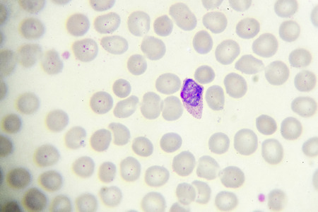
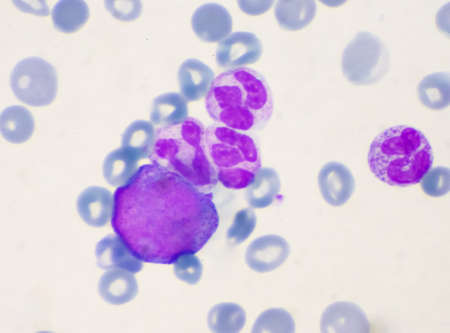
neutrophils. blood smear is often used as a follow-up test to abnormal results on a complete blood count (CBC) to evaluate the different types of blood cells.
Коллекция по умолчанию
Коллекция по умолчанию
Создать новую
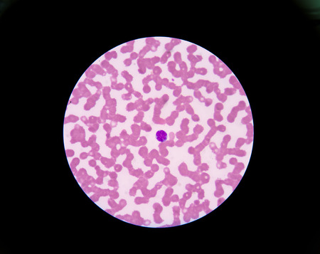
blood films for Malaria parasite
Коллекция по умолчанию
Коллекция по умолчанию
Создать новую
Neutrophil cell
Коллекция по умолчанию
Коллекция по умолчанию
Создать новую

Neutrophil cell (white blood cell) in peripheral blood smear
Коллекция по умолчанию
Коллекция по умолчанию
Создать новую
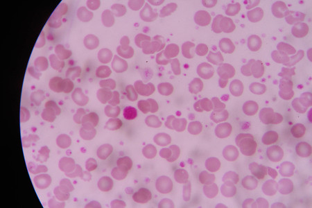
Blood smear with red blood cells in human body, medical background.
Коллекция по умолчанию
Коллекция по умолчанию
Создать новую
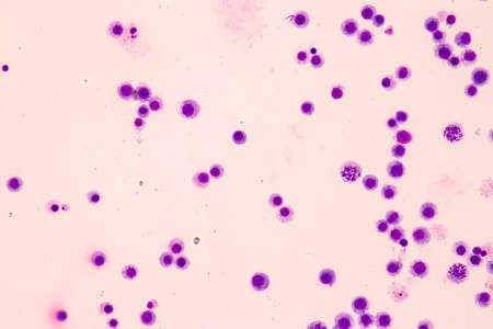
Immature white blood cells in leukemia.Science concept.
Коллекция по умолчанию
Коллекция по умолчанию
Создать новую
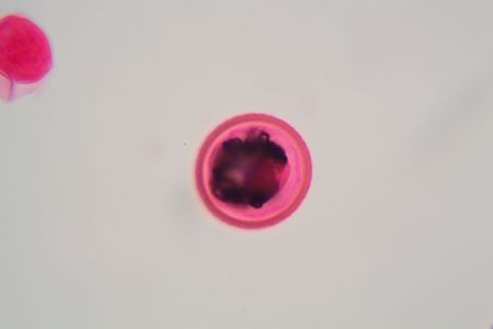
nucleated red cell
Коллекция по умолчанию
Коллекция по умолчанию
Создать новую
Blood cells in human body under microscope view for education in laboratory.
Коллекция по умолчанию
Коллекция по умолчанию
Создать новую

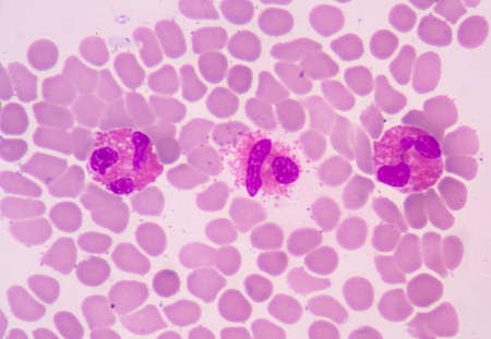
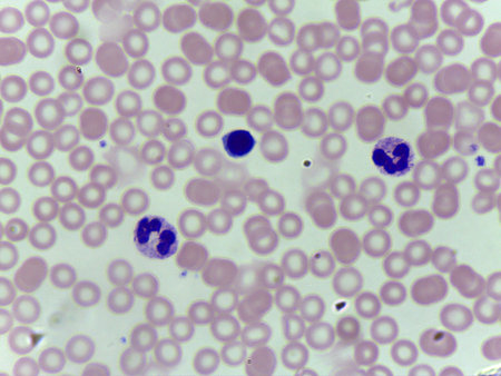
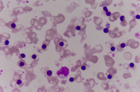
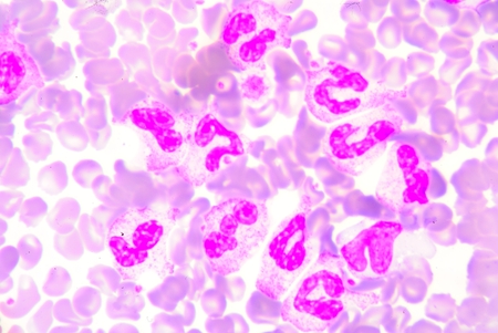
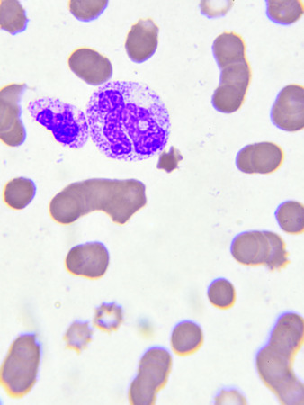
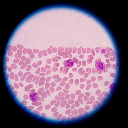
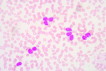
Blood smear showing, in the center, three neutrophil with hypersegmented nucleus. These cells appear in pathological situations such as megaloblastic anemias. Wright stain.
Коллекция по умолчанию
Коллекция по умолчанию
Создать новую
Blood smear showing white and red blood cells
Коллекция по умолчанию
Коллекция по умолчанию
Создать новую
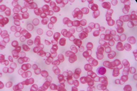
Blood smear with white blood cells and red blood cells. Medical background.
Коллекция по умолчанию
Коллекция по умолчанию
Создать новую
Malaria parasite in blood smear, gemetocyte stage
Коллекция по умолчанию
Коллекция по умолчанию
Создать новую
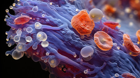
a close up of a colorful structure
Коллекция по умолчанию
Коллекция по умолчанию
Создать новую
Red arrow showing neutrophil with toxic granule active PMN.
Коллекция по умолчанию
Коллекция по умолчанию
Создать новую
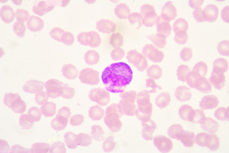
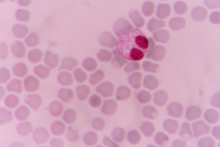
blood smear is often used as a follow-up test to abnormal results on a complete blood count (CBC) to evaluate the different types of blood cells.Atypical lymphocyte.
Коллекция по умолчанию
Коллекция по умолчанию
Создать новую
White blood cells of a human, Eosinophil photomicrograph panorama as seen under the microscope
Коллекция по умолчанию
Коллекция по умолчанию
Создать новую
blood cells with microscope.
Коллекция по умолчанию
Коллекция по умолчанию
Создать новую
complete blood count
Коллекция по умолчанию
Коллекция по умолчанию
Создать новую
complete blood count
Коллекция по умолчанию
Коллекция по умолчанию
Создать новую
Chromosomes Human under the microscope for education.
Коллекция по умолчанию
Коллекция по умолчанию
Создать новую

Picture of acute lymphocytic leukemia or ALL cells in blood smear, analyze by microscope, 400x
Коллекция по умолчанию
Коллекция по умолчанию
Создать новую

Photomicrograph of canine eosinphil
Коллекция по умолчанию
Коллекция по умолчанию
Создать новую
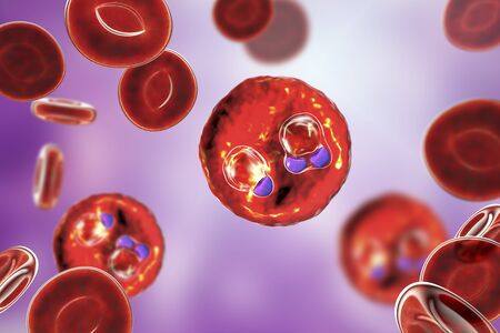
The malaria-infected red blood cells. 3D illustration showing ring-form trophozoites of malaria parasite Plasmodium falciparum inside red blood cells, the causative agent of tropical malaria
Коллекция по умолчанию
Коллекция по умолчанию
Создать новую

Comparison white blood cell Eosinophil and Neutrophil laboratory science concept.
Коллекция по умолчанию
Коллекция по умолчанию
Создать новую
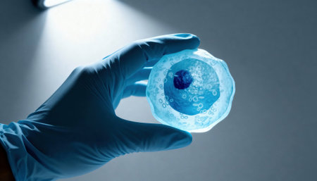
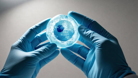
Gloved hand holds a translucent cell model under focused laboratory light with visible nucleus and organelle structures, suggesting scientific research and educational demonstration, with empty background space available for text
Коллекция по умолчанию
Коллекция по умолчанию
Создать новую
Malaria blood parasite infected red blood cells laboratory background.
Коллекция по умолчанию
Коллекция по умолчанию
Создать новую
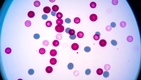
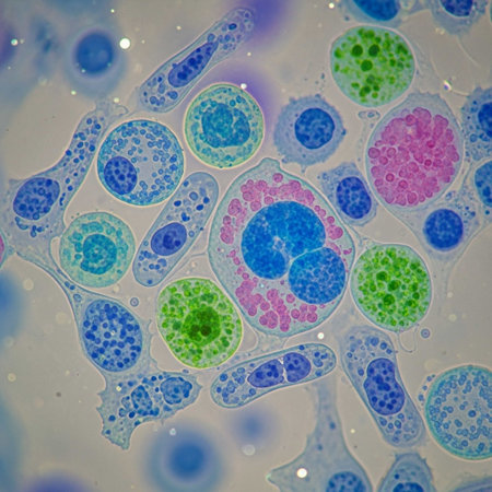
Microscopic close-up of vibrant stained human cells on a blue backdrop
Коллекция по умолчанию
Коллекция по умолчанию
Создать новую
Red blood cells infected with malaria parasite
Коллекция по умолчанию
Коллекция по умолчанию
Создать новую
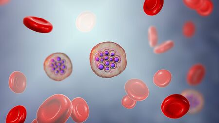
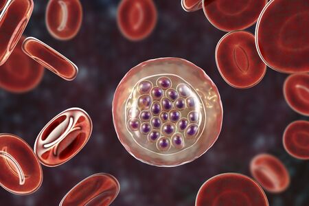
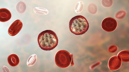
Red blood cells infected with malaria parasite Plasmodium vivax, schizont stage, 3D illustration
Коллекция по умолчанию
Коллекция по умолчанию
Создать новую
nucleated red cell
Коллекция по умолчанию
Коллекция по умолчанию
Создать новую

plasmodium
Коллекция по умолчанию
Коллекция по умолчанию
Создать новую
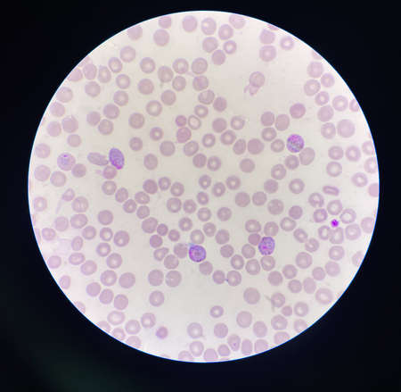
Microscopic View of a Peripheral Blood Smear Showing Red Blood Cells and White blood cell s
Коллекция по умолчанию
Коллекция по умолчанию
Создать новую
Abstract pink cancer cell organism background 3d render digital illustration
Коллекция по умолчанию
Коллекция по умолчанию
Создать новую
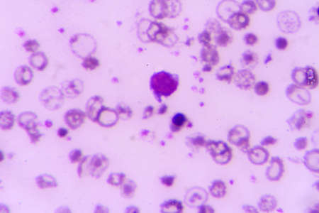
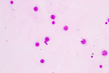
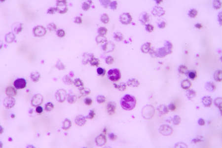
Blast cells in blood smear specimen Leukemia petient.
Коллекция по умолчанию
Коллекция по умолчанию
Создать новую
Leukocytes. Monocyte. White blood cell. Vector medical illustration
Коллекция по умолчанию
Коллекция по умолчанию
Создать новую
Schistosoma mansoni under the microscope. Schistosoma mansoni is human parasite and causes schistosomiasis.
Коллекция по умолчанию
Коллекция по умолчанию
Создать новую
Basophil. Type of white blood cell. Medical education.
Коллекция по умолчанию
Коллекция по умолчанию
Создать новую
The malaria-infected red blood cells. 3D illustration showing malaria parasite Plasmodium falciparum in schizont stage inside red blood cells, the causative agent of tropical malaria
Коллекция по умолчанию
Коллекция по умолчанию
Создать новую
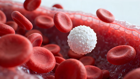
Ultra-detailed 3D render of a white blood cell floating among red blood cells in bloodstream, medical and scientific concept.
Коллекция по умолчанию
Коллекция по умолчанию
Создать новую
Abnormal red blood cells in Blood smear Thalassemia patient.
Коллекция по умолчанию
Коллекция по умолчанию
Создать новую
plasmodium
Коллекция по умолчанию
Коллекция по умолчанию
Создать новую
Red blood cells infected with malaria parasite, 3D illustration showing Plasmodium parasites inside red blood cells in the stage of schizont
Коллекция по умолчанию
Коллекция по умолчанию
Создать новую
Gloved hands hold a translucent cell culture sample with visible cellular structures and bubbles under focused clinical light, indicating laboratory research and microscopy, with available space for text on a neutral background
Коллекция по умолчанию
Коллекция по умолчанию
Создать новую
Promyelocye
Коллекция по умолчанию
Коллекция по умолчанию
Создать новую
Virus cells, 3D illustration. Viruses and bacteria in human body. Viruses in infected organism.
Коллекция по умолчанию
Коллекция по умолчанию
Создать новую
Neutrophils are a type of phagocyte and are normally found in the bloodstream.
Коллекция по умолчанию
Коллекция по умолчанию
Создать новую
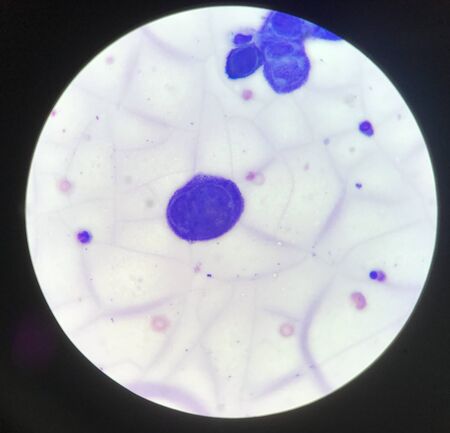
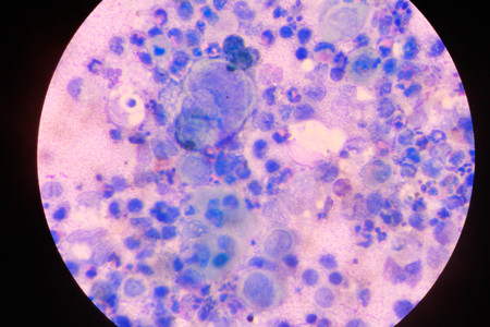
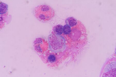
Multinucleated cell in Tzanck test finding with microscope in laboratory.
Коллекция по умолчанию
Коллекция по умолчанию
Создать новую
Science plant cells by light microscope
Коллекция по умолчанию
Коллекция по умолчанию
Создать новую
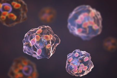
Macrophages infected by Leishmania amastigotes, 3D illustration
Коллекция по умолчанию
Коллекция по умолчанию
Создать новую
Basophil, a white blood cell, 3D illustration. Basophils are granulocytes taking part in inflammatory reactions and allergic diseases
Коллекция по умолчанию
Коллекция по умолчанию
Создать новую

malaria parasite plasmodium falciparum on a thick blood smear reading under a microscope
Коллекция по умолчанию
Коллекция по умолчанию
Создать новую
Cytomegalovirus CMV in human cell, owls eye inclusion in nucleus, multinucleated cell, 3D illustration. It is herpes virus, causes disease in fetus, organ transplant patients, HIV infected people
Коллекция по умолчанию
Коллекция по умолчанию
Создать новую
Eosinophil
Коллекция по умолчанию
Коллекция по умолчанию
Создать новую
Red blood cell in urine under microscopic
Коллекция по умолчанию
Коллекция по умолчанию
Создать новую
Nanoparticles Functionalization Therapeutics, Nanoparticles application in bioiechnology illustration
Коллекция по умолчанию
Коллекция по умолчанию
Создать новую
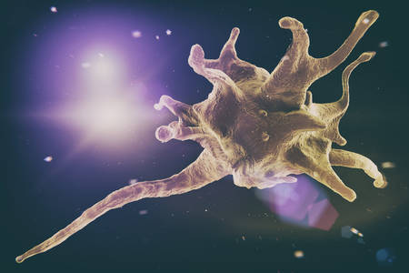
3d rendered medically accurate illustration of a platelet
Коллекция по умолчанию
Коллекция по умолчанию
Создать новую
Immature cells in myeloid serie myelocyte metamyelocyte.
Коллекция по умолчанию
Коллекция по умолчанию
Создать новую

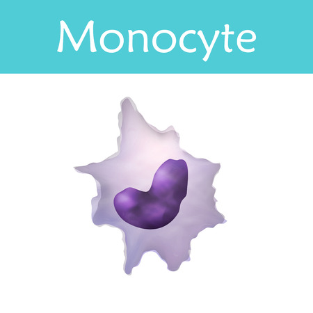
medically accurate illustration of a monocyte
Коллекция по умолчанию
Коллекция по умолчанию
Создать новую
Exploring the microscopic world of cells and tissues, AI generated
Коллекция по умолчанию
Коллекция по умолчанию
Создать новую
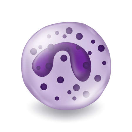
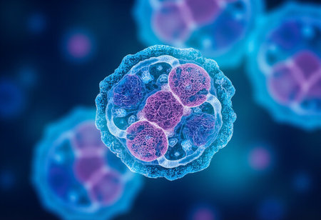
A colorful image of a cell with a purple and blue blob in the center. The image is abstract and has a mood of curiosity and wonder
Коллекция по умолчанию
Коллекция по умолчанию
Создать новую
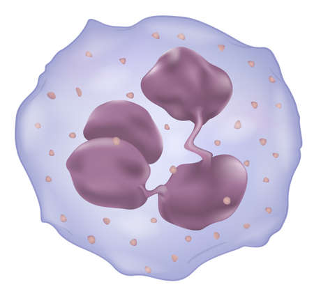
Illustration showing a white blood cell
Коллекция по умолчанию
Коллекция по умолчанию
Создать новую
Delicious glazed donuts covered in colorful sprinkles ready to delight everyone
Коллекция по умолчанию
Коллекция по умолчанию
Создать новую
Chromosomes Human under the microscope for education.
Коллекция по умолчанию
Коллекция по умолчанию
Создать новую
multinucleated giant
Коллекция по умолчанию
Коллекция по умолчанию
Создать новую
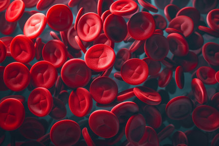
Red blood cells on a dark background. 3d render illustration.
Коллекция по умолчанию
Коллекция по умолчанию
Создать новую
White blood cell in blood smear
Коллекция по умолчанию
Коллекция по умолчанию
Создать новую
neutrophil
Коллекция по умолчанию
Коллекция по умолчанию
Создать новую
Histopathology of human liver under microscope view for medical education.
Коллекция по умолчанию
Коллекция по умолчанию
Создать новую
3d rendered medically accurate illustration of cells
Коллекция по умолчанию
Коллекция по умолчанию
Создать новую
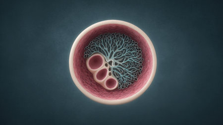
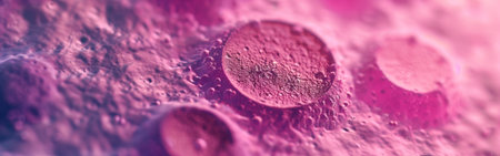
A close up of a pink and blue cell with a tree inside of it. The cell is surrounded by a pink and blue background
Коллекция по умолчанию
Коллекция по умолчанию
Создать новую

3d rendered medically accurate illustration of cells
Коллекция по умолчанию
Коллекция по умолчанию
Создать новую

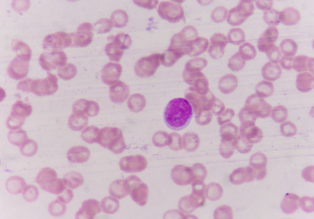
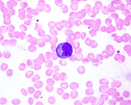
Blood under a microscope. Lymphocyte
Коллекция по умолчанию
Коллекция по умолчанию
Создать новую
The malaria-infected red blood cells. 3D illustration showing malaria parasite Plasmodium falciparum in schizont stage inside red blood cells, the causative agent of tropical malaria
Коллекция по умолчанию
Коллекция по умолчанию
Создать новую
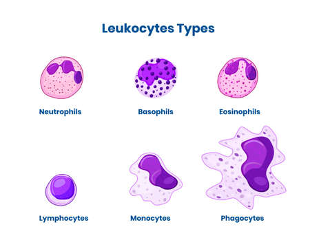
Types of the white blood cells. Leucocyte isolated on white vector illustration
Коллекция по умолчанию
Коллекция по умолчанию
Создать новую
Human blood smear showing a monocyte with a basophilic cytoplasm in an infectious mononucleosis. It is the largest leukocyte (compare with red blood cell size).
Коллекция по умолчанию
Коллекция по умолчанию
Создать новую
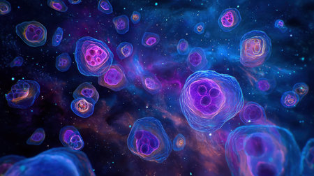
A mesmerizing abstract illustration of cells floating in an enchanting cosmic space, featuring vivid colors and intricate details that inspire imagination.
Коллекция по умолчанию
Коллекция по умолчанию
Создать новую
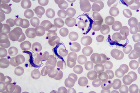
Trypanosoma gambiense blood smear viewed under a microscope at 1250 power.
Коллекция по умолчанию
Коллекция по умолчанию
Создать новую
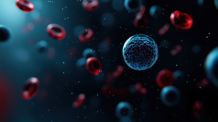
This image showcases a vibrant blue cell delicately surrounded by red blood cells in a dark, captivating space, illustrating fundamental biological concepts.
Коллекция по умолчанию
Коллекция по умолчанию
Создать новую
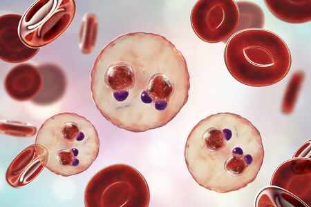
The malaria-infected red blood cells. 3D illustration showing ring-form trophozoites of malaria parasite Plasmodium falciparum inside red blood cells, the causative agent of tropical malaria
Коллекция по умолчанию
Коллекция по умолчанию
Создать новую
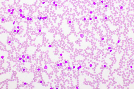
Picture of acute lymphocytic leukemia or ALL cells in blood smear, analyze by microscope, 400x
Коллекция по умолчанию
Коллекция по умолчанию
Создать новую
Red blood cells model with virus molecules on white background
Коллекция по умолчанию
Коллекция по умолчанию
Создать новую
Activatet platelet cell, Thrombocyte are a component of blood whose function is to react to bleeding from blood vessel injury by clumping, thereby initiating a blood clot. 3d illustration
Коллекция по умолчанию
Коллекция по умолчанию
Создать новую
blood films for Malaria parasite.show malaria pigment.
Коллекция по умолчанию
Коллекция по умолчанию
Создать новую
Microscopic view of human cells under microscope.
Коллекция по умолчанию
Коллекция по умолчанию
Создать новую
White blood cells in blood smear, analyze by microscope
Коллекция по умолчанию
Коллекция по умолчанию
Создать новую

Meningococcal meningitis, cerebrospinal fluid smear containing neutrophils with and without bacteria Neisseria meningitidis
Коллекция по умолчанию
Коллекция по умолчанию
Создать новую
Cell division under microscope view. Microscopic view of cell.
Коллекция по умолчанию
Коллекция по умолчанию
Создать новую
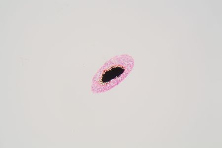
Eggs of a Taenia tapeworm. Taenia is a genus of tapeworm parasites on livestock and humans.
Коллекция по умолчанию
Коллекция по умолчанию
Создать новую
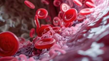
Close-up reveals red blood cells coursing through an artery in macro shot. Ai Generated
Коллекция по умолчанию
Коллекция по умолчанию
Создать новую

Malaria parasites in red blood cells under the microscope 400x
Коллекция по умолчанию
Коллекция по умолчанию
Создать новую
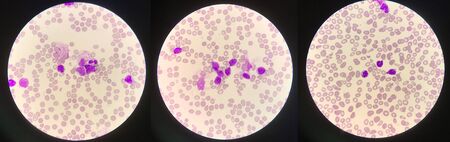
blood smear is often used as a follow-up test to abnormal results on a complete blood count (CBC) to evaluate the different types of blood cells.Medical science background showing blast cells(AML)
Коллекция по умолчанию
Коллекция по умолчанию
Создать новую
blood smear
Коллекция по умолчанию
Коллекция по умолчанию
Создать новую
White blood cells of a human, photomicrograph panorama as seen under the microscope
Коллекция по умолчанию
Коллекция по умолчанию
Создать новую
Eosinophil
Коллекция по умолчанию
Коллекция по умолчанию
Создать новую
microscope lens, viewing Trypanosoma cruzi parasitic protozoan, causer of Chagas disease
Коллекция по умолчанию
Коллекция по умолчанию
Создать новую

Chronic myeloid leukemia cells or CML, analyze by microscope, original magnification 1000x
Коллекция по умолчанию
Коллекция по умолчанию
Создать новую
white blood cells
Коллекция по умолчанию
Коллекция по умолчанию
Создать новую
Legion-Media
Создайте свои проекты на основе качественных стоковых фотографий и видео.


Copyright © Legion-Media.