
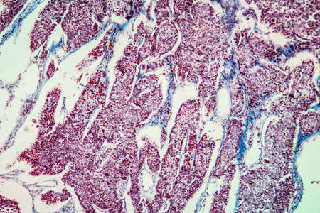
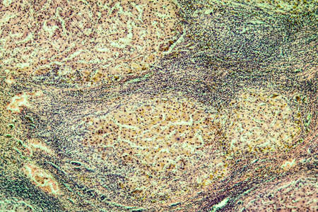
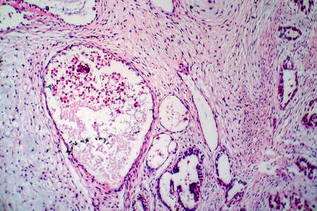
Lungworm under the microscope 100x
Коллекция по умолчанию
Коллекция по умолчанию
Создать новую
Close-up Medical Image: Lipoma Growth on Scalp - Documentary Photography Style
Коллекция по умолчанию
Коллекция по умолчанию
Создать новую

Thyroid follicular carcinoma, light micrograph, photo under microscope
Коллекция по умолчанию
Коллекция по умолчанию
Создать новую
Colon tissue with diverticulum 100x
Коллекция по умолчанию
Коллекция по умолчанию
Создать новую
Squamous cell carcinoma of the uterus, light micrograph, photo under microscope
Коллекция по умолчанию
Коллекция по умолчанию
Создать новую
Hodgkin's lymphoma, light micrograph, photo under microscope. High magnification
Коллекция по умолчанию
Коллекция по умолчанию
Создать новую

Bladder cancer, light micrograph, photo under microscope
Коллекция по умолчанию
Коллекция по умолчанию
Создать новую
Colon inflammation in Crohn's disease 100x
Коллекция по умолчанию
Коллекция по умолчанию
Создать новую
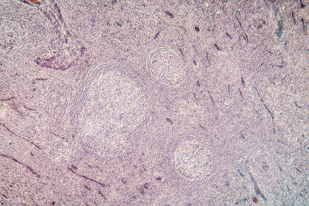
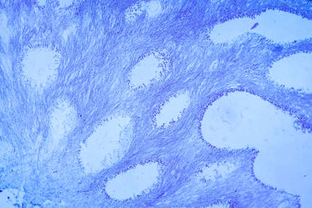
Palatal tonsils transverse 100x under a microscope
Коллекция по умолчанию
Коллекция по умолчанию
Создать новую
Photomicrograph of a neurofibroma tissue sample in neurofibromatosis genetic disease under a microscope, revealing spindle-shaped cells within a myxoid stroma and wavy nuclei.
Коллекция по умолчанию
Коллекция по умолчанию
Создать новую
Breast fibroadenosis, light micrograph, photo under microscope. Common benign hyperplastic process involving breast glands
Коллекция по умолчанию
Коллекция по умолчанию
Создать новую
Wilms tumor, or nephroblastoma, light micrograph, photo under microscope. High magnification
Коллекция по умолчанию
Коллекция по умолчанию
Создать новую

Characteristics of Lichen, hyphae and Symbiotic algae under the microscope for education.
Коллекция по умолчанию
Коллекция по умолчанию
Создать новую

Fibromyoma of the uterus diseased tissue 100x
Коллекция по умолчанию
Коллекция по умолчанию
Создать новую
Fibroepithelium Diseased tissue 100x
Коллекция по умолчанию
Коллекция по умолчанию
Создать новую
Testicular seminoma, light micrograph, photo under microscope. A most common germ cell tumor of the testis
Коллекция по умолчанию
Коллекция по умолчанию
Создать новую
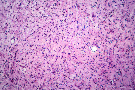
Uterine leiomyoma, also known as fibroids,a benign smooth muscle tumor of the uterus, light micrograph, photo under microscope
Коллекция по умолчанию
Коллекция по умолчанию
Создать новую
Chronic pyelonephritis, light micrograph, photo under microscope
Коллекция по умолчанию
Коллекция по умолчанию
Создать новую

Wound from laser on a face from dermatologist
Коллекция по умолчанию
Коллекция по умолчанию
Создать новую

Growing cancer cell concept image. 3d rendering
Коллекция по умолчанию
Коллекция по умолчанию
Создать новую
3D illustration of a close-up of skin cancer like the malign melanoma inflaming surrounding tissue.
Коллекция по умолчанию
Коллекция по умолчанию
Создать новую
a pimple on the skin of a light-skinned man. a pimple on the skin of a light-skinned man.
Коллекция по умолчанию
Коллекция по умолчанию
Создать новую

Close-up Medical Image: Lipoma Growth on Scalp - Documentary Photography Style
Коллекция по умолчанию
Коллекция по умолчанию
Создать новую

Hodgkins lymphoma, light micrograph, photo under microscope. High magnification
Коллекция по умолчанию
Коллекция по умолчанию
Создать новую
AIDS with fungi 100x infected tissue
Коллекция по умолчанию
Коллекция по умолчанию
Создать новую

Histopathology of interstitial pneumonia, light micrograph, photo under microscope showing diffuse alveolar damage and fibrosis
Коллекция по умолчанию
Коллекция по умолчанию
Создать новую

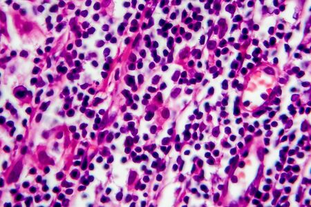
Chronic myeloid leukemia cells or CML, analyze by microscope, original magnification 1000x
Коллекция по умолчанию
Коллекция по умолчанию
Создать новую

Black tongue. A man shows the consequences of an injury, bite or burn of the tongue. Part is damaged. Treatment of internal injuries
Коллекция по умолчанию
Коллекция по умолчанию
Создать новую
Lung adenocarcinoma, light micrograph, photo under microscope
Коллекция по умолчанию
Коллекция по умолчанию
Создать новую
Abstract science background- pyloric division of the stomach of the dog. Cell biology
Коллекция по умолчанию
Коллекция по умолчанию
Создать новую
Testicular seminoma, light micrograph, photo under microscope. A most common germ cell tumor of the testis
Коллекция по умолчанию
Коллекция по умолчанию
Создать новую

diseased liver with cirrhosis 100x under the microscope
Коллекция по умолчанию
Коллекция по умолчанию
Создать новую
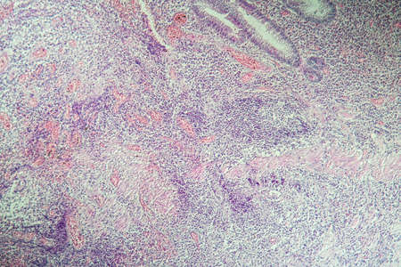
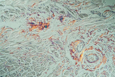
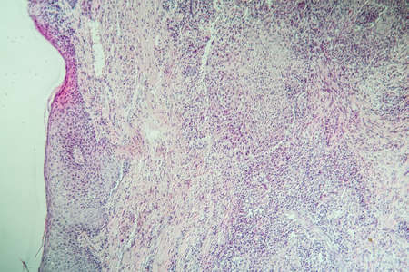
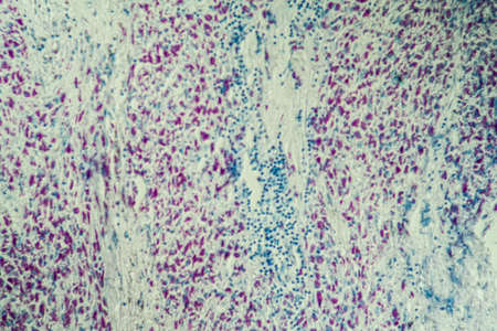
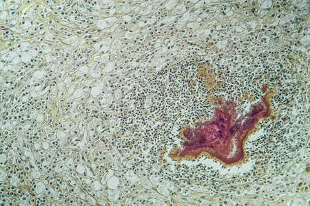
Histopathology of lymph nodal tuberculosis, light micrograph, hematoxylin and eosin staining
Коллекция по умолчанию
Коллекция по умолчанию
Создать новую
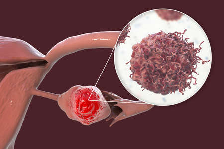
Ovarian cancer, 3D illustration showing malignant tumor in the left ovary and close-up view of cancer cells
Коллекция по умолчанию
Коллекция по умолчанию
Создать новую
hearth with amyloid deposits of sick tissue under the microscope 200x
Коллекция по умолчанию
Коллекция по умолчанию
Создать новую

Villous colon adenocarcinoma, light micrograph, photo under microscope. High magnification
Коллекция по умолчанию
Коллекция по умолчанию
Создать новую
Skin cancer cells, 3D illustration
Коллекция по умолчанию
Коллекция по умолчанию
Создать новую
Wilms tumor, or nephroblastoma, light micrograph, photo under microscope
Коллекция по умолчанию
Коллекция по умолчанию
Создать новую
Bladder transitional cell carcinoma, light micrograph, photo under microscope. High magnification
Коллекция по умолчанию
Коллекция по умолчанию
Создать новую
Wilms tumor, or nephroblastoma, light micrograph, photo under microscope
Коллекция по умолчанию
Коллекция по умолчанию
Создать новую

Renal tuberculosis, light micrograph, photo under microscope
Коллекция по умолчанию
Коллекция по умолчанию
Создать новую
Characteristics of Lichen, hyphae and Symbiotic algae under the microscope for education.
Коллекция по умолчанию
Коллекция по умолчанию
Создать новую
Cervical cancer cells, 3D illustration. Malignant tumor of cervix uteri
Коллекция по умолчанию
Коллекция по умолчанию
Создать новую
Lymph node tissue under the microscope 100x
Коллекция по умолчанию
Коллекция по умолчанию
Создать новую

Scab wound infected on man arm closeup
Коллекция по умолчанию
Коллекция по умолчанию
Создать новую
Photomicrograph of osteosarcoma, a malignant bone tumor, under the microscope revealing atypical osteoblasts producing osteoid matrix.
Коллекция по умолчанию
Коллекция по умолчанию
Создать новую
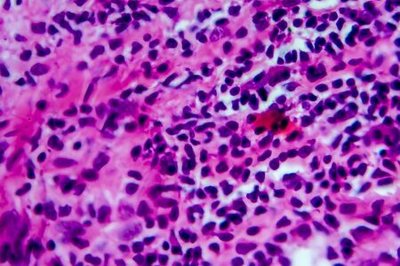
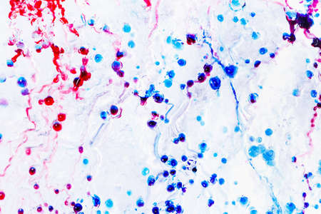
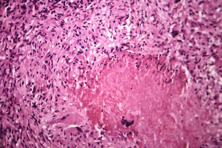
multinucleated giant
Коллекция по умолчанию
Коллекция по умолчанию
Создать новую
Basal cell cancer Diseased tissue 100x
Коллекция по умолчанию
Коллекция по умолчанию
Создать новую
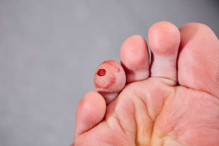
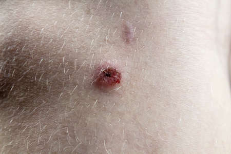
Laser removal of wart from the toe of foot. Cut out verruca.
Коллекция по умолчанию
Коллекция по умолчанию
Создать новую
Skin ulcer carcinoma enlarged 100x
Коллекция по умолчанию
Коллекция по умолчанию
Создать новую
Hodgkins lymphoma, light micrograph, photo under microscope
Коллекция по умолчанию
Коллекция по умолчанию
Создать новую
Histopathology of acute nephritis, light micrograph, photo under microscope
Коллекция по умолчанию
Коллекция по умолчанию
Создать новую
Breast cancer, light micrograph, photo under microscope
Коллекция по умолчанию
Коллекция по умолчанию
Создать новую
Hodgkins lymphoma, light micrograph, photo under microscope. High magnification
Коллекция по умолчанию
Коллекция по умолчанию
Создать новую
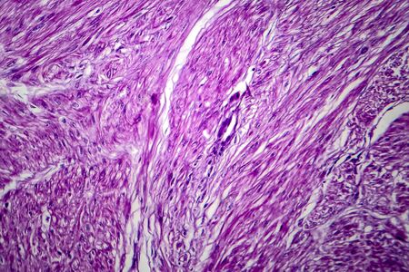
This detailed microscopic image showcases various cellular structures, highlighted in striking purple tones. The intricate patterns and textures reveal the complexity of biological tissues, making it a valuable resource for educational and scientific purposes
Коллекция по умолчанию
Коллекция по умолчанию
Создать новую
destructive mushroom in wood fabric 100x
Коллекция по умолчанию
Коллекция по умолчанию
Создать новую
Chaos ink texture background, ink in water pattern frost. Crystal winter design
Коллекция по умолчанию
Коллекция по умолчанию
Создать новую
AIDS with fungi 200x infected tissue
Коллекция по умолчанию
Коллекция по умолчанию
Создать новую
Hodgkin's lymphoma, light micrograph, photo under microscope. High magnification
Коллекция по умолчанию
Коллекция по умолчанию
Создать новую
macro shot of liver tissue under a microscope, created with generative ai
Коллекция по умолчанию
Коллекция по умолчанию
Создать новую
Actinomyces in the jaw diseased tissue 200x
Коллекция по умолчанию
Коллекция по умолчанию
Создать новую
Breast cancer, light micrograph, photo under microscope
Коллекция по умолчанию
Коллекция по умолчанию
Создать новую
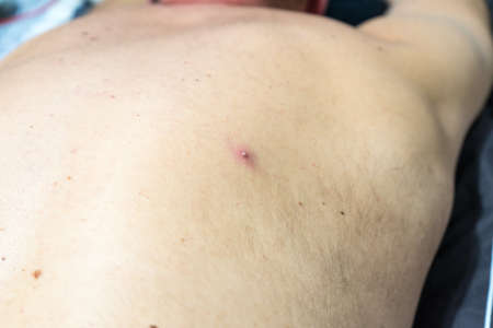
Wound on the knee on the child leg. A Scar on the skin of the child.
Коллекция по умолчанию
Коллекция по умолчанию
Создать новую

Characteristics Tissue of Olfactory epithelium Human under the microscope in Lab.
Коллекция по умолчанию
Коллекция по умолчанию
Создать новую

Ovarian cancer, light micrograph, photo under microscope. Photograph shows a fragment of a cancerous tumor in the female ovary. Selective focus
Коллекция по умолчанию
Коллекция по умолчанию
Создать новую

Histopathology of interstitial nephritis, light micrograph, photo under microscope. High magnification
Коллекция по умолчанию
Коллекция по умолчанию
Создать новую
Liver cirrhosis tissue affected 100x after alcohol abuse
Коллекция по умолчанию
Коллекция по умолчанию
Создать новую
Cervical cancer cells, 3D illustration. Malignant tumor of cervix uteri
Коллекция по умолчанию
Коллекция по умолчанию
Создать новую

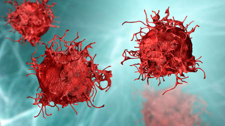
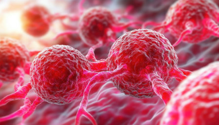
Cancer cells, malignant cells, scientific 3D illustration
Коллекция по умолчанию
Коллекция по умолчанию
Создать новую
Caseous pneumonia, light micrograph, photo under microscope. Tuberculosis pneumonia
Коллекция по умолчанию
Коллекция по умолчанию
Создать новую

Human hyaline cartilage bone under microscope view for education pathology. Human tissue.
Коллекция по умолчанию
Коллекция по умолчанию
Создать новую
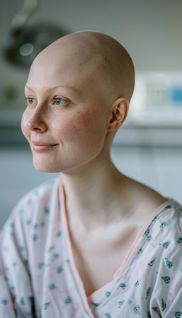
A cheerful woman with a shaved head smiles in a hospital setting, wearing a gown.
Коллекция по умолчанию
Коллекция по умолчанию
Создать новую

Itch mites under the skin cross-section 100x
Коллекция по умолчанию
Коллекция по умолчанию
Создать новую
Cervical cancer cells, 3D illustration. Malignant tumor of cervix uteri
Коллекция по умолчанию
Коллекция по умолчанию
Создать новую
Histopathology of interstitial pneumonia, light micrograph, photo under microscope showing diffuse alveolar damage and fibrosis
Коллекция по умолчанию
Коллекция по умолчанию
Создать новую

Hemosiderosis liver 200x under a microscope
Коллекция по умолчанию
Коллекция по умолчанию
Создать новую

Acute pyelonephritis, light micrograph, photo under microscope. High magnification
Коллекция по умолчанию
Коллекция по умолчанию
Создать новую
Lung adenocarcinoma, light micrograph, photo under microscope
Коллекция по умолчанию
Коллекция по умолчанию
Создать новую
Squamous cell carcinoma of the uterus, light micrograph, photo under microscope
Коллекция по умолчанию
Коллекция по умолчанию
Создать новую
Bladder cancer, light micrograph, photo under microscope
Коллекция по умолчанию
Коллекция по умолчанию
Создать новую
Flu of the lungs Diseased tissue 200x
Коллекция по умолчанию
Коллекция по умолчанию
Создать новую
multinucleated giant
Коллекция по умолчанию
Коллекция по умолчанию
Создать новую
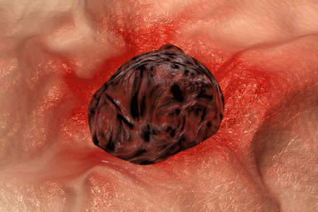
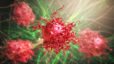
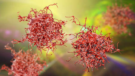
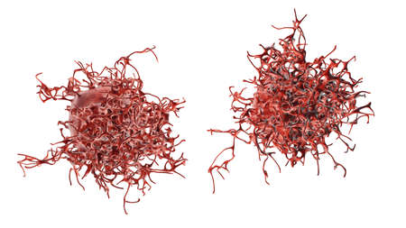
3d illustration of a Cancer Cell
Коллекция по умолчанию
Коллекция по умолчанию
Создать новую

Cancer cells, malignant cells, scientific 3D illustration
Коллекция по умолчанию
Коллекция по умолчанию
Создать новую
Light micrograph of teratoma, a tumor made up of several different types of tissue, such as hair, teeth, muscle, or bone. Teratoma is typically found in the ovary, testicle, or coccyx
Коллекция по умолчанию
Коллекция по умолчанию
Создать новую
Macro shot of purple seaweed on black background with copy space
Коллекция по умолчанию
Коллекция по умолчанию
Создать новую

Actinomyces in the jaw diseased tissue 200x
Коллекция по умолчанию
Коллекция по умолчанию
Создать новую

Yellow ribbon symbolizing sarcoma cancer awareness against white backdrop
Коллекция по умолчанию
Коллекция по умолчанию
Создать новую
Abstract dark purple and gold fluid art alcohol ink pattern with marble texture
Коллекция по умолчанию
Коллекция по умолчанию
Создать новую

Kidney cancer, light micrograph, photo under microscope. High magnification
Коллекция по умолчанию
Коллекция по умолчанию
Создать новую
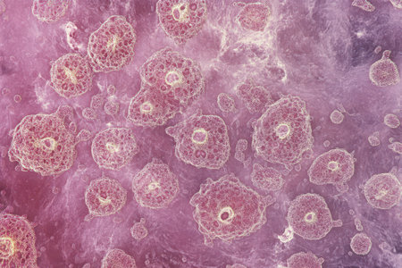
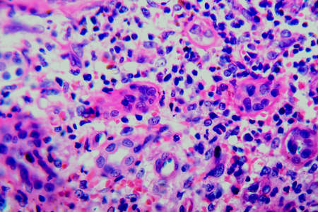
Colorful view of cellular structures captured at a microscopic level showcasing various shapes and sizes in a vibrant environment.
Коллекция по умолчанию
Коллекция по умолчанию
Создать новую
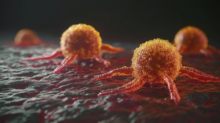
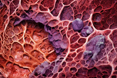
Cancer Cells Under Microscope 3D Visualization, Micro World Render, Pathology Visualization
Коллекция по умолчанию
Коллекция по умолчанию
Создать новую
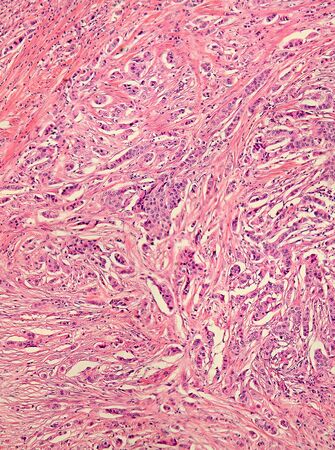
Real photomicrograph of breast adenocarcinoma. Panorama of a slide under the microscope. Some areas will be blurry due to the nature of the tissue.
Коллекция по умолчанию
Коллекция по умолчанию
Создать новую

Chronic glomerulonephritis, light micrograph, photo under microscope
Коллекция по умолчанию
Коллекция по умолчанию
Создать новую
This visual timeline maps the cancer diagnosis journey, from suspicion to confirmation Explore each step, including medical imaging and lab tests This guide simplifies the complex diagnostic process, offering insights for medical professionals and anyone seeking cancer information Understand the progression path to cancer detection AI Generative
Коллекция по умолчанию
Коллекция по умолчанию
Создать новую

Tooth development from human under microscope view for education.
Коллекция по умолчанию
Коллекция по умолчанию
Создать новую

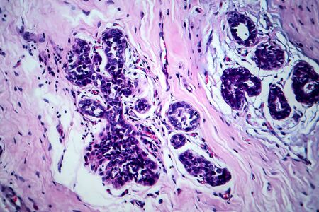
Papillary thyroid carcinoma, light micrograph, photo under microscope. The most common type of thyroid cancer
Коллекция по умолчанию
Коллекция по умолчанию
Создать новую
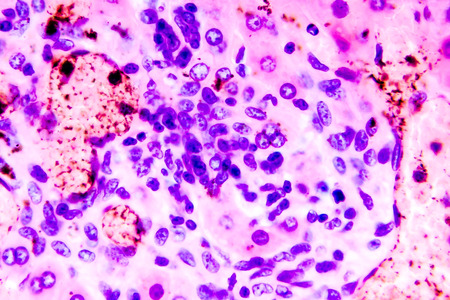
Coccidiosis, coccidia in liver, light micrograph. Micrograph shows bile duct hyperplasia and fibrosis with periductal inflammation, groups of coccidia, large violet cells
Коллекция по умолчанию
Коллекция по умолчанию
Создать новую
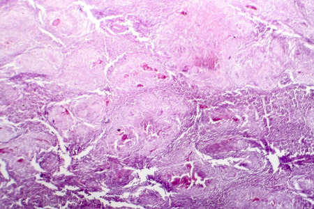
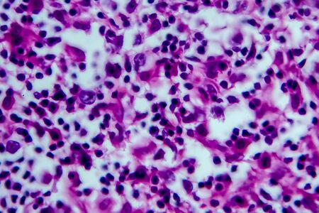
Foreign body granuloma, light micrograph, photo under microscope
Коллекция по умолчанию
Коллекция по умолчанию
Создать новую
A colorful image of a cell with a purple and blue blob in the center. The image is abstract and has a mood of curiosity and wonder
Коллекция по умолчанию
Коллекция по умолчанию
Создать новую
Legion-Media
Создайте свои проекты на основе качественных стоковых фотографий и видео.


Copyright © Legion-Media.