

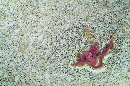
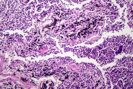
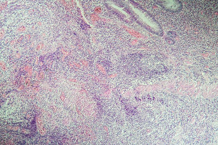
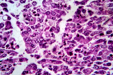
Thyroid follicular carcinoma, light micrograph, photo under microscope
Коллекция по умолчанию
Коллекция по умолчанию
Создать новую
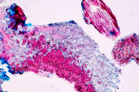
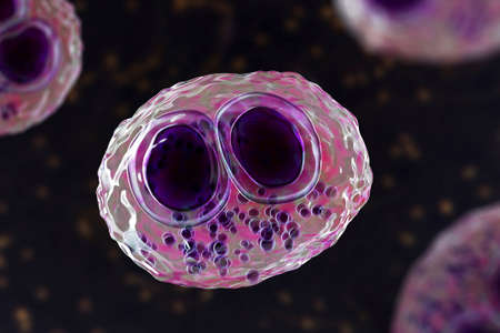
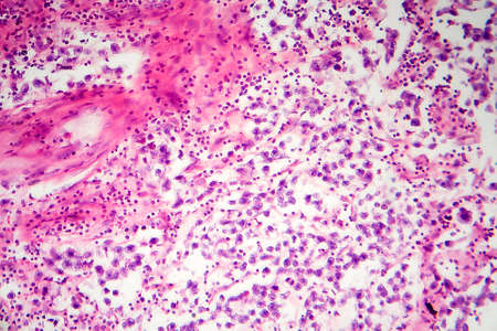
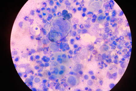
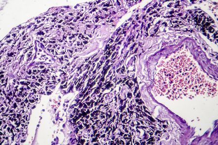
Lungworm under the microscope 100x
Коллекция по умолчанию
Коллекция по умолчанию
Создать новую
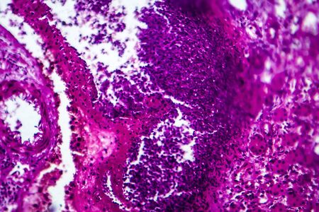
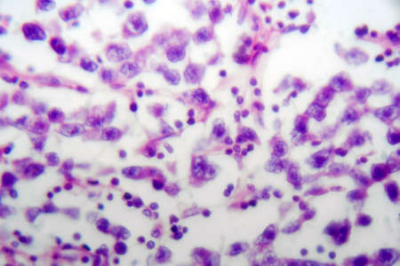
Lung adenocarcinoma, light micrograph, photo under microscope
Коллекция по умолчанию
Коллекция по умолчанию
Создать новую

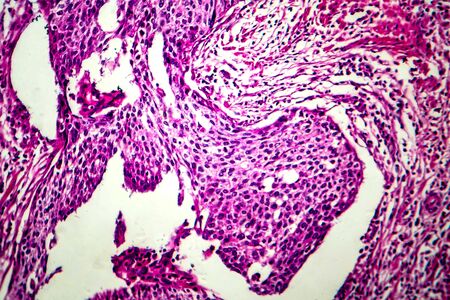
Bladder cancer, light micrograph, photo under microscope
Коллекция по умолчанию
Коллекция по умолчанию
Создать новую

Characteristics of Lichen, hyphae and Symbiotic algae under the microscope for education.
Коллекция по умолчанию
Коллекция по умолчанию
Создать новую
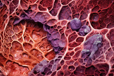
A 3D illustration of the upper half part of a female patient with transparent skin, revealing the condition of smoker's lungs, along with a micrograph image of lungs affected by smoking.
Коллекция по умолчанию
Коллекция по умолчанию
Создать новую
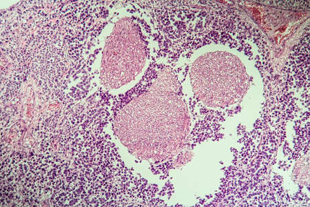
Lymph node tissue under the microscope 100x
Коллекция по умолчанию
Коллекция по умолчанию
Создать новую
Colon tissue with diverticulum 100x
Коллекция по умолчанию
Коллекция по умолчанию
Создать новую
Chaos ink texture background, ink in water pattern frost. Crystal winter design
Коллекция по умолчанию
Коллекция по умолчанию
Создать новую
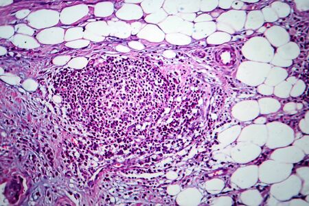
Fibroepithelium Diseased tissue 100x
Коллекция по умолчанию
Коллекция по умолчанию
Создать новую
Human lung pathology under light microscope, The lungs is organs of the respiratory system in humans. Human pathology education. Haematoxylin and eosin staining technique slide.
Коллекция по умолчанию
Коллекция по умолчанию
Создать новую
Actinomyces in the jaw diseased tissue 200x
Коллекция по умолчанию
Коллекция по умолчанию
Создать новую
Characteristics of Lichen, hyphae and Symbiotic algae under the microscope for education.
Коллекция по умолчанию
Коллекция по умолчанию
Создать новую
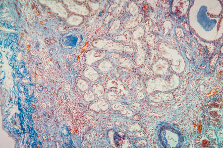
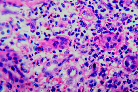
Histopathology of acute nephritis, light micrograph, photo under microscope
Коллекция по умолчанию
Коллекция по умолчанию
Создать новую
Squamous cell carcinoma of the uterus, light micrograph, photo under microscope
Коллекция по умолчанию
Коллекция по умолчанию
Создать новую
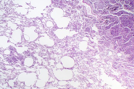
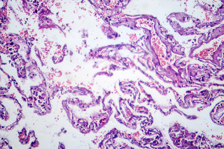
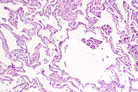
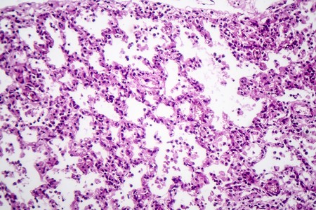
Histopathology of interstitial pneumonia, light micrograph, photo under microscope showing diffuse alveolar damage and fibrosis
Коллекция по умолчанию
Коллекция по умолчанию
Создать новую

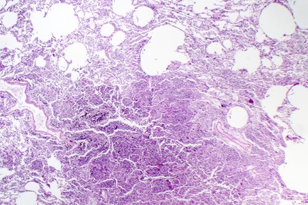
Histopathology of interstitial pneumonia, light micrograph, photo under microscope showing diffuse alveolar damage and fibrosis
Коллекция по умолчанию
Коллекция по умолчанию
Создать новую
Atrophy kidney tissue under the microscope 100x
Коллекция по умолчанию
Коллекция по умолчанию
Создать новую
Flu of the lungs Diseased tissue 200x
Коллекция по умолчанию
Коллекция по умолчанию
Создать новую
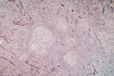
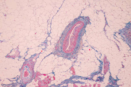
Palatal tonsils transverse 100x under a microscope
Коллекция по умолчанию
Коллекция по умолчанию
Создать новую

Itch mites under the skin cross-section 100x
Коллекция по умолчанию
Коллекция по умолчанию
Создать новую
Cytomegalovirus CMV in a human cell, owl's eye inclusion in nucleus, multinucleated cell, 3D illustration. It is herpes virus, causes diseases in fetus, organ transplant patients, HIV infected people
Коллекция по умолчанию
Коллекция по умолчанию
Создать новую
Breast fibroadenosis, light micrograph, photo under microscope. Common benign hyperplastic process involving breast glands
Коллекция по умолчанию
Коллекция по умолчанию
Создать новую
AIDS with fungi 100x infected tissue
Коллекция по умолчанию
Коллекция по умолчанию
Создать новую
Histopathology of interstitial pneumonia, light micrograph, photo under microscope showing diffuse alveolar damage and fibrosis
Коллекция по умолчанию
Коллекция по умолчанию
Создать новую
Colon inflammation in Crohn's disease 100x
Коллекция по умолчанию
Коллекция по умолчанию
Создать новую
water flowing over boulders, rocks, and stones
Коллекция по умолчанию
Коллекция по умолчанию
Создать новую
Breast cancer, light micrograph, photo under microscope
Коллекция по умолчанию
Коллекция по умолчанию
Создать новую

Characteristics Tissue of Olfactory epithelium Human under the microscope in Lab.
Коллекция по умолчанию
Коллекция по умолчанию
Создать новую
Blue and pink refill ink spilled onto the white washbasin and the ink mixed into abstract blobs and patterns.
Коллекция по умолчанию
Коллекция по умолчанию
Создать новую
Testicular seminoma, light micrograph, photo under microscope. A most common germ cell tumor of the testis
Коллекция по умолчанию
Коллекция по умолчанию
Создать новую
macro shot of liver tissue under a microscope, created with generative ai
Коллекция по умолчанию
Коллекция по умолчанию
Создать новую
Testicular seminoma, light micrograph, photo under microscope. A most common germ cell tumor of the testis
Коллекция по умолчанию
Коллекция по умолчанию
Создать новую
Chronic pyelonephritis, light micrograph, photo under microscope
Коллекция по умолчанию
Коллекция по умолчанию
Создать новую
Histopathology of interstitial nephritis, light micrograph, photo under microscope
Коллекция по умолчанию
Коллекция по умолчанию
Создать новую
Basal cell cancer Diseased tissue 100x
Коллекция по умолчанию
Коллекция по умолчанию
Создать новую
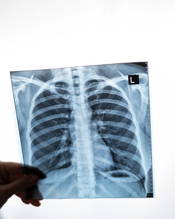
X-ray picture in the hands of a doctor on a white background, a medical worker looks at a picture of an X-ray of the human lungs.
Коллекция по умолчанию
Коллекция по умолчанию
Создать новую
Hodgkin's lymphoma, light micrograph, photo under microscope. High magnification
Коллекция по умолчанию
Коллекция по умолчанию
Создать новую
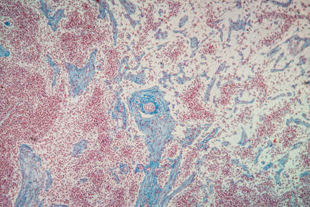
hearth with amyloid deposits of sick tissue under the microscope 200x
Коллекция по умолчанию
Коллекция по умолчанию
Создать новую
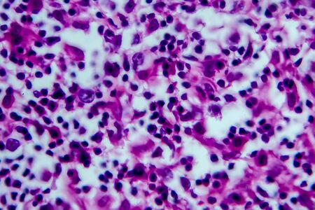
AIDS with fungi 200x infected tissue
Коллекция по умолчанию
Коллекция по умолчанию
Создать новую
Tissue of Stomach Human under the microscope in Lab.
Коллекция по умолчанию
Коллекция по умолчанию
Создать новую
A man's hand holds the CT scan. Transverse view or axial plain of CT chest showing normal study of heart, lungs, spine, rib, other. Selective focuse
Коллекция по умолчанию
Коллекция по умолчанию
Создать новую
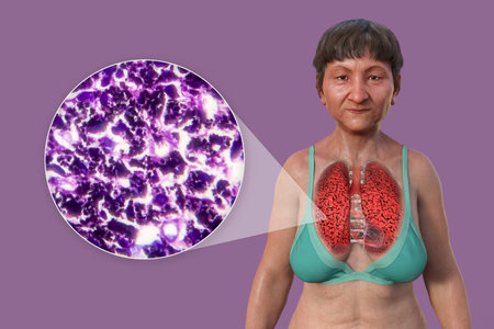
Photomicrograph of smoker's lung, revealing characteristic changes and damage associated with long-term smoking habits.
Коллекция по умолчанию
Коллекция по умолчанию
Создать новую
A closeup examination of a cultured cell on a nanoengineered substrate with bright staining revealing cellular morphology and interaction with the surface at the nanoscale
Коллекция по умолчанию
Коллекция по умолчанию
Создать новую
Lung tissue as dust lung under the microscope 100x
Коллекция по умолчанию
Коллекция по умолчанию
Создать новую

Poly nephritis, tissue under the microscope 100x
Коллекция по умолчанию
Коллекция по умолчанию
Создать новую
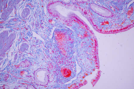
Columnar epithelium of human gall bladder under the microscope in Lab.
Коллекция по умолчанию
Коллекция по умолчанию
Создать новую
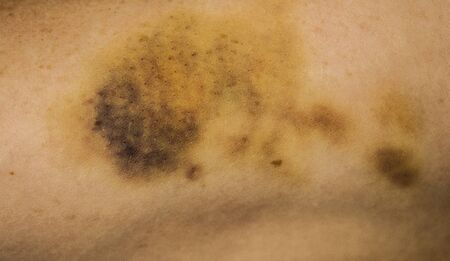
Violence victim with a bruise on her arm
Коллекция по умолчанию
Коллекция по умолчанию
Создать новую

Host cells with spores (mold) are inside wood under the microscope for education.
Коллекция по умолчанию
Коллекция по умолчанию
Создать новую
Abstract view of semiprecious resin surface with vivid grape-like purple clusters scattered across cloudy blue and white texture.
Коллекция по умолчанию
Коллекция по умолчанию
Создать новую
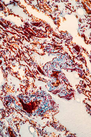
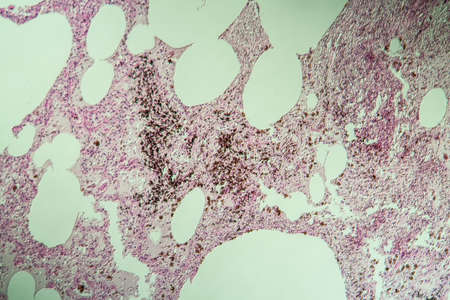
Chronic pulmonary congestion and edema, light micrograph
Коллекция по умолчанию
Коллекция по умолчанию
Создать новую
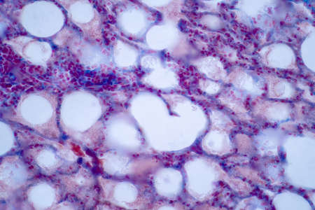
Hemosiderosis liver 200x under a microscope
Коллекция по умолчанию
Коллекция по умолчанию
Создать новую
Tongue Tissue with taste buds across 200x
Коллекция по умолчанию
Коллекция по умолчанию
Создать новую

Ovarian cancer, light micrograph, photo under microscope. Photograph shows a fragment of a cancerous tumor in the female ovary. Selective focus
Коллекция по умолчанию
Коллекция по умолчанию
Создать новую
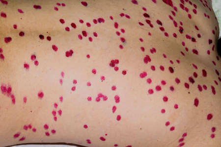
Papules on the skin of a patient with chickenpox close-up. Varicella Zoster virus.
Коллекция по умолчанию
Коллекция по умолчанию
Создать новую
Bacterial dysentery, light micrograph, photo under microscope showing accumulation of inflammatory cells, changes in structure of intestinal epithelium
Коллекция по умолчанию
Коллекция по умолчанию
Создать новую

Hemosiderosis liver 200x under a microscope
Коллекция по умолчанию
Коллекция по умолчанию
Создать новую

Histopathology of interstitial nephritis, light micrograph, photo under microscope. High magnification
Коллекция по умолчанию
Коллекция по умолчанию
Создать новую
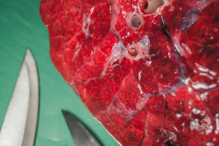
A piece of beef lung close-up. Meat for cooking. Selective focus
Коллекция по умолчанию
Коллекция по умолчанию
Создать новую

Human hyaline cartilage bone under microscope view for education pathology. Human tissue.
Коллекция по умолчанию
Коллекция по умолчанию
Создать новую

Hodgkins lymphoma, light micrograph, photo under microscope. High magnification
Коллекция по умолчанию
Коллекция по умолчанию
Создать новую
Coccidiosis, coccidia in liver, light micrograph. Micrograph shows bile duct hyperplasia and fibrosis with periductal inflammation, groups of coccidia, large violet cells
Коллекция по умолчанию
Коллекция по умолчанию
Создать новую
Liver cirrhosis tissue affected 100x after alcohol abuse
Коллекция по умолчанию
Коллекция по умолчанию
Создать новую
Wilms tumor, or nephroblastoma, light micrograph, photo under microscope
Коллекция по умолчанию
Коллекция по умолчанию
Создать новую
Wilms tumor, or nephroblastoma, light micrograph, photo under microscope. High magnification
Коллекция по умолчанию
Коллекция по умолчанию
Создать новую
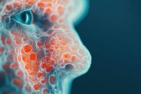
Abstract representation of a human face with colorful textures and patterns, highlighting emotions and features in a surreal style
Коллекция по умолчанию
Коллекция по умолчанию
Создать новую
Chronic pyelonephritis, light micrograph, photo under microscope
Коллекция по умолчанию
Коллекция по умолчанию
Создать новую
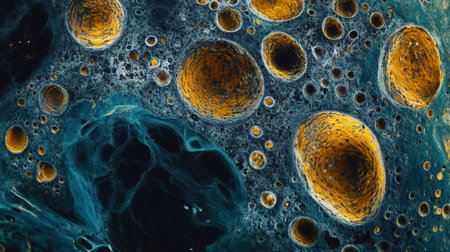
This view reveals colorful cells and structures, highlighting their intricate patterns and vibrant hues under a microscope.
Коллекция по умолчанию
Коллекция по умолчанию
Создать новую

Chronic glomerulonephritis, light micrograph, photo under microscope
Коллекция по умолчанию
Коллекция по умолчанию
Создать новую
Hodgkins lymphoma, light micrograph, photo under microscope. High magnification
Коллекция по умолчанию
Коллекция по умолчанию
Создать новую
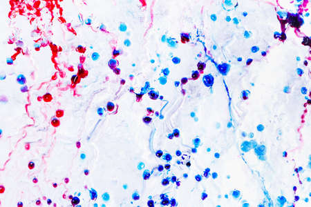
multinucleated giant
Коллекция по умолчанию
Коллекция по умолчанию
Создать новую

diseased liver with cirrhosis 100x under the microscope
Коллекция по умолчанию
Коллекция по умолчанию
Создать новую
Chronic pulmonary congestion, light micrograph. Photo under microscope
Коллекция по умолчанию
Коллекция по умолчанию
Создать новую
A colorful image of a cell with a purple and blue blob in the center. The image is abstract and has a mood of curiosity and wonder
Коллекция по умолчанию
Коллекция по умолчанию
Создать новую
Abstract science background- pyloric division of the stomach of the dog. Cell biology
Коллекция по умолчанию
Коллекция по умолчанию
Создать новую
Microscopic view of human cells under microscope.
Коллекция по умолчанию
Коллекция по умолчанию
Создать новую
Spinal cord tissue section under the microscope 100x
Коллекция по умолчанию
Коллекция по умолчанию
Создать новую

Scab wound infected on man arm closeup
Коллекция по умолчанию
Коллекция по умолчанию
Создать новую
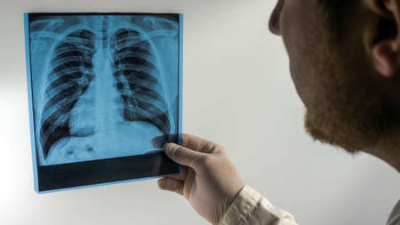
Doctor showing x-ray of patients lungs.
Коллекция по умолчанию
Коллекция по умолчанию
Создать новую

Wound from laser on a face from dermatologist
Коллекция по умолчанию
Коллекция по умолчанию
Создать новую

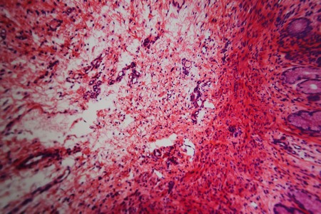
Histopathology of alcoholic hepatitis, light micrograph, photo under microscope. High magnification
Коллекция по умолчанию
Коллекция по умолчанию
Создать новую
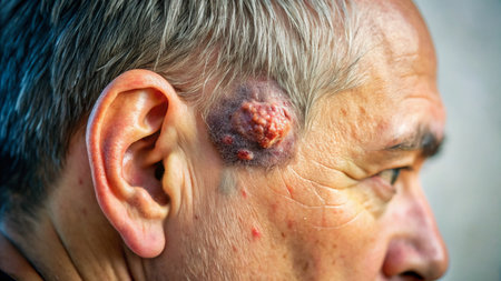
Close-up Medical Image: Lipoma Growth on Scalp - Documentary Photography Style
Коллекция по умолчанию
Коллекция по умолчанию
Создать новую
Smokers lung, histopathology, light micrograph showing accumulation of carbon particles in lung tissue
Коллекция по умолчанию
Коллекция по умолчанию
Создать новую
A piece of beef lung close-up. Meat for cooking. Selective focus
Коллекция по умолчанию
Коллекция по умолчанию
Создать новую
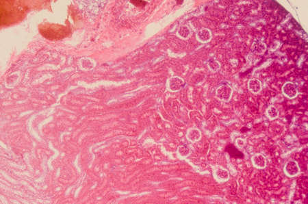
science medical anthropotomy physiology microscopic section of human kidney tissue background
Коллекция по умолчанию
Коллекция по умолчанию
Создать новую

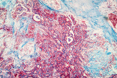
Villous colon adenocarcinoma, light micrograph, photo under microscope. High magnification
Коллекция по умолчанию
Коллекция по умолчанию
Создать новую
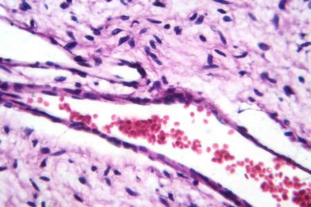
A small blood vessel with red blood cells in neurofibroma tissue sample, light photomicrograph.
Коллекция по умолчанию
Коллекция по умолчанию
Создать новую
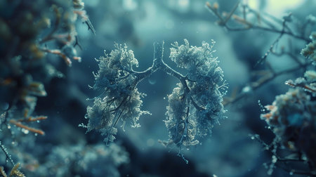
Bronchi represented as delicate underwater formations, showing the beauty of nature's design
Коллекция по умолчанию
Коллекция по умолчанию
Создать новую
Endometrial adenocarcinoma, light micrograph, photo under microscope
Коллекция по умолчанию
Коллекция по умолчанию
Создать новую
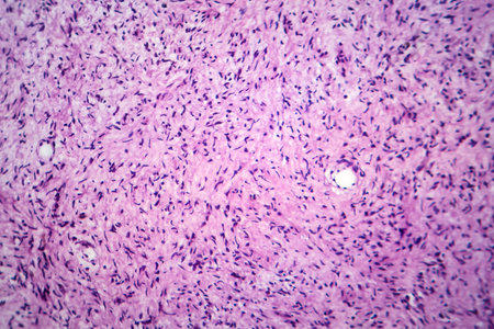
Photomicrograph of a neurofibroma tissue sample in neurofibromatosis genetic disease under a microscope, revealing spindle-shaped cells within a myxoid stroma and wavy nuclei.
Коллекция по умолчанию
Коллекция по умолчанию
Создать новую
Metastases tumor diseased tissue 100x
Коллекция по умолчанию
Коллекция по умолчанию
Создать новую
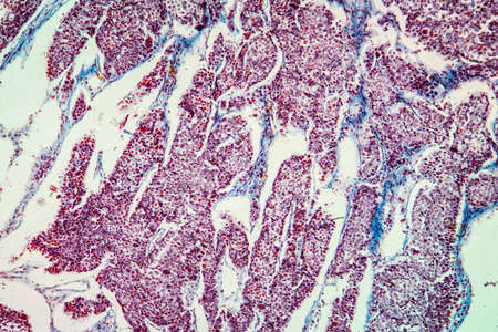
Histopathology of bronchopneumonia, light micrograph, photo under microscope
Коллекция по умолчанию
Коллекция по умолчанию
Создать новую

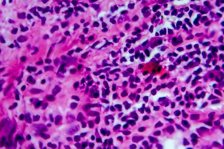
Chronic myeloid leukemia cells or CML, analyze by microscope, original magnification 1000x
Коллекция по умолчанию
Коллекция по умолчанию
Создать новую
Histopathology of interstitial pneumonia, light micrograph, photo under microscope showing diffuse alveolar damage and fibrosis
Коллекция по умолчанию
Коллекция по умолчанию
Создать новую
Scalp and hair follicles of human under the microscope in Lab.
Коллекция по умолчанию
Коллекция по умолчанию
Создать новую
Hodgkin's lymphoma, light micrograph, photo under microscope. High magnification
Коллекция по умолчанию
Коллекция по умолчанию
Создать новую
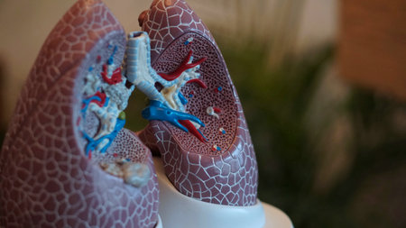
Lungs of a child. Close-up.
Коллекция по умолчанию
Коллекция по умолчанию
Создать новую
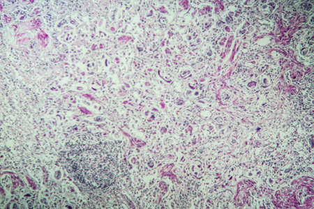
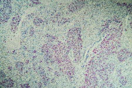
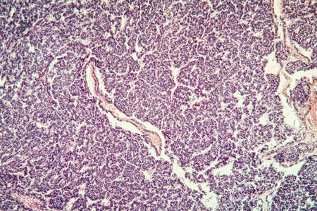
Histopathology of lymph nodal tuberculosis, light micrograph, hematoxylin and eosin staining
Коллекция по умолчанию
Коллекция по умолчанию
Создать новую

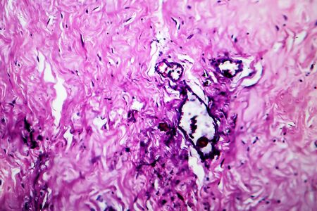
Fibromyoma of the uterus diseased tissue 100x
Коллекция по умолчанию
Коллекция по умолчанию
Создать новую
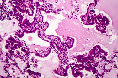
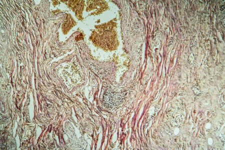
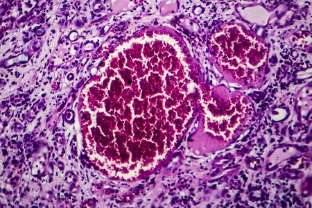
Goiter colloid goiter disease 100x
Коллекция по умолчанию
Коллекция по умолчанию
Создать новую
Legion-Media
Создайте свои проекты на основе качественных стоковых фотографий и видео.


Copyright © Legion-Media.