

Thyroid follicular carcinoma, light micrograph, photo under microscope
Коллекция по умолчанию
Коллекция по умолчанию
Создать новую
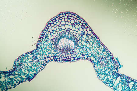
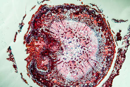
Linguster leaf cross section under the microscope 100x
Коллекция по умолчанию
Коллекция по умолчанию
Создать новую
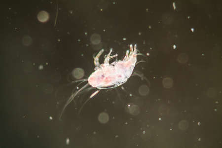
House dust mite under the microscope 100x
Коллекция по умолчанию
Коллекция по умолчанию
Создать новую
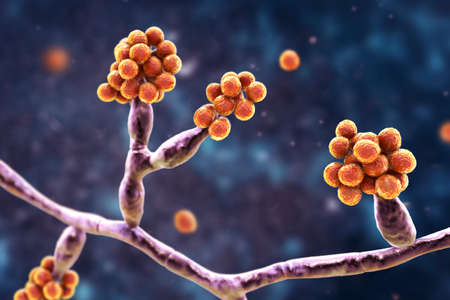
Microscopic fungi Cunninghamella, scientific 3D illustration. Pathogenic fungi from the order Mucorales, cause sinopulmonary and disseminated infections, one of the causative agents of mucormycosis
Коллекция по умолчанию
Коллекция по умолчанию
Создать новую

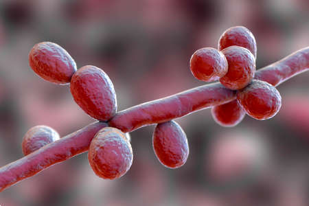
Candida tropicalis yeasts, microscopic fungi that cause infections in immunocompromised patients. Scientific 3D illustration showing pseudohyphae and blastoconidia formed singly or in small groups
Коллекция по умолчанию
Коллекция по умолчанию
Создать новую

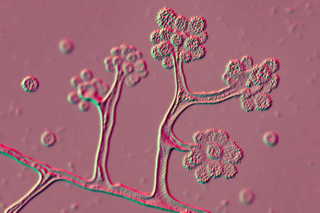
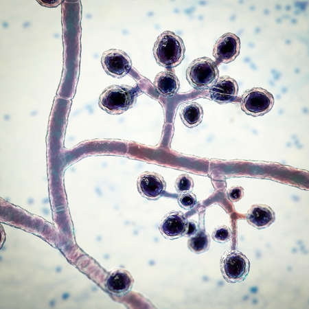
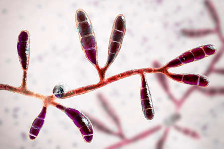
Fungi Trichophyton mentagrophytes, 3D illustration showing macroconidia, branched conidiophores bearing spherical conidia, septate and spiral hyphae. Causes ringworm, hair and nail infections
Коллекция по умолчанию
Коллекция по умолчанию
Создать новую
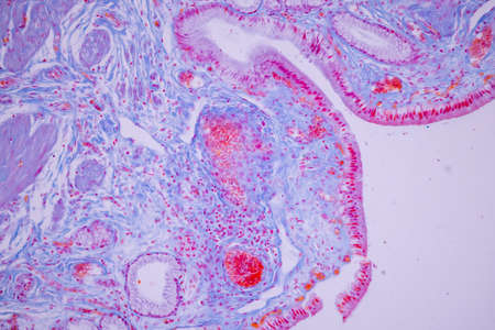
Columnar epithelium of human gall bladder under the microscope in Lab.
Коллекция по умолчанию
Коллекция по умолчанию
Создать новую
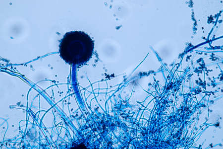
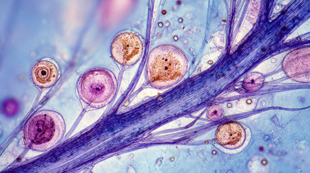
Aspergillus niger and Aspergillus oryzae (mold) under microscope for Microbiology in Lab.
Коллекция по умолчанию
Коллекция по умолчанию
Создать новую

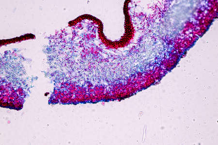
Characteristics of Lichen, hyphae and Symbiotic algae under the microscope for education.
Коллекция по умолчанию
Коллекция по умолчанию
Создать новую
Fungi Trichophyton mentagrophytes, 3D illustration showing branched conidiophores bearing spherical microconidia. Causes skin infection (ringworm), hair and nail infections
Коллекция по умолчанию
Коллекция по умолчанию
Создать новую

Bladder cancer, light micrograph, photo under microscope
Коллекция по умолчанию
Коллекция по умолчанию
Создать новую
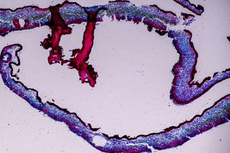
Colon tissue with diverticulum 100x
Коллекция по умолчанию
Коллекция по умолчанию
Создать новую
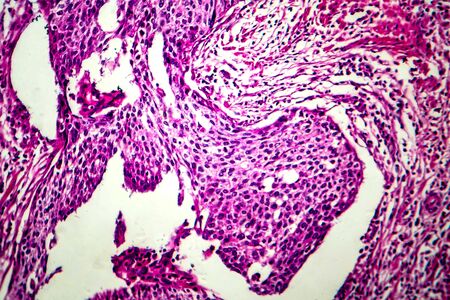
Squamous cell carcinoma of the uterus, light micrograph, photo under microscope
Коллекция по умолчанию
Коллекция по умолчанию
Создать новую
destructive mushroom in wood fabric 100x
Коллекция по умолчанию
Коллекция по умолчанию
Создать новую
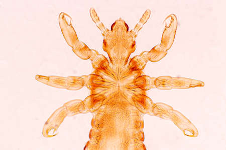
The head louse (Pediculus humanus capitis) is a parasite Live on the body, person or animal and live by sucking blood into food.
Коллекция по умолчанию
Коллекция по умолчанию
Создать новую
Characteristics of Lichen, hyphae and Symbiotic algae under the microscope for education.
Коллекция по умолчанию
Коллекция по умолчанию
Создать новую

Apple pollen from a blossom in spring under the microscope
Коллекция по умолчанию
Коллекция по умолчанию
Создать новую
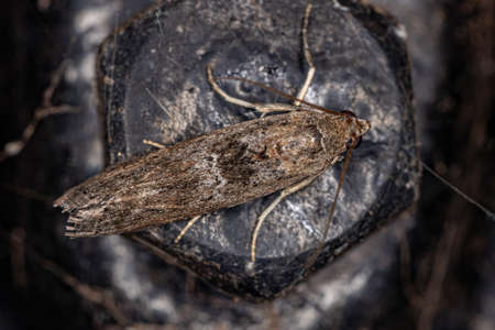
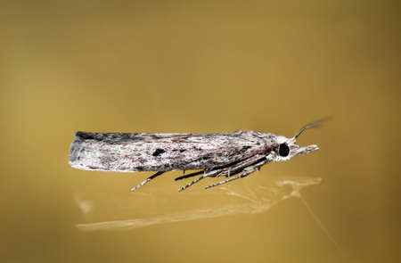
Adult Pyralid Snout Moth of the Family Pyralidae
Коллекция по умолчанию
Коллекция по умолчанию
Создать новую
An intricate digital representation of bacteria displaying vibrant colors and glowing elements, highlighting the fascinating world of microscopic life in a stunning abstract setting.
Коллекция по умолчанию
Коллекция по умолчанию
Создать новую

Scalp and hair follicles of human under the microscope in Lab.
Коллекция по умолчанию
Коллекция по умолчанию
Создать новую
Rotifers as microscopic plankton in drops of water
Коллекция по умолчанию
Коллекция по умолчанию
Создать новую
Characteristics of Lichen, hyphae and Symbiotic algae under the microscope for education.
Коллекция по умолчанию
Коллекция по умолчанию
Создать новую

Proglottid (body unit) of tapeworm Taenia saginata, 3D illustration. A flatworm parasitizing animal and human intestine. Proglottid contains uterus with 12-30 primary lateral branches filled with eggs
Коллекция по умолчанию
Коллекция по умолчанию
Создать новую
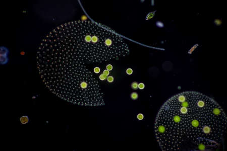
Volvox in drop of water under the microscope for classroom education.
Коллекция по умолчанию
Коллекция по умолчанию
Создать новую
Trumpet animal as a microscopic plankton animal in drops of water
Коллекция по умолчанию
Коллекция по умолчанию
Создать новую
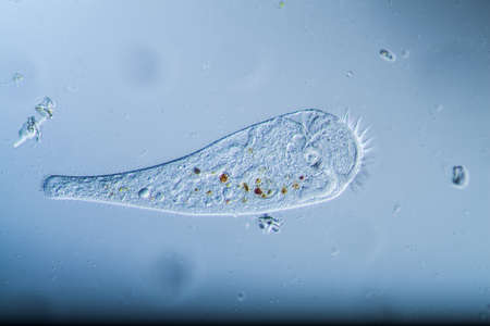
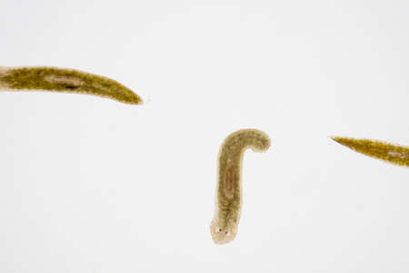
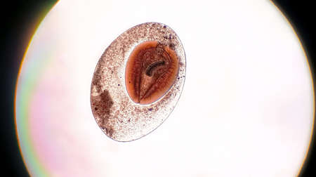
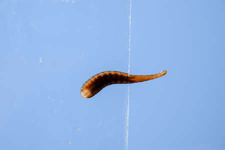
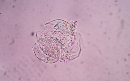
Planarian parasite (flatworm) under microscope view.
Коллекция по умолчанию
Коллекция по умолчанию
Создать новую
sporangia of a microscopic organism, microbiology concept
Коллекция по умолчанию
Коллекция по умолчанию
Создать новую
Microscopic view of unspecified eggs in tube shaped packet on Common duckweed (Lemna minor) root. Rheinberg illumination.
Коллекция по умолчанию
Коллекция по умолчанию
Создать новую
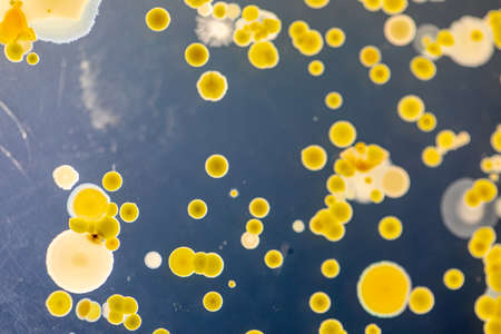
Backgrounds of Characteristics and Different shaped Colony of Bacteria and Mold growing on agar plates from Soil samples for education in Microbiology laboratory.
Коллекция по умолчанию
Коллекция по умолчанию
Создать новую

Microscopic fungi Microsporum audouinii, 3D illustration. Anthropophilic dermatophyte fungus, causes infections of scalp (tinea capitis), body skin (tinea corporis) mainly in children
Коллекция по умолчанию
Коллекция по умолчанию
Создать новую
Vibrant cross-section of a developing seed under UV light, highlighting the embryo and endosperm in bright colors
Коллекция по умолчанию
Коллекция по умолчанию
Создать новую

Ovarian cancer, light micrograph, photo under microscope. Photograph shows a fragment of a cancerous tumor in the female ovary. Selective focus
Коллекция по умолчанию
Коллекция по умолчанию
Создать новую

Stomach tissue under the microscope 100x
Коллекция по умолчанию
Коллекция по умолчанию
Создать новую
Fibroepithelium Diseased tissue 100x
Коллекция по умолчанию
Коллекция по умолчанию
Создать новую
Juicy Algae Extract in Nature's Garden
Коллекция по умолчанию
Коллекция по умолчанию
Создать новую
Planarian parasite (flatworm) under microscope view.
Коллекция по умолчанию
Коллекция по умолчанию
Создать новую
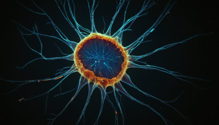
Radiant Neuron Structure
Коллекция по умолчанию
Коллекция по умолчанию
Создать новую
Microscopic fungi Trichosporon, 3D illustration shows septate hyphae, pseudohyphae, blastoconidia singly or in short chains, arthroconidia. Cause white piedra, superficial and invasive infections
Коллекция по умолчанию
Коллекция по умолчанию
Создать новую
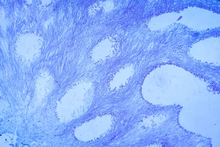
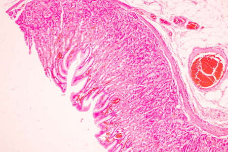
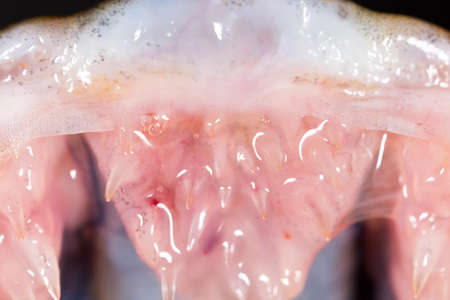
Palatal tonsils transverse 100x under a microscope
Коллекция по умолчанию
Коллекция по умолчанию
Создать новую
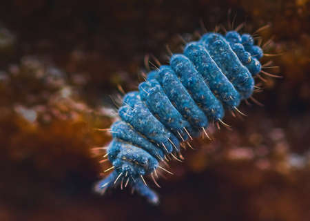
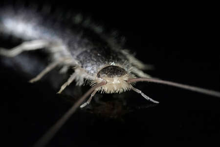
Close up of Moss springtail - Neanura muscorum
Коллекция по умолчанию
Коллекция по умолчанию
Создать новую

Bee compound eye enlarged 100x along
Коллекция по умолчанию
Коллекция по умолчанию
Создать новую
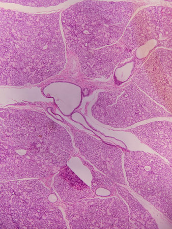
Lymph node tissue under the microscope 100x
Коллекция по умолчанию
Коллекция по умолчанию
Создать новую

Fungus Sporothrix schenckii, the causative agent of sporotrichosis, especially common in florists and gardeners. 3D illustration showing fungal hyphae and spores
Коллекция по умолчанию
Коллекция по умолчанию
Создать новую
Tissue of Stomach Human under the microscope in Lab.
Коллекция по умолчанию
Коллекция по умолчанию
Создать новую
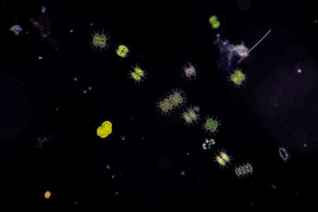
Protozoa and Green Algae in waste water under the microscope.
Коллекция по умолчанию
Коллекция по умолчанию
Создать новую

Blood vessel with flowing blood cells, 3D illustration. Small blood vessels, capillaries
Коллекция по умолчанию
Коллекция по умолчанию
Создать новую

Backgrounds of Characteristics and Different shaped Colony of Bacteria and Mold growing on agar plates from Soil samples for education in Microbiology laboratory.
Коллекция по умолчанию
Коллекция по умолчанию
Создать новую

Tick crawling on gray background.
Коллекция по умолчанию
Коллекция по умолчанию
Создать новую
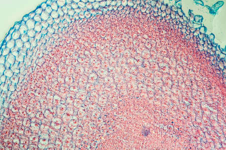
Oak tree with root across 100x
Коллекция по умолчанию
Коллекция по умолчанию
Создать новую
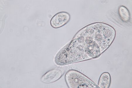
Paramecium caudatum is a genus of unicellular ciliated protozoan and Bacterium under the microscope.
Коллекция по умолчанию
Коллекция по умолчанию
Создать новую

drain fly
Коллекция по умолчанию
Коллекция по умолчанию
Создать новую
Moniliformis dubius in the Intestine of rat, intermediate host
Коллекция по умолчанию
Коллекция по умолчанию
Создать новую
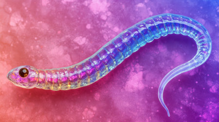
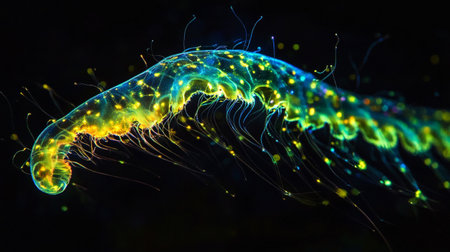
A stunning close-up of a colorful, transparent snake-like creature set against a vibrant pink and purple background. This image highlights the fascinating anatomy and beauty of wildlife.
Коллекция по умолчанию
Коллекция по умолчанию
Создать новую
Microscopic fungi Epidermophyton floccosum, scientific 3D illustration. A filamentous fungus, causes skin and nail infections, such as athlete's foot, tinea cruris, tinea corporis and onychomycosis
Коллекция по умолчанию
Коллекция по умолчанию
Создать новую
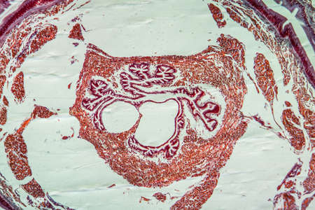
Gladiolus with root tip across 100x
Коллекция по умолчанию
Коллекция по умолчанию
Создать новую
serous gland tissue under the microscope 100x
Коллекция по умолчанию
Коллекция по умолчанию
Создать новую

Vegetation cone of the water pest plant 100x
Коллекция по умолчанию
Коллекция по умолчанию
Создать новую
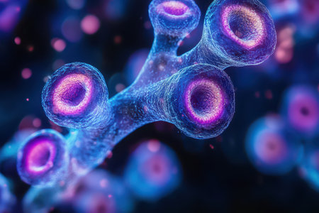
Explore a stunning microscopic view featuring colorful cells and thin filaments against a vibrant blue background, showcasing scientific beauty in detail.
Коллекция по умолчанию
Коллекция по умолчанию
Создать новую
This detailed microscopic image showcases various cellular structures, highlighted in striking purple tones. The intricate patterns and textures reveal the complexity of biological tissues, making it a valuable resource for educational and scientific purposes
Коллекция по умолчанию
Коллекция по умолчанию
Создать новую

Photo Picture of Some Jellyfish Dangerous Poisonous Medusa
Коллекция по умолчанию
Коллекция по умолчанию
Создать новую
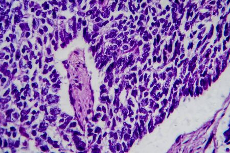
Wilms tumor, or nephroblastoma, light micrograph, photo under microscope. High magnification
Коллекция по умолчанию
Коллекция по умолчанию
Создать новую
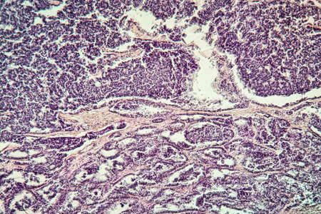
Metastases tumor diseased tissue 100x
Коллекция по умолчанию
Коллекция по умолчанию
Создать новую
Red planaria flatworms - Convolutriloba retrogemma
Коллекция по умолчанию
Коллекция по умолчанию
Создать новую
Neon glowing jellyfish isolated on black background. 3d illustration
Коллекция по умолчанию
Коллекция по умолчанию
Создать новую
Leech on the glass. Bloodsucking animal. subclass of ringworms from the belt-type class. Hirudotherapy.
Коллекция по умолчанию
Коллекция по умолчанию
Создать новую
teeth pike fish. super macro
Коллекция по умолчанию
Коллекция по умолчанию
Создать новую
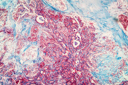
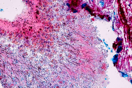
A microscopic view of tissue showing pink-stained cells and structures, indicative of biological samples, possibly related to histology or pathology
Коллекция по умолчанию
Коллекция по умолчанию
Создать новую
Mould fungi Madurella, 3D illustration. The microscopic fungus that causes black-grain mycetoma, or maduromycosis, an infection of human extremities and nervous system found in tropical areas
Коллекция по умолчанию
Коллекция по умолчанию
Создать новую
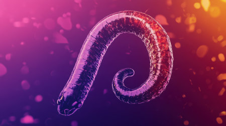
This vibrant abstract composition features a stylized worm design, showcasing smooth curves and a colorful gradient that captivates the viewer's attention.
Коллекция по умолчанию
Коллекция по умолчанию
Создать новую

Euglena is a genus of single-celled flagellate Eukaryotes under microscopic view for education.
Коллекция по умолчанию
Коллекция по умолчанию
Создать новую
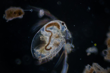
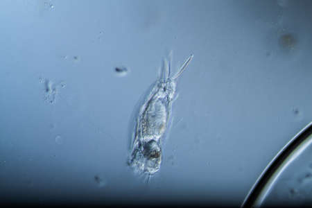
Water flea (Moina macrocopa) under microscope view
Коллекция по умолчанию
Коллекция по умолчанию
Создать новую
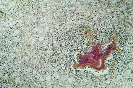
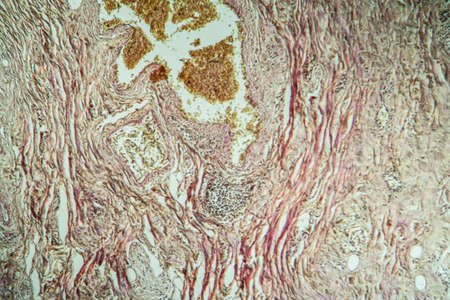
Actinomyces in the jaw diseased tissue 200x
Коллекция по умолчанию
Коллекция по умолчанию
Создать новую

Condyloma acuminatum, also known as genital warts. Light micrograph, photo under microscope
Коллекция по умолчанию
Коллекция по умолчанию
Создать новую
Candida tropicalis yeasts, microscopic fungi that cause infections in immunocompromised patients. Scientific 3D illustration showing pseudohyphae and blastoconidia formed singly or in small groups
Коллекция по умолчанию
Коллекция по умолчанию
Создать новую
microscope slide with detailed view of plant stem, complete with cells and minutiae, created with generative ai
Коллекция по умолчанию
Коллекция по умолчанию
Создать новую

Microphoto of a larva
Коллекция по умолчанию
Коллекция по умолчанию
Создать новую
Creative ebru art background abstract pint. Marbling texture pattern
Коллекция по умолчанию
Коллекция по умолчанию
Создать новую

cells biology- endosperm weevil rye
Коллекция по умолчанию
Коллекция по умолчанию
Создать новую
Lungworm under the microscope 100x
Коллекция по умолчанию
Коллекция по умолчанию
Создать новую
Leech on the glass. Bloodsucking animal. subclass of ringworms from the belt-type class. Hirudotherapy.
Коллекция по умолчанию
Коллекция по умолчанию
Создать новую
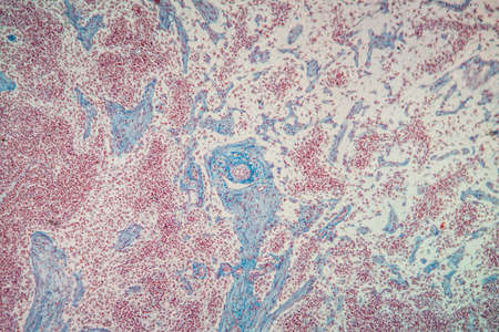
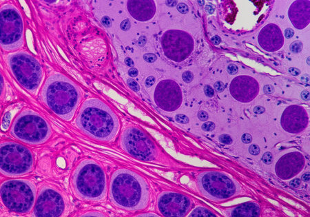
Goiter colloid goiter disease 100x
Коллекция по умолчанию
Коллекция по умолчанию
Создать новую
Characteristics of Lichen, hyphae and Symbiotic algae under the microscope for education.
Коллекция по умолчанию
Коллекция по умолчанию
Создать новую
Cytomegalovirus CMV in a human cell, owl's eye inclusion in nucleus, multinucleated cell, 3D illustration. It is herpes virus, causes diseases in fetus, organ transplant patients, HIV infected people
Коллекция по умолчанию
Коллекция по умолчанию
Создать новую
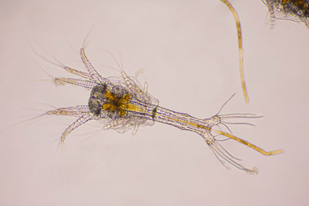
Closeup zoea stage of Vannamei shrimp in light microscope, Shrimp larvae under a microscope, Shrimp, White shrimp, Nauplius, zoea, Larvae. Background.
Коллекция по умолчанию
Коллекция по умолчанию
Создать новую

microscopy micrograph plant tissue, corn embryo
Коллекция по умолчанию
Коллекция по умолчанию
Создать новую
Characteristics of Lichen, hyphae and Symbiotic algae under the microscope for education.
Коллекция по умолчанию
Коллекция по умолчанию
Создать новую

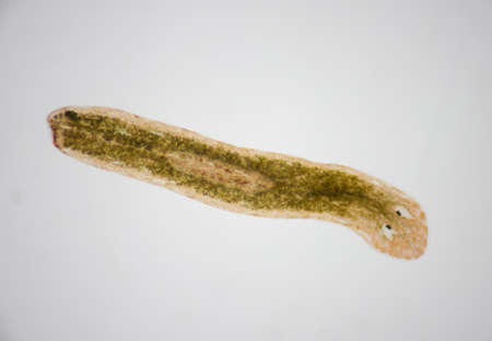
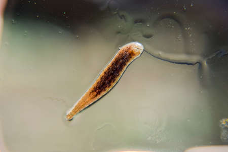
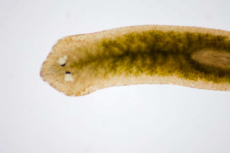
Planaria flatworm, under microscope view.(Soft focus)
Коллекция по умолчанию
Коллекция по умолчанию
Создать новую
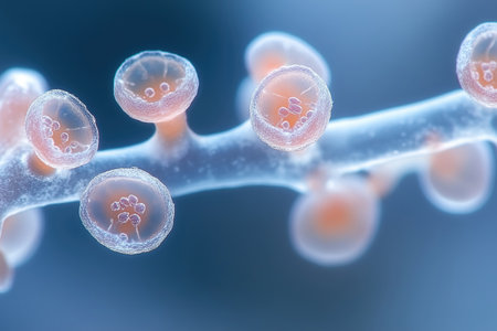
This vivid depiction shows coral polyps extending from their skeletons during high tide in a vibrant marine ecosystem.
Коллекция по умолчанию
Коллекция по умолчанию
Создать новую

X chromosome . 3D render
Коллекция по умолчанию
Коллекция по умолчанию
Создать новую
squamous cell
Коллекция по умолчанию
Коллекция по умолчанию
Создать новую

Protozoa and Green Algae in waste water under the microscope.
Коллекция по умолчанию
Коллекция по умолчанию
Создать новую
Colorful cells are illuminated under a microscope showcasing intricate structures and details illustrating biological processes in action.
Коллекция по умолчанию
Коллекция по умолчанию
Создать новую
Macro photo of a Silverfish (Lepisma saccharina) on a black studio background
Коллекция по умолчанию
Коллекция по умолчанию
Создать новую
A macro of a moth sitting on a glass pane
Коллекция по умолчанию
Коллекция по умолчанию
Создать новую

Rare image of Ghost flatworm - Maricola (Planarian) triclad flatworms in reef aquarium glass
Коллекция по умолчанию
Коллекция по умолчанию
Создать новую
Planarian parasite (flatworm) under microscope view.
Коллекция по умолчанию
Коллекция по умолчанию
Создать новую

Blastomyces dermatitidis fungi, the causative agent of the disease blastomycosis affecting lungs, more rarely skin, bones, other organs, 3D illustration. Filamentous form
Коллекция по умолчанию
Коллекция по умолчанию
Создать новую
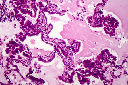
Lung adenocarcinoma, light micrograph, photo under microscope
Коллекция по умолчанию
Коллекция по умолчанию
Создать новую
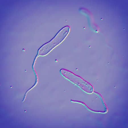
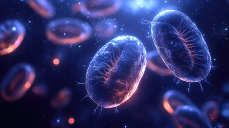
Vibrio cholerae bacteria, 3D illustration. Bacterium which causes cholera disease and is transmitted by contaminated water
Коллекция по умолчанию
Коллекция по умолчанию
Создать новую

Fungus Trichophyton rubrum, 3D illustration showing macroconidia, microconidia and septate hyphae. Infects skin and nails causing dermatophytosis, especially on feet (tinea pedis), and onychomycosis
Коллекция по умолчанию
Коллекция по умолчанию
Создать новую
Legion-Media
Создайте свои проекты на основе качественных стоковых фотографий и видео.


Copyright © Legion-Media.