
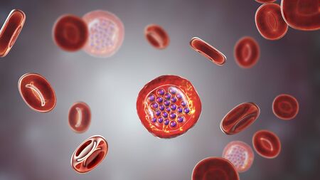
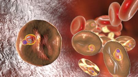
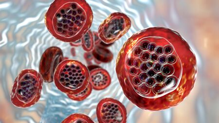
The malaria-infected red blood cells. 3D illustration showing ring-form trophozoites of malaria parasite Plasmodium falciparum inside red blood cells, the causative agent of tropical malaria
Коллекция по умолчанию
Коллекция по умолчанию
Создать новую
Malaria blood parasite infected red blood cells laboratory background.
Коллекция по умолчанию
Коллекция по умолчанию
Создать новую
Cytomegalovirus CMV in a human cell, owl's eye inclusion in nucleus, multinucleated cell, 3D illustration. It is herpes virus, causes diseases in fetus, organ transplant patients, HIV infected people
Коллекция по умолчанию
Коллекция по умолчанию
Создать новую
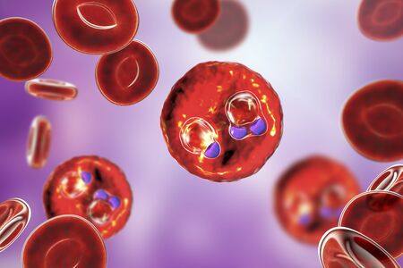
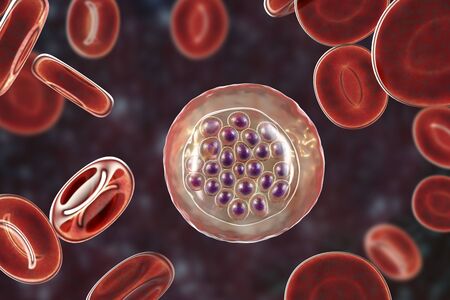
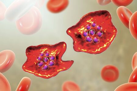
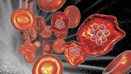
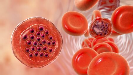
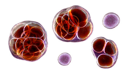
Red blood cells infected with malaria parasite, 3D illustration showing Plasmodium parasites inside red blood cells in the stage of schizont
Коллекция по умолчанию
Коллекция по умолчанию
Создать новую
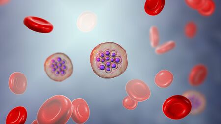
Red blood cells infected with malaria parasite Plasmodium vivax, schizont stage, 3D illustration
Коллекция по умолчанию
Коллекция по умолчанию
Создать новую
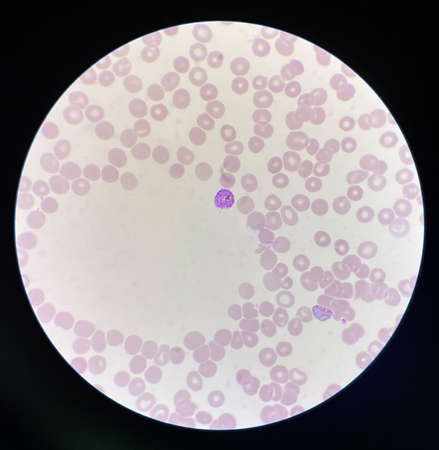
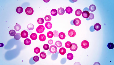
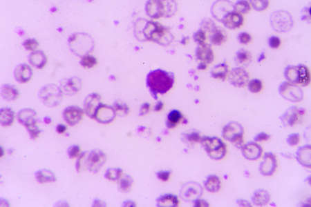
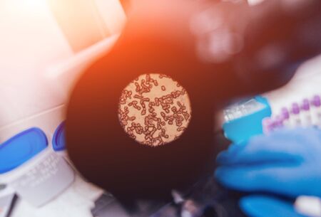
blood films for Malaria parasite
Коллекция по умолчанию
Коллекция по умолчанию
Создать новую
Neutrophil show in blood smear CBC test find with microscope.
Коллекция по умолчанию
Коллекция по умолчанию
Создать новую
Immature white blood cells in leukemia.Science concept.
Коллекция по умолчанию
Коллекция по умолчанию
Создать новую
The malaria-infected red blood cells. 3D illustration showing malaria parasite Plasmodium falciparum in schizont stage inside red blood cells, the causative agent of tropical malaria
Коллекция по умолчанию
Коллекция по умолчанию
Создать новую
The malaria-infected red blood cells. 3D illustration showing malaria parasite Plasmodium falciparum in schizont stage inside red blood cells, the causative agent of tropical malaria
Коллекция по умолчанию
Коллекция по умолчанию
Создать новую
The malaria-infected red blood cells. 3D illustration showing ring-form trophozoites of malaria parasite Plasmodium falciparum inside red blood cells, the causative agent of tropical malaria
Коллекция по умолчанию
Коллекция по умолчанию
Создать новую
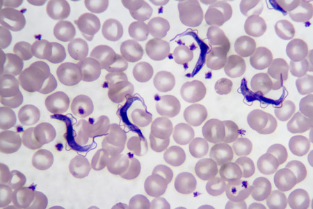
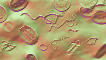
Trypanosoma gambiense blood smear viewed under a microscope at 1250 power.
Коллекция по умолчанию
Коллекция по умолчанию
Создать новую

Trypanosoma cruzi parasite, 3D illustration. A protozoan that causes Chagas disease transmitted to humans by the bite of triatomine bug
Коллекция по умолчанию
Коллекция по умолчанию
Создать новую
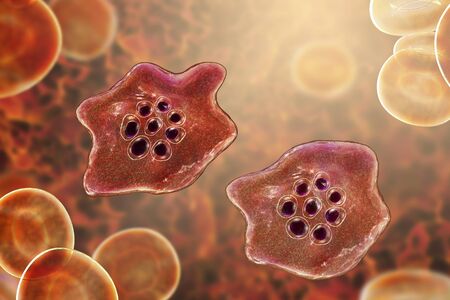
Babesia parasites inside red blood cell, the causative agent of babesiosis. 3D illustration showing classic tetrad-forms of Babesia merozoites so-called Maltese cross formation
Коллекция по умолчанию
Коллекция по умолчанию
Создать новую
Histopathology of human liver under microscope view for medical education.
Коллекция по умолчанию
Коллекция по умолчанию
Создать новую
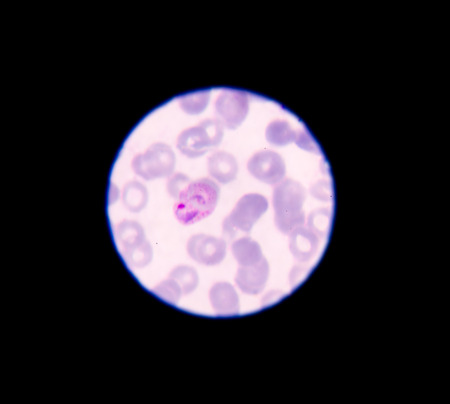
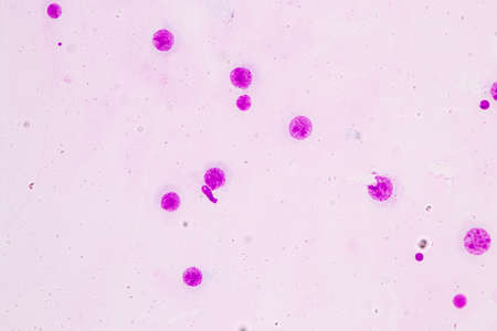
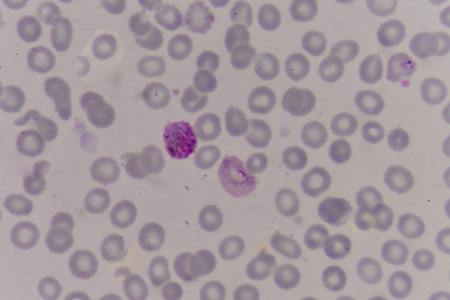
Malaria parasite in blood smear, gemetocyte stage
Коллекция по умолчанию
Коллекция по умолчанию
Создать новую
Blood smear with red blood cells in human body, medical background.
Коллекция по умолчанию
Коллекция по умолчанию
Создать новую
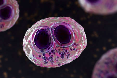
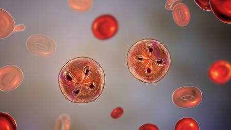
The malaria-infected red blood cell. 3D illustration showing malaria parasite Plasmodium ovale in the stage of schizont
Коллекция по умолчанию
Коллекция по умолчанию
Создать новую
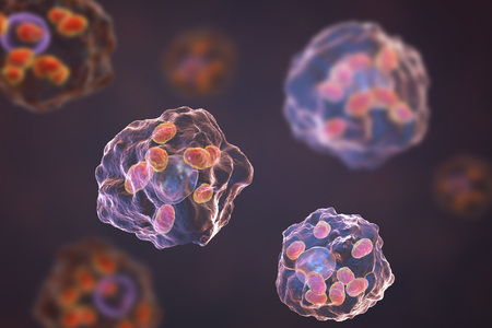
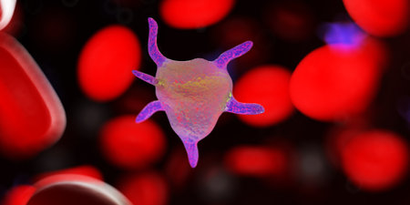
Macrophages infected by Leishmania amastigotes, 3D illustration
Коллекция по умолчанию
Коллекция по умолчанию
Создать новую

The malaria-infected red blood cell. 3D illustration showing malaria parasite Plasmodium ovale in the stage of schizont
Коллекция по умолчанию
Коллекция по умолчанию
Создать новую

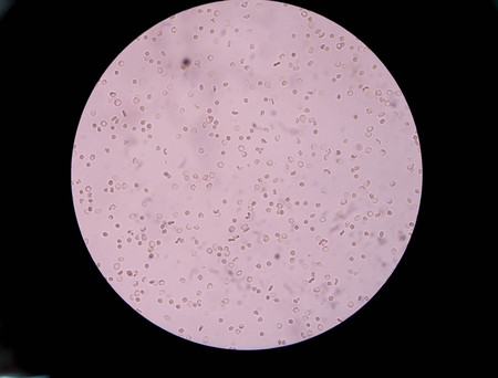
malaria parasite plasmodium falciparum on a thick blood smear reading under a microscope
Коллекция по умолчанию
Коллекция по умолчанию
Создать новую
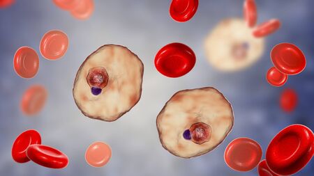
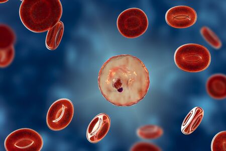
The malaria-infected red blood cell. 3D illustration showing malaria parasite Plasmodium ovale in the stage of ring-form trophozoite
Коллекция по умолчанию
Коллекция по умолчанию
Создать новую
Red blood cells infected with malaria parasite
Коллекция по умолчанию
Коллекция по умолчанию
Создать новую
Babesia parasites inside red blood cell, the causative agent of babesiosis. 3D illustration showing classic tetrad-forms of Babesia merozoites so-called Maltese cross formation
Коллекция по умолчанию
Коллекция по умолчанию
Создать новую
The malaria-infected red blood cell. 3D illustration showing malaria parasite Plasmodium ovale in the stage of schizont
Коллекция по умолчанию
Коллекция по умолчанию
Создать новую

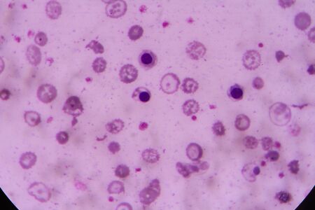
Picture of acute lymphocytic leukemia or ALL cells in blood smear, analyze by microscope, 400x
Коллекция по умолчанию
Коллекция по умолчанию
Создать новую
Blood cells in human body under microscope view for education in laboratory.
Коллекция по умолчанию
Коллекция по умолчанию
Создать новую
Chromosomes Human under the microscope for education.
Коллекция по умолчанию
Коллекция по умолчанию
Создать новую
Borrelia bacteria in blood, 3D illustration. The causative agent of Lyme disease and relapsing fever. Borrelia recurrentis, B. burgdorferi in blood smear under microscope
Коллекция по умолчанию
Коллекция по умолчанию
Создать новую

virus ebola and blood for sci and medical content 3d rendering
Коллекция по умолчанию
Коллекция по умолчанию
Создать новую
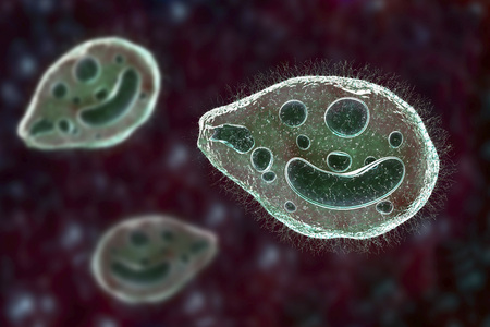
Balantidium coli protozoan, 3D illustration. Ciliated intestinal parasite that causes balantidiasis
Коллекция по умолчанию
Коллекция по умолчанию
Создать новую
3d rendered medically accurate illustration of a platelet
Коллекция по умолчанию
Коллекция по умолчанию
Создать новую
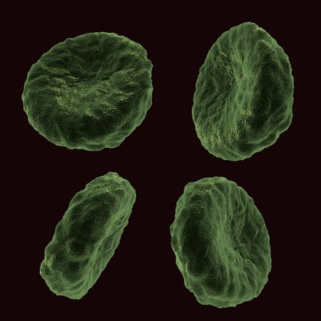
Four blood human cells in different positions
Коллекция по умолчанию
Коллекция по умолчанию
Создать новую
nucleated red cell
Коллекция по умолчанию
Коллекция по умолчанию
Создать новую
The malaria-infected red blood cells. 3D illustration showing parasite Plasmodium falciparum in schizont stage inside red blood cells, the causative agent of tropical malaria
Коллекция по умолчанию
Коллекция по умолчанию
Создать новую
The malaria-infected red blood cell. 3D illustration showing parasite Plasmodium malariae in the stage of ring-form trophozoite
Коллекция по умолчанию
Коллекция по умолчанию
Создать новую
The malaria-infected red blood cell. 3D illustration showing malaria parasite Plasmodium ovale in the stage of schizont
Коллекция по умолчанию
Коллекция по умолчанию
Создать новую
Microscope with metal lens at laboratory. Medical equipment.
Коллекция по умолчанию
Коллекция по умолчанию
Создать новую
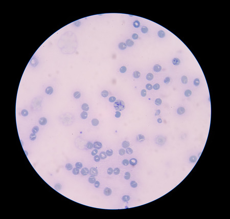
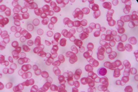
Blood smear showing white and red blood cells
Коллекция по умолчанию
Коллекция по умолчанию
Создать новую
blood cells with microscope.
Коллекция по умолчанию
Коллекция по умолчанию
Создать новую

Parasitic protozoans Toxoplasma gondii, the causative agent of toxoplasmosis in tachyzoite stage, 3D illustration
Коллекция по умолчанию
Коллекция по умолчанию
Создать новую
Electron micrograph of blood cells, showing intricate cellular structures in rich detail
Коллекция по умолчанию
Коллекция по умолчанию
Создать новую
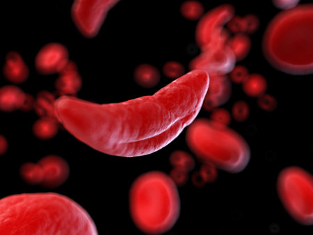
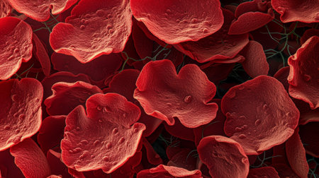
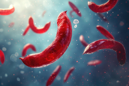
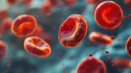
3d rendered medically accurate illustration of sickle cells
Коллекция по умолчанию
Коллекция по умолчанию
Создать новую
chia seeds in water
Коллекция по умолчанию
Коллекция по умолчанию
Создать новую
Malaria blood parasite infected red blood cells laboratory background.
Коллекция по умолчанию
Коллекция по умолчанию
Создать новую

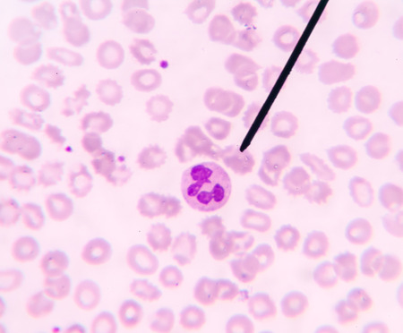
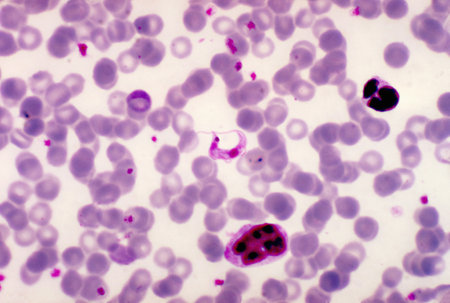
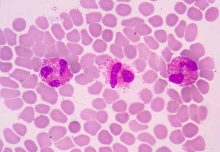
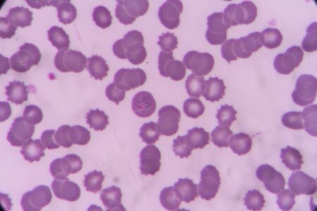
Blood smear showing, in the center, three neutrophil with hypersegmented nucleus. These cells appear in pathological situations such as megaloblastic anemias. Wright stain.
Коллекция по умолчанию
Коллекция по умолчанию
Создать новую
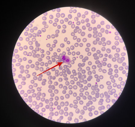
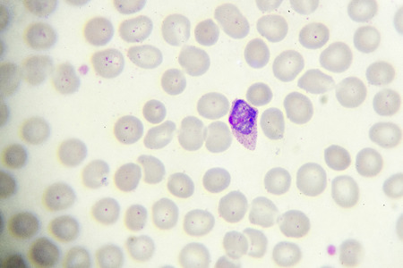
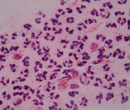
Red arrow showing neutrophil with toxic granule active PMN.
Коллекция по умолчанию
Коллекция по умолчанию
Создать новую
nucleated red cell
Коллекция по умолчанию
Коллекция по умолчанию
Создать новую
Trypanosoma lewisi parasites
Коллекция по умолчанию
Коллекция по умолчанию
Создать новую
Probiotics and prebiotics. The scientist examines useful microorganisms under a magnifying glass. Gram-positive bacteria under a magnifying glass. Healthy bacteria.
Коллекция по умолчанию
Коллекция по умолчанию
Создать новую
nucleated red cell
Коллекция по умолчанию
Коллекция по умолчанию
Создать новую
microscope lens, viewing Trypanosoma cruzi parasitic protozoan, causer of Chagas disease
Коллекция по умолчанию
Коллекция по умолчанию
Создать новую

Hydatid cyst of Echinococcus granulosus, 3D illustration. Echinococcus is a parasitic worm that causes of echinococcosis. It produces cysts in liver, lungs and other organs in humans
Коллекция по умолчанию
Коллекция по умолчанию
Создать новую
Microscopic close-up of vibrant stained human cells on a blue backdrop
Коллекция по умолчанию
Коллекция по умолчанию
Создать новую

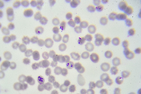
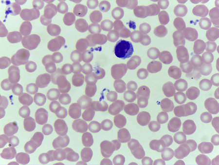
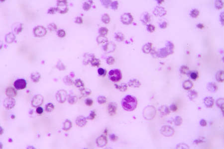
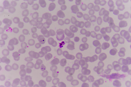
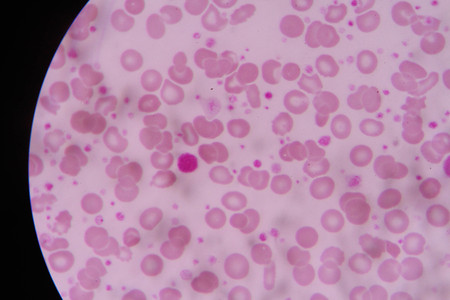
Malaria parasites in red blood cells under the microscope 400x
Коллекция по умолчанию
Коллекция по умолчанию
Создать новую

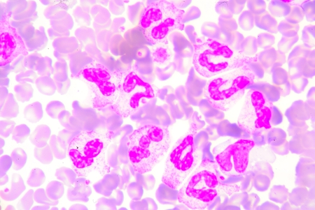
neutrophils. blood smear is often used as a follow-up test to abnormal results on a complete blood count (CBC) to evaluate the different types of blood cells.
Коллекция по умолчанию
Коллекция по умолчанию
Создать новую
Microscopic Field View of a Putrefaction Sample, Revealing Fungal Spores, Microbial Debris, and Protozoan Life Forms
Коллекция по умолчанию
Коллекция по умолчанию
Создать новую
Histopathology of human liver under microscope view for education in laboratory.
Коллекция по умолчанию
Коллекция по умолчанию
Создать новую
Fasciola hepatica, or liver fluke, 3D illustration. A parasitic trematode worm that causes fasciolosis, an infection of liver
Коллекция по умолчанию
Коллекция по умолчанию
Создать новую
Neutrophils are a type of phagocyte and are normally found in the bloodstream.
Коллекция по умолчанию
Коллекция по умолчанию
Создать новую
Close up blue bacteria cells with microscope.
Коллекция по умолчанию
Коллекция по умолчанию
Создать новую

Medicine - Human Blood Cells photographed using a microscope.
Коллекция по умолчанию
Коллекция по умолчанию
Создать новую
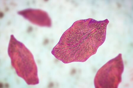
Vibrant Group of Red Peppers Flying Through the Air, An image representing the sickle-shaped blood cells found in sickle cell anemia, AI Generated
Коллекция по умолчанию
Коллекция по умолчанию
Создать новую
White blood cells of a human, Eosinophil photomicrograph panorama as seen under the microscope
Коллекция по умолчанию
Коллекция по умолчанию
Создать новую

Cytomegalovirus CMV in a human cell, owl's eye inclusion in nucleus, multinucleated cell, 3D illustration. It is herpes virus, causes diseases in fetus, organ transplant patients, HIV infected people
Коллекция по умолчанию
Коллекция по умолчанию
Создать новую

Abstract macro image of particles looking like bacteria, macro shot, microbiology theme
Коллекция по умолчанию
Коллекция по умолчанию
Создать новую
Cancer Cell in human showing abnormal cells.
Коллекция по умолчанию
Коллекция по умолчанию
Создать новую
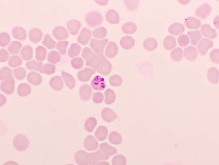
Malaria parasite in red blood cells, ring form stage of Plasmodium falciparum, original magnification 1000x
Коллекция по умолчанию
Коллекция по умолчанию
Создать новую

Pathogen, Bacteria and bacterium cells in microscopic as a medical illustration of bacterial disease infection in a human body
Коллекция по умолчанию
Коллекция по умолчанию
Создать новую
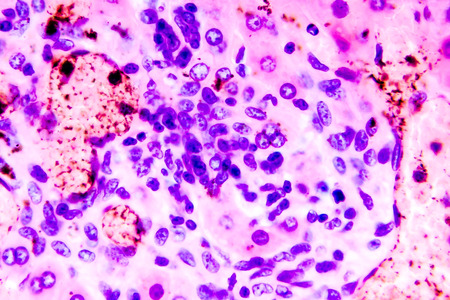
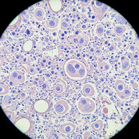
Coccidiosis, coccidia in liver, light micrograph. Micrograph shows bile duct hyperplasia and fibrosis with periductal inflammation, groups of coccidia, large violet cells
Коллекция по умолчанию
Коллекция по умолчанию
Создать новую
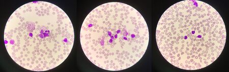
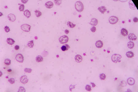
blood films for Malaria parasite.show malaria pigment.
Коллекция по умолчанию
Коллекция по умолчанию
Создать новую
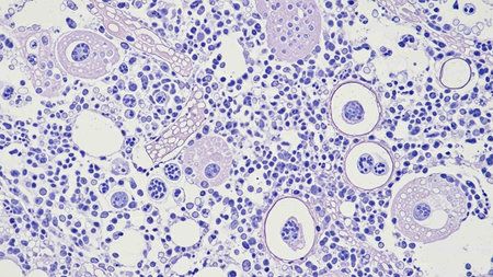
Histopathology of kidney failure, light micrograph, photo under microscope
Коллекция по умолчанию
Коллекция по умолчанию
Создать новую

Parasitic protozoans Toxoplasma gondii in the bloodstream, the causative agent of toxoplasmosis in tachyzoite stage, 3D illustration
Коллекция по умолчанию
Коллекция по умолчанию
Создать новую
Education anatomy and Histological sample of Human under the microscope.
Коллекция по умолчанию
Коллекция по умолчанию
Создать новую

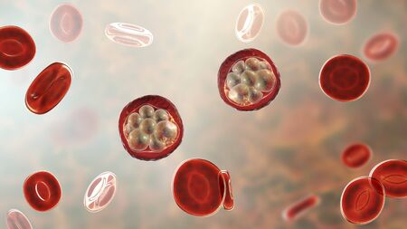
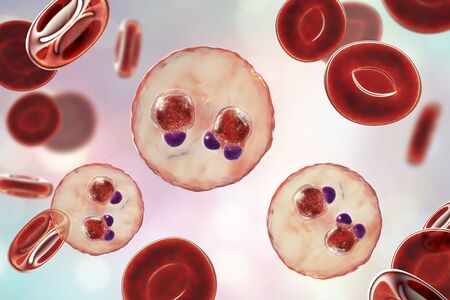
The malaria-infected red blood cell. 3D illustration showing parasite Plasmodium malariae in the schizont stage
Коллекция по умолчанию
Коллекция по умолчанию
Создать новую
nucleated red cell
Коллекция по умолчанию
Коллекция по умолчанию
Создать новую
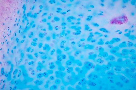
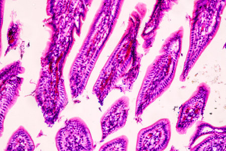
Hyaline cartilage, Elastic cartilage and Bone Human under the microscope in Lab.
Коллекция по умолчанию
Коллекция по умолчанию
Создать новую
Dynamic flow of blood cells in a medical background illustrating circulatory system functionality
Коллекция по умолчанию
Коллекция по умолчанию
Создать новую

plasmodium
Коллекция по умолчанию
Коллекция по умолчанию
Создать новую
Anatomy and Histological Ovary, Testis and Sperm human cells under microscope.
Коллекция по умолчанию
Коллекция по умолчанию
Создать новую
Reticulocytes develop and mature in the bone marrow and then circulate for about a day in the blood stream before developing into mature red blood cells.
Коллекция по умолчанию
Коллекция по умолчанию
Создать новую

Euglena is a genus of single-celled flagellate Eukaryotes under microscopic view for education.
Коллекция по умолчанию
Коллекция по умолчанию
Создать новую
Moderate Red cell on center Mycobacterium Tuberculosis bacteria.
Коллекция по умолчанию
Коллекция по умолчанию
Создать новую
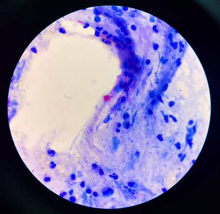
complete blood count
Коллекция по умолчанию
Коллекция по умолчанию
Создать новую

Ascaris lumbricoides, a large roundworm, fertilized egg, 3D illustration
Коллекция по умолчанию
Коллекция по умолчанию
Создать новую

Human hyaline cartilage bone under microscope view for education pathology. Human tissue.
Коллекция по умолчанию
Коллекция по умолчанию
Создать новую
complete blood count
Коллекция по умолчанию
Коллекция по умолчанию
Создать новую

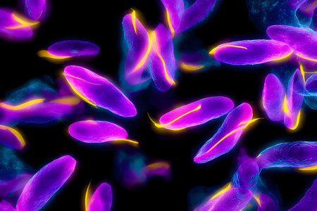
Sleeping sickness parasites, 3D illustration. Trypanosoma parasites transmitted by tse-tse fly and causing African sleeping sickness
Коллекция по умолчанию
Коллекция по умолчанию
Создать новую
Red blood cell in urine under microscopic
Коллекция по умолчанию
Коллекция по умолчанию
Создать новую
Colorful microscopic view of fluorescent protozoa or bacteria with glowing purple and yellow cell structures
Коллекция по умолчанию
Коллекция по умолчанию
Создать новую
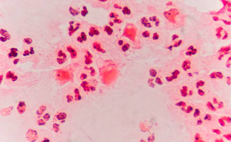
Moderate red white blood cells with gram negative diplococci intracellular Gram-negative coffee bean-shaped diplococci bacteria responsible for the sexually transmitted infection gonorrhea
Коллекция по умолчанию
Коллекция по умолчанию
Создать новую
plasmodium
Коллекция по умолчанию
Коллекция по умолчанию
Создать новую
Red Blood Cells Flowing in human circulatory system
Коллекция по умолчанию
Коллекция по умолчанию
Создать новую
The malaria-infected red blood cells. 3D illustration showing parasite Plasmodium falciparum in schizont stage inside red blood cells, the causative agent of tropical malaria
Коллекция по умолчанию
Коллекция по умолчанию
Создать новую
Characteristics of Lichen, hyphae and Symbiotic algae under the microscope for education.
Коллекция по умолчанию
Коллекция по умолчанию
Создать новую
Erythrocytes red blood cells floating against dark backdrop. Medical illustration. Hematology concept. Generative AI.
Коллекция по умолчанию
Коллекция по умолчанию
Создать новую
Prototheca wickerhamii green algae that cause protothecosis, clinically seen as skin nodules and elbow bursitis. 3D illustration shows sporangia with endospores and nonviable ghostlike forms.
Коллекция по умолчанию
Коллекция по умолчанию
Создать новую

Host cells with spores (mold) are inside wood under the microscope for education.
Коллекция по умолчанию
Коллекция по умолчанию
Создать новую

Blood vessel with flowing blood cells, 3D illustration. Small blood vessels, capillaries
Коллекция по умолчанию
Коллекция по умолчанию
Создать новую
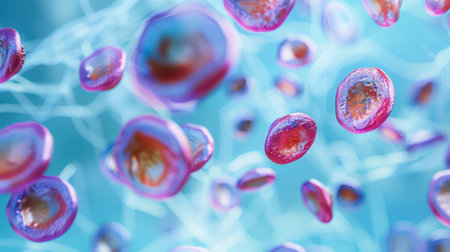
A microscopic image shows a cluster of pink and orange cells, with light refracting off their surfaces. The cells are suspended in a blue liquid, with a fine network of pale blue lines extending throughout the background.
Коллекция по умолчанию
Коллекция по умолчанию
Создать новую
Legion-Media
Создайте свои проекты на основе качественных стоковых фотографий и видео.


Copyright © Legion-Media.