

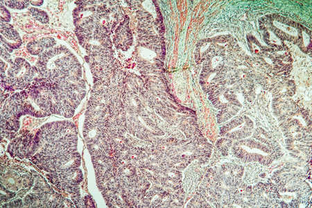
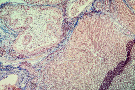
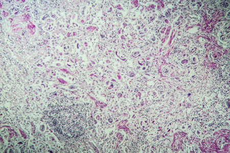
Ovarian cancer, light micrograph, photo under microscope. Photograph shows a fragment of a cancerous tumor in the female ovary. Selective focus
Коллекция по умолчанию
Коллекция по умолчанию
Создать новую
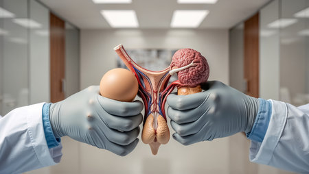
The guy holds his hands together in gloves. Close plan.
Коллекция по умолчанию
Коллекция по умолчанию
Создать новую

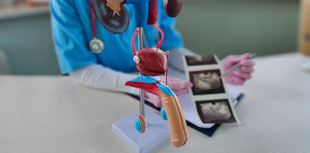
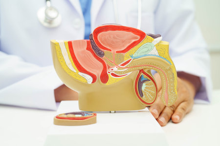
A doctor showing the anatomical model of the bladder with a prostate to the patient and explaining about it. The photo might be used for any articles about anatomical model of the bladder or for stude
Коллекция по умолчанию
Коллекция по умолчанию
Создать новую
Education anatomy and Histological sample of Human under the microscope.
Коллекция по умолчанию
Коллекция по умолчанию
Создать новую
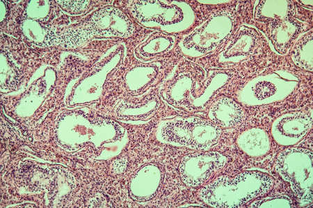
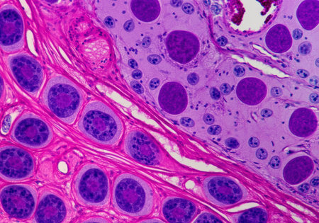
Inguinal testicles gonadically diseased tissue 100x
Коллекция по умолчанию
Коллекция по умолчанию
Создать новую
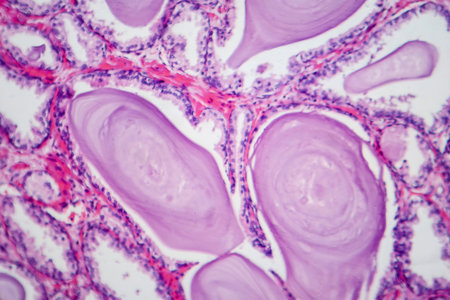
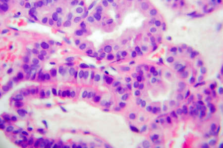
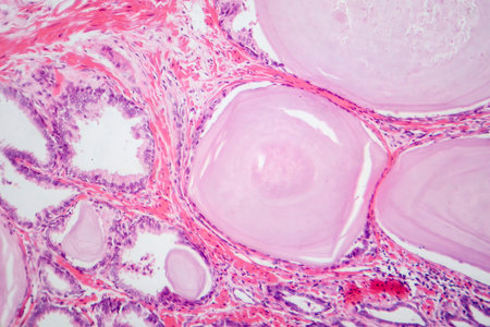
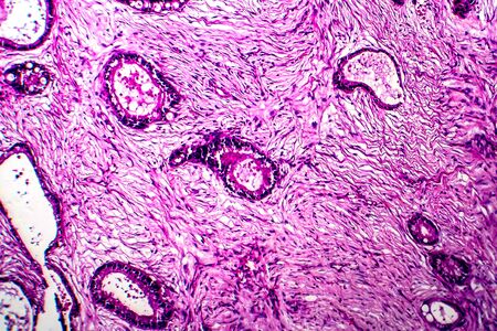
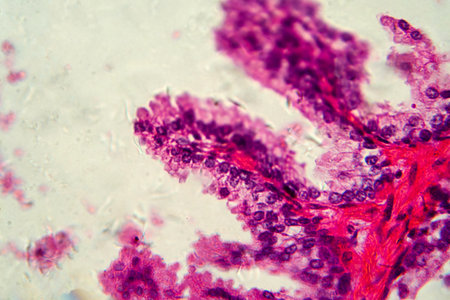
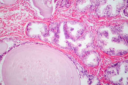
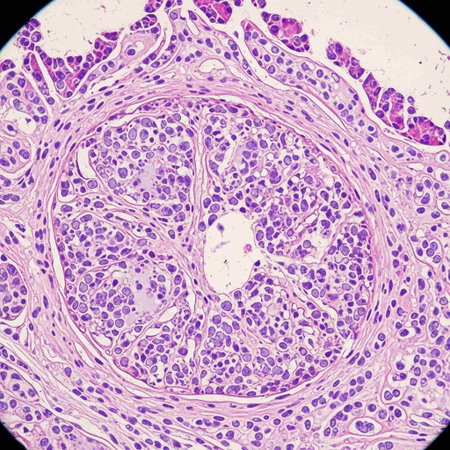
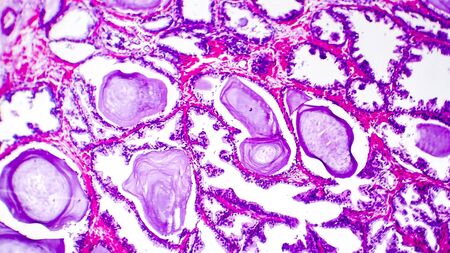
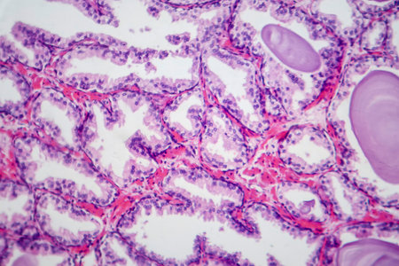
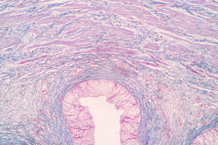
Photomicrograph showing histological features of benign prostatic hyperplasia. Enlarged prostate gland with nodular proliferation of glandular and stromal components.
Коллекция по умолчанию
Коллекция по умолчанию
Создать новую
Papillary thyroid carcinoma, light micrograph, photo under microscope. The most common type of thyroid cancer
Коллекция по умолчанию
Коллекция по умолчанию
Создать новую

Papillary thyroid carcinoma, light micrograph, photo under microscope. The most common type of thyroid cancer
Коллекция по умолчанию
Коллекция по умолчанию
Создать новую
Columnar epithelium of human gall bladder under the microscope in Lab.
Коллекция по умолчанию
Коллекция по умолчанию
Создать новую
science medical anthropotomy physiology microscopic section of lymph gland tissue background
Коллекция по умолчанию
Коллекция по умолчанию
Создать новую

Thyroid follicular carcinoma, light micrograph, photo under microscope
Коллекция по умолчанию
Коллекция по умолчанию
Создать новую

Stomach tissue under the microscope 100x
Коллекция по умолчанию
Коллекция по умолчанию
Создать новую
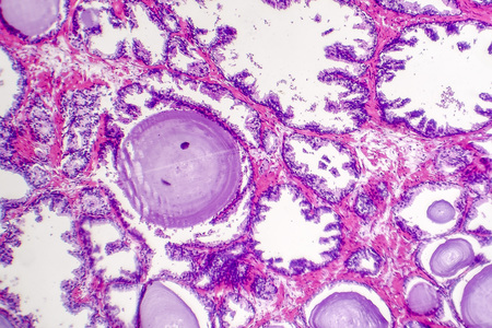
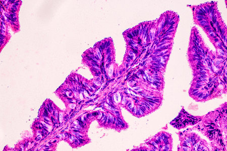
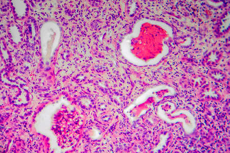
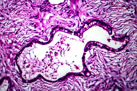
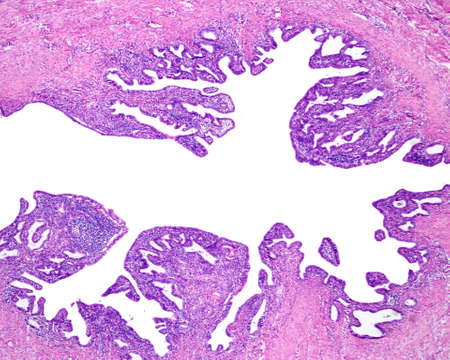
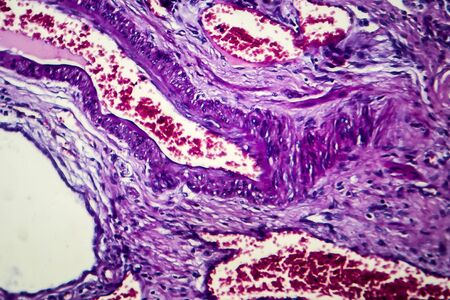
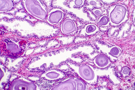
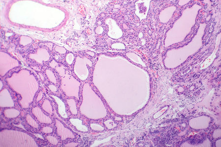
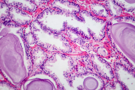
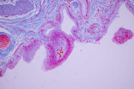
Benign prostatic hyperplasia. Micrograph shows dilated glands, papillary projections inside the lumen of the glands, cystic dilatation with accumulation of secretory material. Photo under microscope
Коллекция по умолчанию
Коллекция по умолчанию
Создать новую
Photomicrograph showing histological features of benign prostatic hyperplasia. Enlarged prostate gland with nodular proliferation of glandular and stromal components.
Коллекция по умолчанию
Коллекция по умолчанию
Создать новую
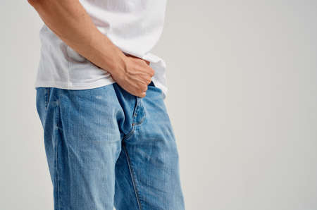
man with groin pain health problems impotence urology
Коллекция по умолчанию
Коллекция по умолчанию
Создать новую
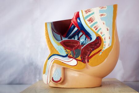
Male reproductive system model in half-cut perspective for education.
Коллекция по умолчанию
Коллекция по умолчанию
Создать новую
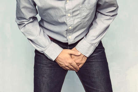
Man wants to pee and is holding his bladder.
Коллекция по умолчанию
Коллекция по умолчанию
Создать новую
Uterine cancer, light micrograph, photo under microscope
Коллекция по умолчанию
Коллекция по умолчанию
Создать новую
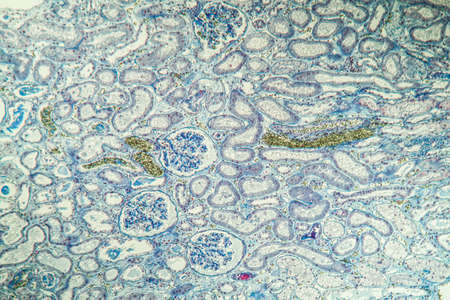
Chronic pyelonephritis, light micrograph, photo under microscope
Коллекция по умолчанию
Коллекция по умолчанию
Создать новую

Papillary thyroid carcinoma, light micrograph, photo under microscope. The most common type of thyroid cancer
Коллекция по умолчанию
Коллекция по умолчанию
Создать новую
Columnar epithelium of human gall bladder under the microscope in Lab.
Коллекция по умолчанию
Коллекция по умолчанию
Создать новую
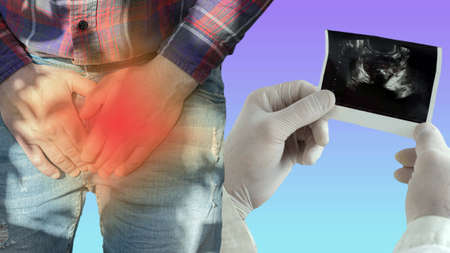
The male prostate gland is examined by an ultrasound scan at the doctor's hands, the prostate adenoma is analyzed by a urologist, a person with pain in the genitourinary system.
Коллекция по умолчанию
Коллекция по умолчанию
Создать новую
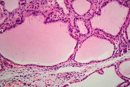
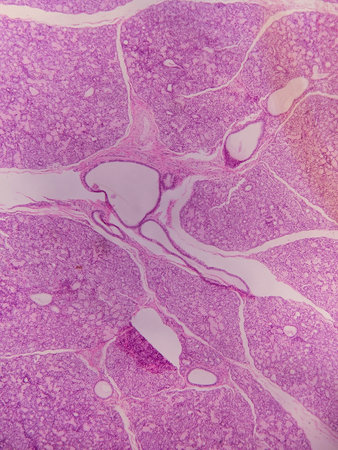
Endemic goiter, light micrograph, abnormal enlargement of the thyroid gland due to dietary iodine deficiency. Photomicrograph shows follicles of varying size, abundant colloid, lymphocytic infiltrate
Коллекция по умолчанию
Коллекция по умолчанию
Создать новую
Uterine cancer, light micrograph, photo under microscope
Коллекция по умолчанию
Коллекция по умолчанию
Создать новую
Photomicrograph showing histological features of benign prostatic hyperplasia. Enlarged prostate gland with nodular proliferation of glandular and stromal components. High-resolution histology image.
Коллекция по умолчанию
Коллекция по умолчанию
Создать новую
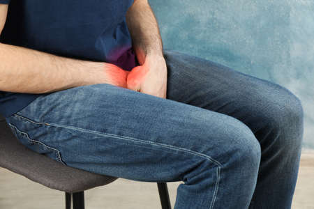
Man holds his crotch and sitting on chair. Urology
Коллекция по умолчанию
Коллекция по умолчанию
Создать новую
businessman open hand
Коллекция по умолчанию
Коллекция по умолчанию
Создать новую
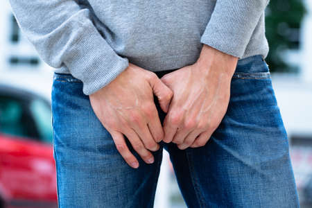
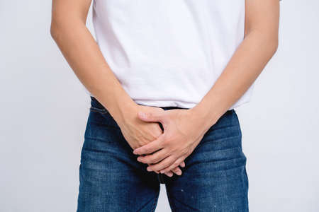
A man holds his groin with both hands, an attack of urological pain in the bladder
Коллекция по умолчанию
Коллекция по умолчанию
Создать новую

Influence of smoking on male potency and urologist. Smoking and sexual function harms of smoking and effects of smoking
Коллекция по умолчанию
Коллекция по умолчанию
Создать новую

Male doctors and patients, sexual problems are discussing the symptoms and treatment methods.
Коллекция по умолчанию
Коллекция по умолчанию
Создать новую
Colon carcinoma arising from adenoma, 100x
Коллекция по умолчанию
Коллекция по умолчанию
Создать новую
Close-up Of A Man's Hand On His Crotch
Коллекция по умолчанию
Коллекция по умолчанию
Создать новую
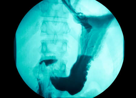
picture of intestinal abdominal xray
Коллекция по умолчанию
Коллекция по умолчанию
Создать новую
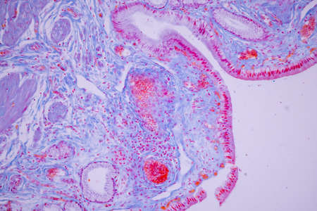
Human fallopian tube affected by chronic salpingitis. The fallopian tube lumen is enlarged, the mucosal folds widened and shortened, and the lamina propria shows chronic inflammatory infiltrates.
Коллекция по умолчанию
Коллекция по умолчанию
Создать новую

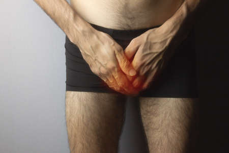
Pain on penis concept : Man use his hands to press on his penis on grey background
Коллекция по умолчанию
Коллекция по умолчанию
Создать новую

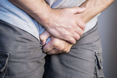
Bowel Incontinence Pain. Man Hand Holding Crotch
Коллекция по умолчанию
Коллекция по умолчанию
Создать новую

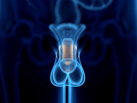
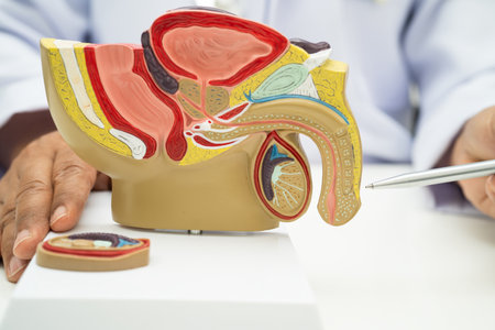
Male genitalia on the human model. Incision of the penis, testicles, bladder, prostate
Коллекция по умолчанию
Коллекция по умолчанию
Создать новую

X-ray of the pelvic bones of a man. A doctor radiologist is studying an x-ray examination. A hip joint is placed on the patient’s body
Коллекция по умолчанию
Коллекция по умолчанию
Создать новую
Cancer swollen Parotid diseased tissue 100x
Коллекция по умолчанию
Коллекция по умолчанию
Создать новую
Education anatomy and Histological sample of Human under the microscope.
Коллекция по умолчанию
Коллекция по умолчанию
Создать новую
Man holds his crotch on blue background, close up
Коллекция по умолчанию
Коллекция по умолчанию
Создать новую
Chronic nephritis, light micrograph, photo under microscope
Коллекция по умолчанию
Коллекция по умолчанию
Создать новую
Photomicrograph showing histological features of benign prostatic hyperplasia. Enlarged prostate gland with nodular proliferation of glandular and stromal components.
Коллекция по умолчанию
Коллекция по умолчанию
Создать новую

Characteristics of Lichen, hyphae and Symbiotic algae under the microscope for education.
Коллекция по умолчанию
Коллекция по умолчанию
Создать новую
Prostate cancer, light micrograph, photo under microscope
Коллекция по умолчанию
Коллекция по умолчанию
Создать новую

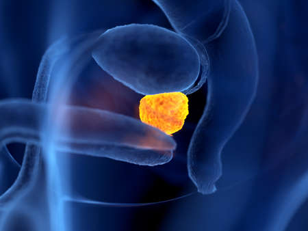
3d rendered medically accurate illustration of prostate cancer
Коллекция по умолчанию
Коллекция по умолчанию
Создать новую
Коллекция по умолчанию
Коллекция по умолчанию
Создать новую
Doctor makes diagnosis and gives advice on treating male erectile dysfunction. Man health problems and pills
Коллекция по умолчанию
Коллекция по умолчанию
Создать новую

Characteristics Tissue of Olfactory epithelium Human under the microscope in Lab.
Коллекция по умолчанию
Коллекция по умолчанию
Создать новую
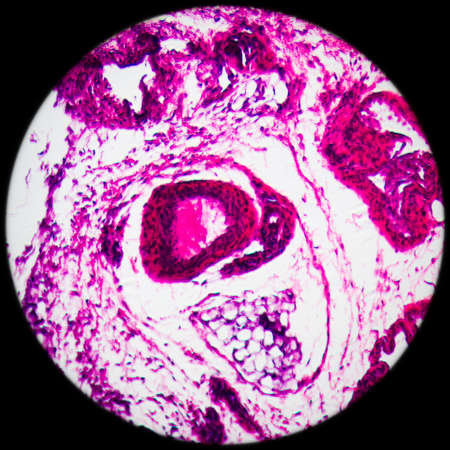
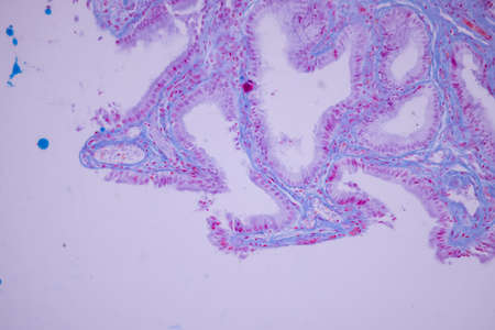
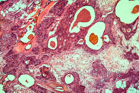
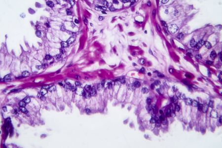
Benign prostatic hyperplasia. Micrograph shows dilated glands, papillary projections inside the lumen of the glands, cystic dilatation with accumulation of secretory material. Photo under microscope
Коллекция по умолчанию
Коллекция по умолчанию
Создать новую
Histopathology of cirrhosis, light micrograph, photo under microscope
Коллекция по умолчанию
Коллекция по умолчанию
Создать новую
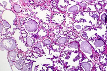
Benign prostatic hyperplasia. Micrograph shows dilated glands, papillary projections inside the lumen of the glands, cystic dilatation with accumulation of secretory material. Photo under microscope
Коллекция по умолчанию
Коллекция по умолчанию
Создать новую
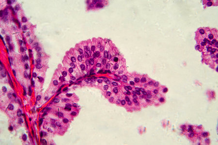
Photomicrograph showing histological features of benign prostatic hyperplasia. Enlarged prostate gland with nodular proliferation of glandular and stromal components. High-resolution histology image.
Коллекция по умолчанию
Коллекция по умолчанию
Создать новую
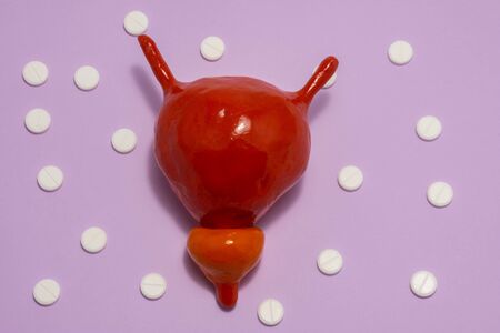
Model or figure of urinary bladder and prostate, which corresponds to anatomical original is located in purple background surrounded by white pills ornamented in polka dots. Photo for use in urology
Коллекция по умолчанию
Коллекция по умолчанию
Создать новую

Photomicrograph showing histological features of benign prostatic hyperplasia. Enlarged prostate gland with nodular proliferation of glandular and stromal components.
Коллекция по умолчанию
Коллекция по умолчанию
Создать новую
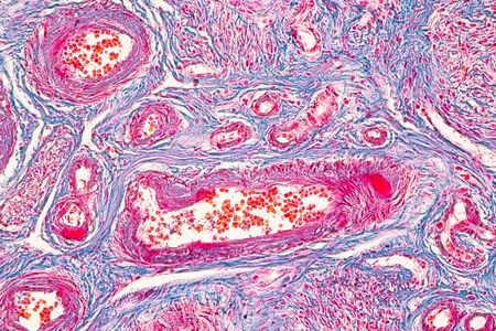
serous gland tissue under the microscope 100x
Коллекция по умолчанию
Коллекция по умолчанию
Создать новую
3d rendered illustration of the corpus cavernosum
Коллекция по умолчанию
Коллекция по умолчанию
Создать новую
Man holding his urethra in pain on gray background
Коллекция по умолчанию
Коллекция по умолчанию
Создать новую
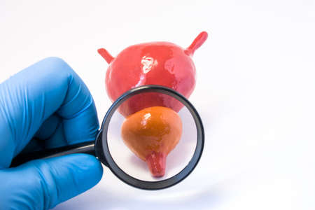
Realistic anatomical model of urine bladder and prostate gland is in hand of doctor, health medical professional or scientist wearing blue medical glove. Concept photo of studying anatomy of urinary
Коллекция по умолчанию
Коллекция по умолчанию
Создать новую

Endemic goiter, light micrograph, abnormal enlargement of the thyroid gland due to dietary iodine deficiency. Photomicrograph shows follicles of varying size, abundant colloid, lymphocytic infiltrate
Коллекция по умолчанию
Коллекция по умолчанию
Создать новую
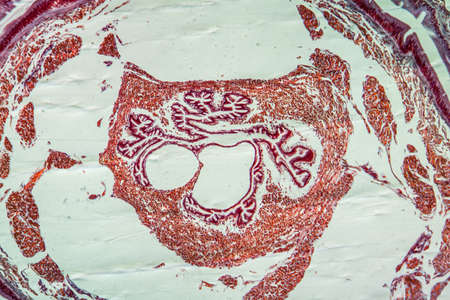
Histopathology of prostate gland hyperplasia, light micrograph, photo under microscope
Коллекция по умолчанию
Коллекция по умолчанию
Создать новую

Prostate cancer, light micrograph, photo under microscope
Коллекция по умолчанию
Коллекция по умолчанию
Создать новую
Coccidiosis of liver tissue under the microscope 100x
Коллекция по умолчанию
Коллекция по умолчанию
Создать новую
Abstract background of acrylic paint in aquamarine and red tones
Коллекция по умолчанию
Коллекция по умолчанию
Создать новую
Male Reproductive System Anatomy Model In Doctors Office
Коллекция по умолчанию
Коллекция по умолчанию
Создать новую

Endemic goiter, light micrograph, abnormal enlargement of the thyroid gland due to dietary iodine deficiency. Photomicrograph shows follicles of varying size, abundant colloid, lymphocytic infiltrate
Коллекция по умолчанию
Коллекция по умолчанию
Создать новую
The urinary system in man palm hand on blue background
Коллекция по умолчанию
Коллекция по умолчанию
Создать новую
Dentists instruments with shallow depth of field.
Коллекция по умолчанию
Коллекция по умолчанию
Создать новую
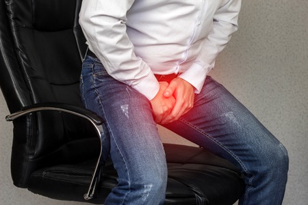
A man sits in an office chair and holds on to the groin, crotch, prostatitis, average plan, adenoma
Коллекция по умолчанию
Коллекция по умолчанию
Создать новую
Man suffering from prostate cancer isolated on a white background.
Коллекция по умолчанию
Коллекция по умолчанию
Создать новую

3D Illustration Concept of Human Urinary System Bladder Anatomy
Коллекция по умолчанию
Коллекция по умолчанию
Создать новую
Photomicrograph showing histological features of benign prostatic hyperplasia. Enlarged prostate gland with nodular proliferation of glandular and stromal components.
Коллекция по умолчанию
Коллекция по умолчанию
Создать новую
Man holds his crotch on blue background, close up
Коллекция по умолчанию
Коллекция по умолчанию
Создать новую

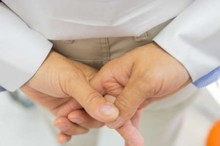
A mans intertwined fingers, a close-up of his hands folded in thought.
Коллекция по умолчанию
Коллекция по умолчанию
Создать новую
Prostate disease and treatment. Male reproductive system anatomical model in doctors hands close-up during consultation of male patient with suspected bacterial prostatitis
Коллекция по умолчанию
Коллекция по умолчанию
Создать новую

illustration of bladder and kydneys detox with highlighted organ and contrast hands on dark background. Low key photo with copy space toned in dark green colors. Medical concept design template
Коллекция по умолчанию
Коллекция по умолчанию
Создать новую
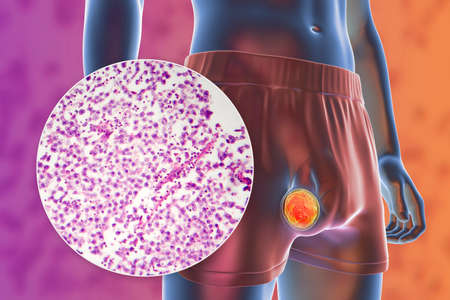
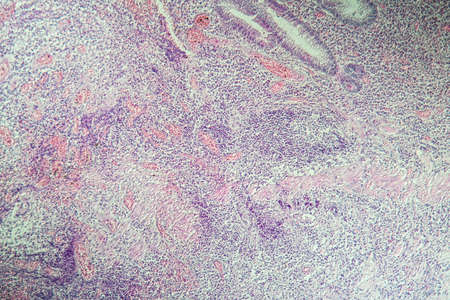
Testicular cancer, testicular seminoma, medical 3D illustration and light micrograph. Malignant tumor of the testis
Коллекция по умолчанию
Коллекция по умолчанию
Создать новую
Male reproductive system, doctor holding human anatomy model for study diagnosis and treatment in hospital.
Коллекция по умолчанию
Коллекция по умолчанию
Создать новую

The Bladder 3D rendered anatomical illustration
Коллекция по умолчанию
Коллекция по умолчанию
Создать новую
Woman suffering from pain in the stomach.
Коллекция по умолчанию
Коллекция по умолчанию
Создать новую
Photomicrograph of an endemic goiter tissue sample under a microscope, revealing thyroid gland abnormalities, including thyroid follicular cell hyperplasia and colloid-filled follicles.
Коллекция по умолчанию
Коллекция по умолчанию
Создать новую
Fibrin deposits in the kidney, microscopy 100x
Коллекция по умолчанию
Коллекция по умолчанию
Создать новую
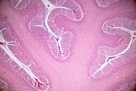
Cross section of the Cerebellum and Nerve human under the microscope for education in Lab.
Коллекция по умолчанию
Коллекция по умолчанию
Создать новую

Ovarian cancer, light micrograph, photo under microscope. Photograph shows a fragment of a cancerous tumor in the female ovary. Selective focus
Коллекция по умолчанию
Коллекция по умолчанию
Создать новую

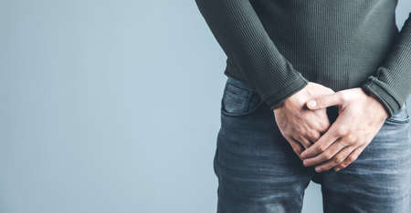
Man holding his groin, isolated on white background. Men's health
Коллекция по умолчанию
Коллекция по умолчанию
Создать новую
This detailed microscopic image showcases various cellular structures, highlighted in striking purple tones. The intricate patterns and textures reveal the complexity of biological tissues, making it a valuable resource for educational and scientific purposes
Коллекция по умолчанию
Коллекция по умолчанию
Создать новую
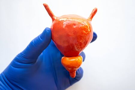
Doctor's hands in rubber gloves holding a model of the prostate
Коллекция по умолчанию
Коллекция по умолчанию
Создать новую
3d rendered illustration of the human prostate
Коллекция по умолчанию
Коллекция по умолчанию
Создать новую
Histological Uterus human, Uterine tube human, Placenta human and Umbilical cord Human under the microscope for education.
Коллекция по умолчанию
Коллекция по умолчанию
Создать новую

Man with prostate problem visiting urologist in clinic, closeup
Коллекция по умолчанию
Коллекция по умолчанию
Создать новую
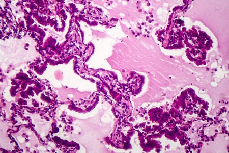
Lung adenocarcinoma, light micrograph, photo under microscope
Коллекция по умолчанию
Коллекция по умолчанию
Создать новую
A microscopic view of tissue showing pink-stained cells and structures, indicative of biological samples, possibly related to histology or pathology
Коллекция по умолчанию
Коллекция по умолчанию
Создать новую
Colon inflammation in Crohn's disease 100x
Коллекция по умолчанию
Коллекция по умолчанию
Создать новую
Photomicrograph showing histological features of benign prostatic hyperplasia. Enlarged prostate gland with nodular proliferation of glandular and stromal components.
Коллекция по умолчанию
Коллекция по умолчанию
Создать новую
Male reproductive system, doctor holding human anatomy model for study diagnosis and treatment in hospital.
Коллекция по умолчанию
Коллекция по умолчанию
Создать новую
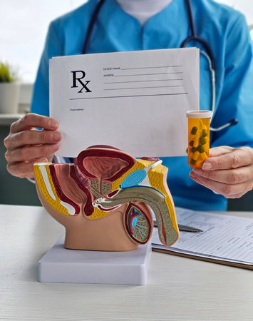
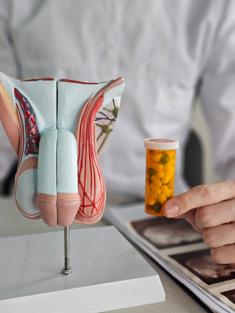
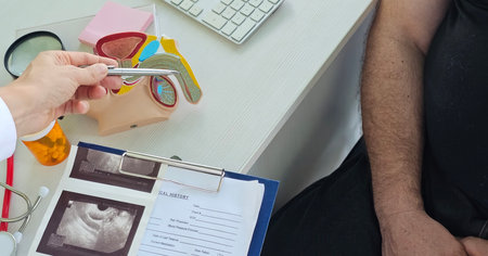
Medical professional holds prescription and anatomical model of male reproductive system during a consultation
Коллекция по умолчанию
Коллекция по умолчанию
Создать новую
Columnar epithelium of human gall bladder under the microscope in Lab.
Коллекция по умолчанию
Коллекция по умолчанию
Создать новую
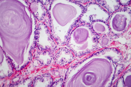
Photomicrograph showing histological features of benign prostatic hyperplasia. Enlarged prostate gland with nodular proliferation of glandular and stromal components.
Коллекция по умолчанию
Коллекция по умолчанию
Создать новую
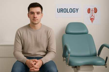
Young man sitting nervously in a doctor's office waiting room, anticipating an upcoming urology appointment and feeling anxious about his health
Коллекция по умолчанию
Коллекция по умолчанию
Создать новую
Lungworm under the microscope 100x
Коллекция по умолчанию
Коллекция по умолчанию
Создать новую
Legion-Media
Создайте свои проекты на основе качественных стоковых фотографий и видео.


Copyright © Legion-Media.