
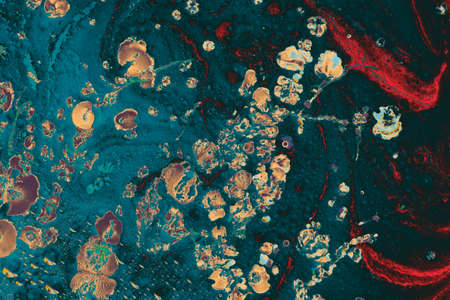
Painting acrylic paint- abstract drawing. Texture background
Коллекция по умолчанию
Коллекция по умолчанию
Создать новую

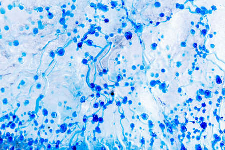
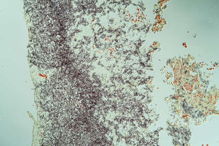
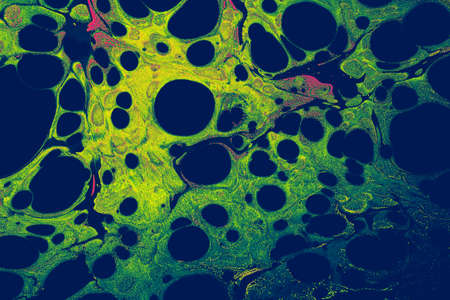
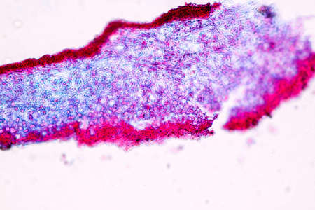
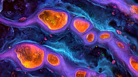
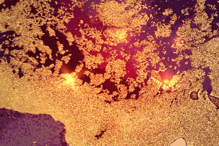
Abstract macro image of particles looking like bacteria, macro shot, microbiology theme
Коллекция по умолчанию
Коллекция по умолчанию
Создать новую
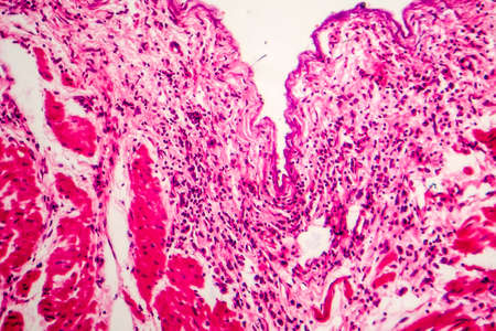
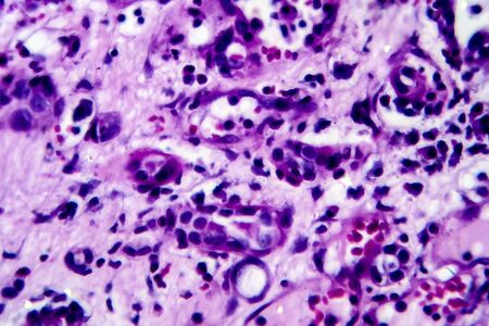
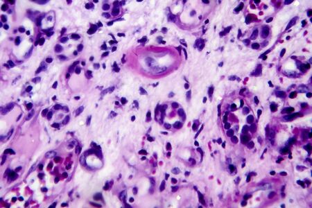
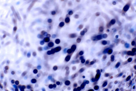
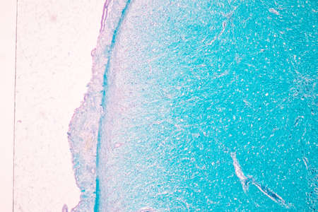
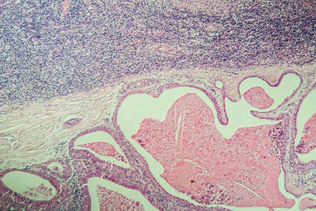
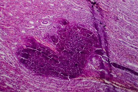
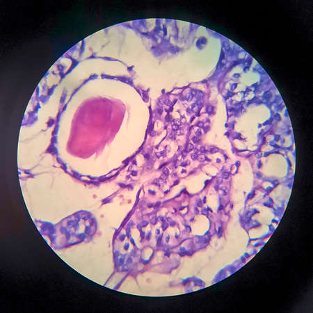
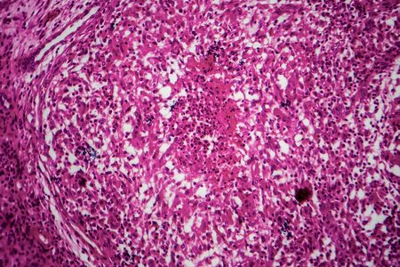
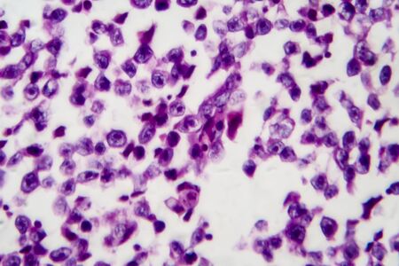
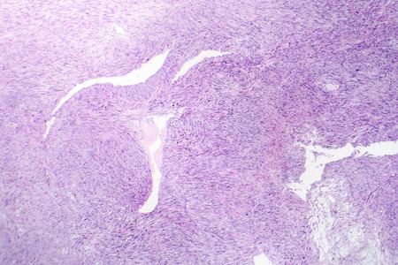
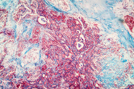
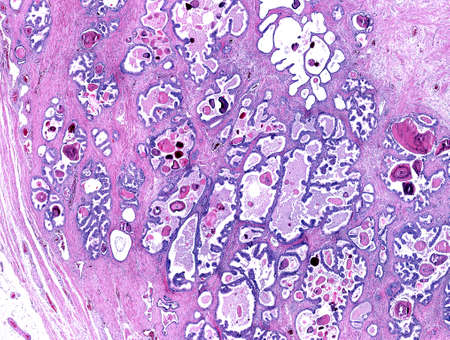
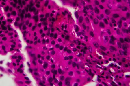
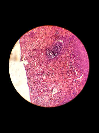
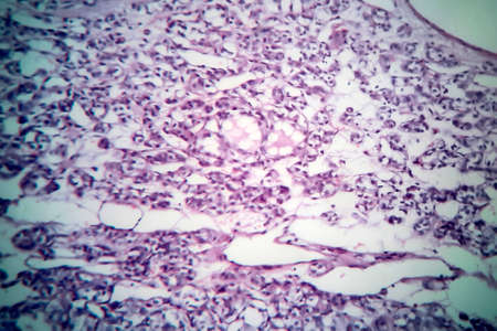
Signet ring cell carcinoma of the stomach, light micrograph, photo under microscope
Коллекция по умолчанию
Коллекция по умолчанию
Создать новую
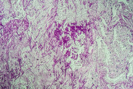
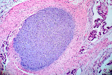
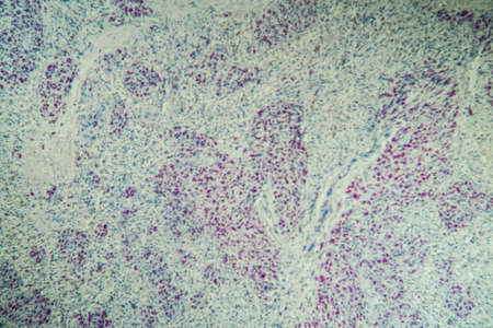
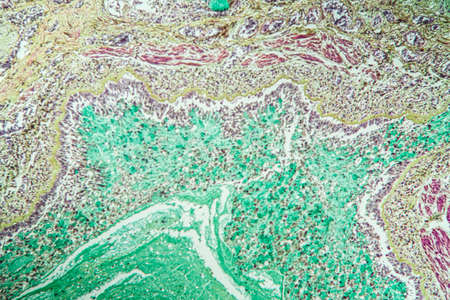
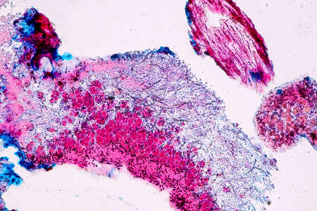
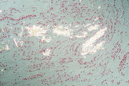
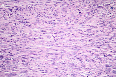
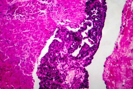
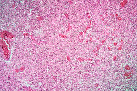
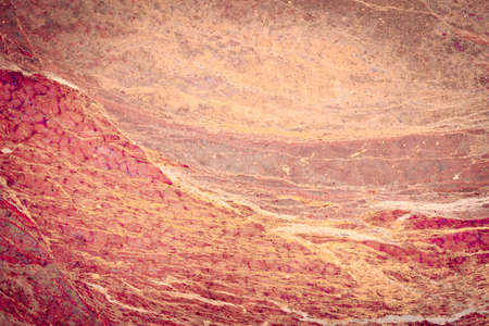
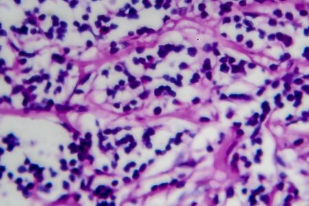
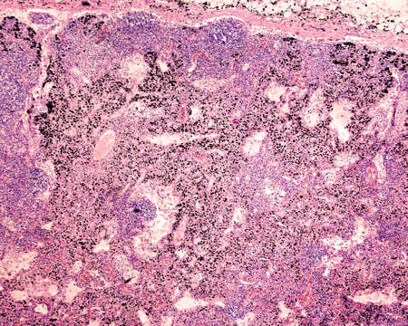
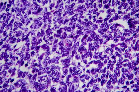
lungs Infection with Candida and Aspergillus in AIDS patients 200x
Коллекция по умолчанию
Коллекция по умолчанию
Создать новую
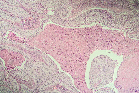
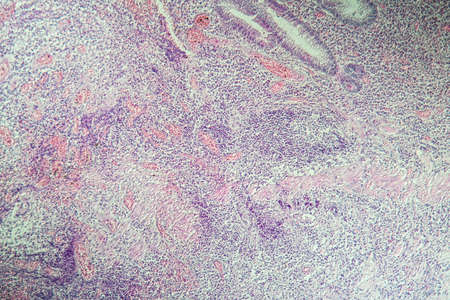
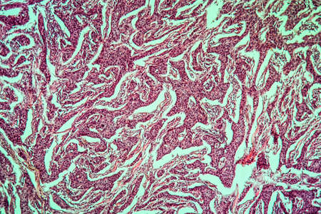
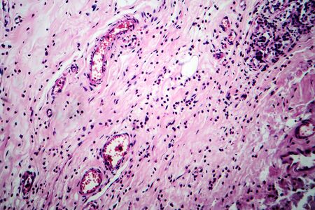
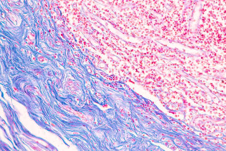
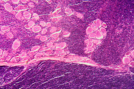
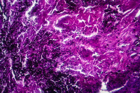
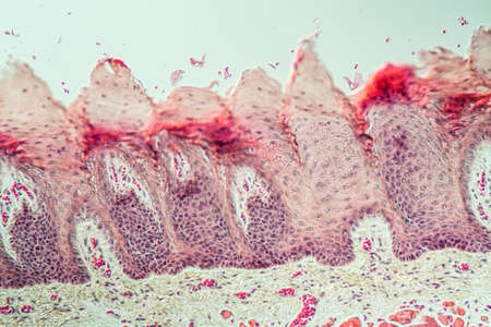
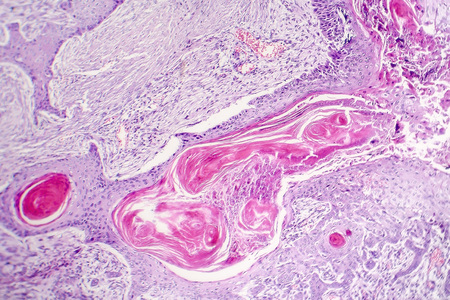
Squamous cell carcinoma diseased tissue under the microscope 100x
Коллекция по умолчанию
Коллекция по умолчанию
Создать новую
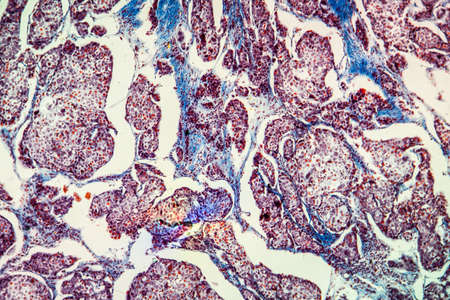
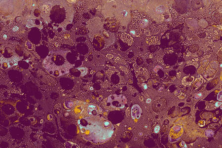
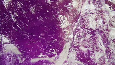
Histopathology of silicosis, the most prevalent chronic occupational disease. Light micrograph, photo under microscope
Коллекция по умолчанию
Коллекция по умолчанию
Создать новую
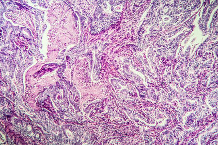
Histopathology of interstitial nephritis, light micrograph, photo under microscope. High magnification
Коллекция по умолчанию
Коллекция по умолчанию
Создать новую
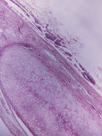
Light micrograph of teratoma, a tumor made up of several different types of tissue, such as hair, teeth, muscle, or bone. Teratoma is typically found in the ovary, testicle, or coccyx
Коллекция по умолчанию
Коллекция по умолчанию
Создать новую
Colon inflammation in Crohn's disease 100x
Коллекция по умолчанию
Коллекция по умолчанию
Создать новую

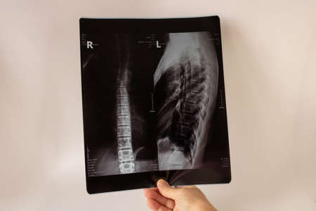
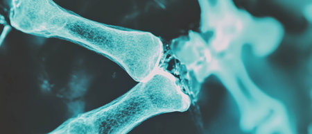
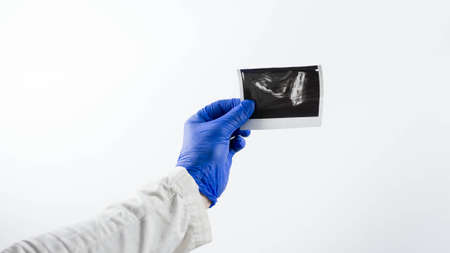
The doctor examines an X-ray of a patient with a knee injury on a white background with a place for text.
Коллекция по умолчанию
Коллекция по умолчанию
Создать новую

Characteristics of Lichen, hyphae and Symbiotic algae under the microscope for education.
Коллекция по умолчанию
Коллекция по умолчанию
Создать новую
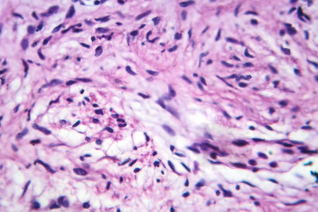
Breast cancer, light micrograph, photo under microscope
Коллекция по умолчанию
Коллекция по умолчанию
Создать новую
Bowen's Disease Tumor under the microscope 100x
Коллекция по умолчанию
Коллекция по умолчанию
Создать новую
Liver cirrhosis tissue affected 100x after alcohol abuse
Коллекция по умолчанию
Коллекция по умолчанию
Создать новую
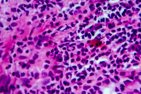
Hodgkin's lymphoma, light micrograph, photo under microscope. High magnification
Коллекция по умолчанию
Коллекция по умолчанию
Создать новую
AIDS with fungi 100x infected tissue
Коллекция по умолчанию
Коллекция по умолчанию
Создать новую
Breast cancer of the woman diseased tissue 100x
Коллекция по умолчанию
Коллекция по умолчанию
Создать новую
Basal cell cancer Diseased tissue 100x
Коллекция по умолчанию
Коллекция по умолчанию
Создать новую
Gastric carcinoma in tissue section 100x
Коллекция по умолчанию
Коллекция по умолчанию
Создать новую
Histopathology of interstitial nephritis, light micrograph, photo under microscope. High magnification
Коллекция по умолчанию
Коллекция по умолчанию
Создать новую
Asthma of the lungs diseased tissue under the microscope 100x
Коллекция по умолчанию
Коллекция по умолчанию
Создать новую
Medical X-ray of the teenagers back.The concept of medical healthcare
Коллекция по умолчанию
Коллекция по умолчанию
Создать новую
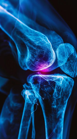
Blue x-ray photograph of knee pain, pain glows red. Trauma concept. Medical checkup. Generative AI.
Коллекция по умолчанию
Коллекция по умолчанию
Создать новую
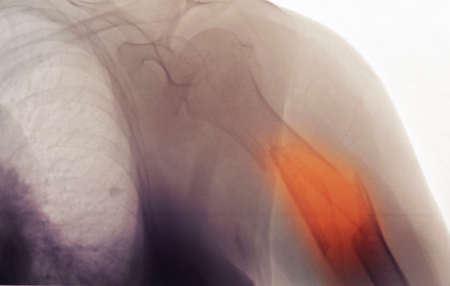
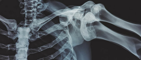
x-ray showing a comminuted fracture of the humerus
Коллекция по умолчанию
Коллекция по умолчанию
Создать новую

Lung adenocarcinoma, light micrograph, photo under microscope
Коллекция по умолчанию
Коллекция по умолчанию
Создать новую
Characteristics of Lichen, hyphae and Symbiotic algae under the microscope for education.
Коллекция по умолчанию
Коллекция по умолчанию
Создать новую
abstract background
Коллекция по умолчанию
Коллекция по умолчанию
Создать новую

Cells of a human spleen with chronic myelogenous leukemia, under the microscope.
Коллекция по умолчанию
Коллекция по умолчанию
Создать новую
Photomicrograph of chondrosarcoma, a malignant cartilage tumor, revealing chondrocytes with atypical nuclei and an abundant chondroid matrix.
Коллекция по умолчанию
Коллекция по умолчанию
Создать новую

Tissue of Small intestine (Duodenum) and Vermiform appendix Human under the microscope in Lab.
Коллекция по умолчанию
Коллекция по умолчанию
Создать новую

Cancer Cell in blood cells human showing abnormal cells.
Коллекция по умолчанию
Коллекция по умолчанию
Создать новую
Ice texture background, ink in water pattern frost. Crystal winter design
Коллекция по умолчанию
Коллекция по умолчанию
Создать новую
Cliated epithelium of human under the microscope in Lab.
Коллекция по умолчанию
Коллекция по умолчанию
Создать новую

Blue spots from the dye in the white tub dissolves in water
Коллекция по умолчанию
Коллекция по умолчанию
Создать новую
Cerebellum, Thalamus, Medulla oblongata, Spinal cord and Motor Neuron human under the microscope in Lab.
Коллекция по умолчанию
Коллекция по умолчанию
Создать новую
A microscopic view of tissue with pink and purple staining, showing cellular structures and patterns
Коллекция по умолчанию
Коллекция по умолчанию
Создать новую
Salivary gland swollen diseased tissue under the microscope 100x
Коллекция по умолчанию
Коллекция по умолчанию
Создать новую
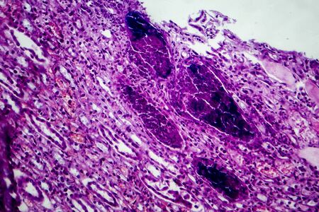
Gout crystals deposits in the kidney tissue under the microscope 100x
Коллекция по умолчанию
Коллекция по умолчанию
Создать новую

Histopathology of interstitial nephritis, light micrograph, photo under microscope
Коллекция по умолчанию
Коллекция по умолчанию
Создать новую
Breast fibroadenosis, light micrograph, photo under microscope. Common benign hyperplastic process involving breast glands
Коллекция по умолчанию
Коллекция по умолчанию
Создать новую
Abstract creative marbling pattern templat for fabric, design background texture
Коллекция по умолчанию
Коллекция по умолчанию
Создать новую
diseased ear tissue infected with Aspergillus 200x
Коллекция по умолчанию
Коллекция по умолчанию
Создать новую
Acute pyelonephritis, light micrograph, photo under microscope
Коллекция по умолчанию
Коллекция по умолчанию
Создать новую
Leiomyosarcoma, a malignant cancerous smooth muscle tumor, light micrograph, photo under microscope
Коллекция по умолчанию
Коллекция по умолчанию
Создать новую

Cancer Cell in human showing abnormal cells.Medical science background concept.
Коллекция по умолчанию
Коллекция по умолчанию
Создать новую
Tissue of Small intestine (Duodenum) and Vermiform appendix Human under the microscope in Lab.
Коллекция по умолчанию
Коллекция по умолчанию
Создать новую
Tissue diseased with alcoholism 100x
Коллекция по умолчанию
Коллекция по умолчанию
Создать новую
Thyroid follicular carcinoma, light micrograph, photo under microscope
Коллекция по умолчанию
Коллекция по умолчанию
Создать новую
X-ray showing details of a broken bone with sharp fragments in medical diagnosis
Коллекция по умолчанию
Коллекция по умолчанию
Создать новую
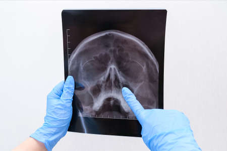
X-ray of the skull in the hand of a female doctor. With copy space. Diagnosis of sinusitis
Коллекция по умолчанию
Коллекция по умолчанию
Создать новую
Renal tuberculosis, light micrograph, photo under microscope
Коллекция по умолчанию
Коллекция по умолчанию
Создать новую
Papillary serous ovarian adenocarcinoma, cancer of ovary, light micrograph, photo under microscope
Коллекция по умолчанию
Коллекция по умолчанию
Создать новую
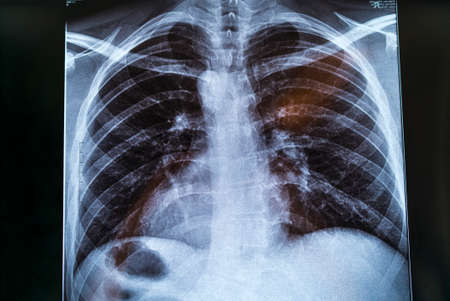
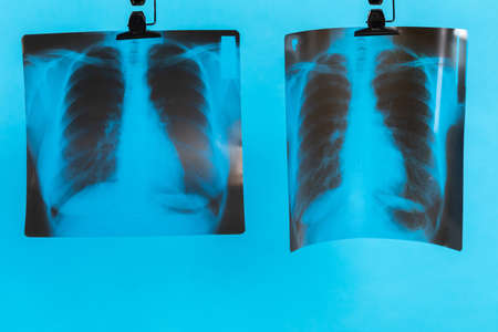
Chest X-ray image for physician's examination
Коллекция по умолчанию
Коллекция по умолчанию
Создать новую

Ice texture background, ink in water pattern frost. Crystal winter design
Коллекция по умолчанию
Коллекция по умолчанию
Создать новую
Abstract creative marbling pattern templat for fabric, design background texture
Коллекция по умолчанию
Коллекция по умолчанию
Создать новую
Bilberry stains on white paper. Abstract background.
Коллекция по умолчанию
Коллекция по умолчанию
Создать новую
Testicular seminoma, light micrograph, photo under microscope. A most common germ cell tumor of the testis
Коллекция по умолчанию
Коллекция по умолчанию
Создать новую
Glioma tumor with diseased tissue 100x
Коллекция по умолчанию
Коллекция по умолчанию
Создать новую

Histopathology of interstitial nephritis, light micrograph, photo under microscope. High magnification
Коллекция по умолчанию
Коллекция по умолчанию
Создать новую
A longitudinal section of human spinal ganglion cells under the microscope.
Коллекция по умолчанию
Коллекция по умолчанию
Создать новую
Leiomyosarcoma, a malignant cancerous smooth muscle tumor, light micrograph, photo under microscope
Коллекция по умолчанию
Коллекция по умолчанию
Создать новую
Colorful grunge stone texture background, creative abstract marble backdrop in red color.
Коллекция по умолчанию
Коллекция по умолчанию
Создать новую

Bowen's Disease Tumor under the microscope 100x
Коллекция по умолчанию
Коллекция по умолчанию
Создать новую

Chronic myeloid leukemia cells or CML, analyze by microscope, original magnification 400x
Коллекция по умолчанию
Коллекция по умолчанию
Создать новую
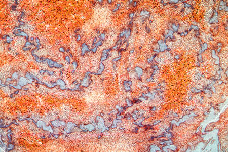
Photomicrograph of lung tissue depicting silicosis pathology under a microscope, revealing silica particle accumulation in alveoli and fibrosis.
Коллекция по умолчанию
Коллекция по умолчанию
Создать новую
Ice on purple background. Abstract background.
Коллекция по умолчанию
Коллекция по умолчанию
Создать новую
Fibroepithelium Diseased tissue 100x
Коллекция по умолчанию
Коллекция по умолчанию
Создать новую
Hodgkins lymphoma, light micrograph, photo under microscope. High magnification
Коллекция по умолчанию
Коллекция по умолчанию
Создать новую
Human pancreas under microscope view. Histological sample for human biology.
Коллекция по умолчанию
Коллекция по умолчанию
Создать новую
Tongue Tissue with taste buds across 200x
Коллекция по умолчанию
Коллекция по умолчанию
Создать новую
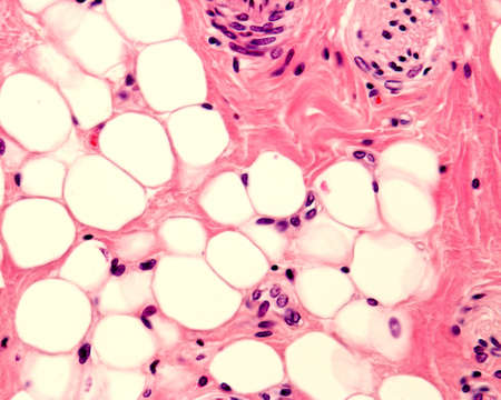
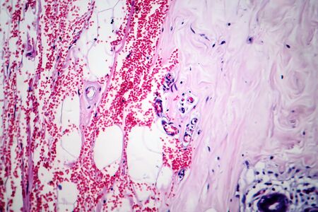
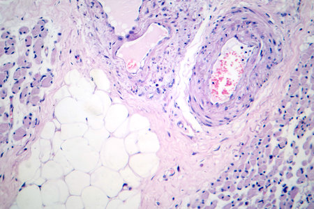
Small adipocyte lobule located in a connective tissue. A small nerve is located in the upper right corner. Light micrograph. H&E stain.
Коллекция по умолчанию
Коллекция по умолчанию
Создать новую
Charcoal dust lung tissue under the microscope 100x
Коллекция по умолчанию
Коллекция по умолчанию
Создать новую
Characteristics of Lichen, hyphae and Symbiotic algae under the microscope for education.
Коллекция по умолчанию
Коллекция по умолчанию
Создать новую
Painting acrylic paint- abstract drawing. Texture background
Коллекция по умолчанию
Коллекция по умолчанию
Создать новую
Microscopic view of tissue section showing cellular structures and layers, stained for examination
Коллекция по умолчанию
Коллекция по умолчанию
Создать новую
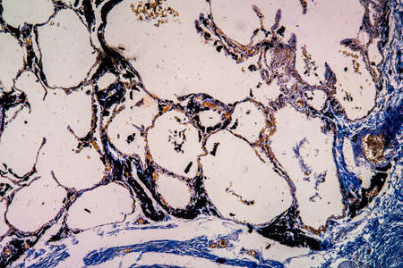
Anthracotic lymph node. Accumulation of carbon is most commonly found in intrapulmonary lymph nodes, due to coal dust, smoke or pollution. H & E stain
Коллекция по умолчанию
Коллекция по умолчанию
Создать новую
Intricate patterns observed in X-ray texture revealing detailed skeletal structure
Коллекция по умолчанию
Коллекция по умолчанию
Создать новую
Doctor demonstrates an X-ray of the male prostate gland, to diagnose the X-ray picture of the prostate, on a white background.
Коллекция по умолчанию
Коллекция по умолчанию
Создать новую
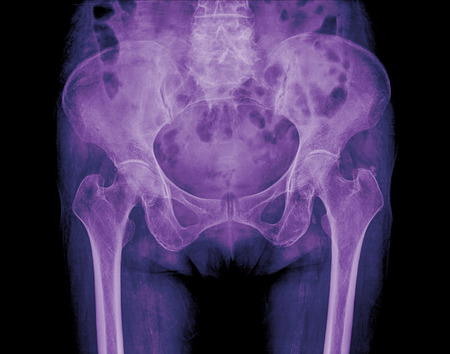
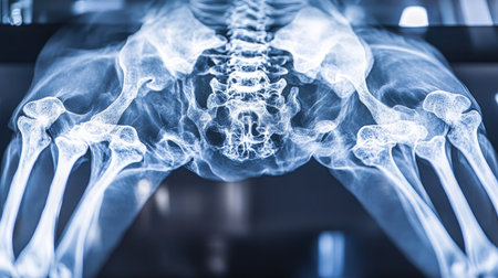
x-ray of hip
Коллекция по умолчанию
Коллекция по умолчанию
Создать новую
Acute pyelonephritis, light micrograph, photo under microscope
Коллекция по умолчанию
Коллекция по умолчанию
Создать новую
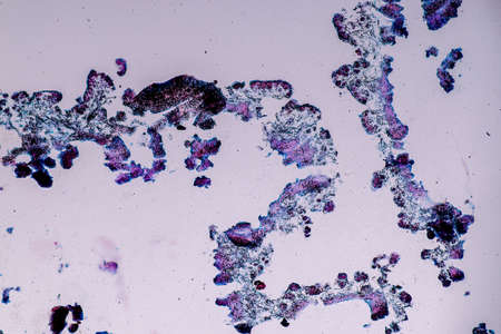
picture of histology human tissue with microscope from laboratory (not Illustration Designation)
Коллекция по умолчанию
Коллекция по умолчанию
Создать новую
Fat heart tissue under the microscope 100x
Коллекция по умолчанию
Коллекция по умолчанию
Создать новую

Foam texture
Коллекция по умолчанию
Коллекция по умолчанию
Создать новую
Characteristics of Lichen, hyphae and Symbiotic algae under the microscope for education.
Коллекция по умолчанию
Коллекция по умолчанию
Создать новую
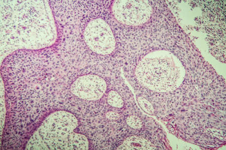
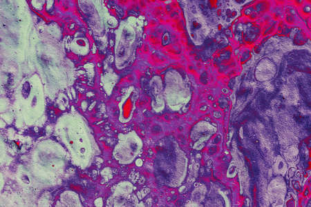
Low magnification of a human prostate gland in a 70-year-old man. The prostate gland appears with dilated alveoli, which contains many corpora amylacea (prostatic concretions) in their lumen. Light microscope micrograph. Hematoxylin & eosin stain.
Коллекция по умолчанию
Коллекция по умолчанию
Создать новую
Wilms tumor, or nephroblastoma, light micrograph, photo under microscope. High magnification
Коллекция по умолчанию
Коллекция по умолчанию
Создать новую
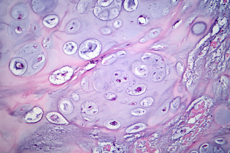
Squamous cell carcinoma, light micrograph, photo under microscope showing typical keratinous pearls
Коллекция по умолчанию
Коллекция по умолчанию
Создать новую
Bladder cancer, light micrograph, photo under microscope. High magnification
Коллекция по умолчанию
Коллекция по умолчанию
Создать новую
See vibrant neurons ignite during creative challenges! Stunning visualizations reveal enhanced brain function, showcasing improved focus, concentration, and problem-solving Witness the beauty of neurological processes under pressure as cognitive skills are amplified AI Generative
Коллекция по умолчанию
Коллекция по умолчанию
Создать новую
Beautiful abstract art of Ebru marbling painting techniques on water
Коллекция по умолчанию
Коллекция по умолчанию
Создать новую
Skeletal muscle atrophy, photomicrograph showing decreased fiber size with increased spacing between them, reduced myofibrils, increased endomysial connective tissue with fatty infiltration.
Коллекция по умолчанию
Коллекция по умолчанию
Создать новую
Lungs on a black and white photo taken on a photographic film of a special device.
Коллекция по умолчанию
Коллекция по умолчанию
Создать новую
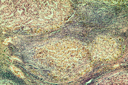
Biological histological fixed colored preparation of the spleen - a secondary organ of the immune system
Коллекция по умолчанию
Коллекция по умолчанию
Создать новую
Dark gold and purple abstract pattern with marble texture and sparkles
Коллекция по умолчанию
Коллекция по умолчанию
Создать новую
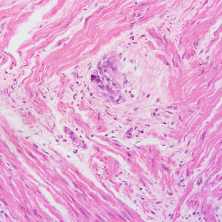
Photomicrograph of a neurofibroma tissue sample in neurofibromatosis genetic disease under a microscope, revealing spindle-shaped cells within a myxoid stroma and wavy nuclei.
Коллекция по умолчанию
Коллекция по умолчанию
Создать новую
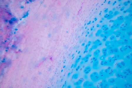
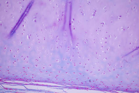
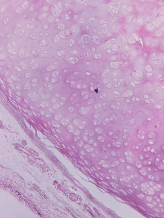
Hyaline cartilage, Elastic cartilage and Bone Human under the microscope in Lab.
Коллекция по умолчанию
Коллекция по умолчанию
Создать новую
X-ray image of a abdomen, showing bones and internal organs on a veterinary clinic screen.
Коллекция по умолчанию
Коллекция по умолчанию
Создать новую
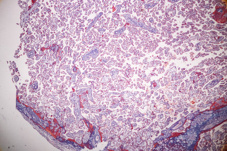
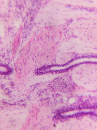
Histological Uterus human, Uterine tube human, Placenta human and Umbilical cord Human under the microscope for education.
Коллекция по умолчанию
Коллекция по умолчанию
Создать новую
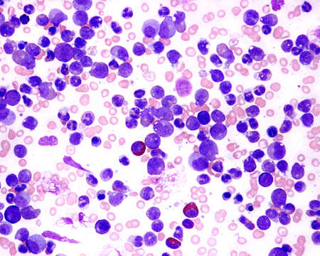
Blood smear. Chronic myelogenous or granulocytic leukemia. Abundant immature myeloid cells. Wright stain.
Коллекция по умолчанию
Коллекция по умолчанию
Создать новую
Thyroid follicular carcinoma, light micrograph, photo under microscope
Коллекция по умолчанию
Коллекция по умолчанию
Создать новую
Legion-Media
Создайте свои проекты на основе качественных стоковых фотографий и видео.


Copyright © Legion-Media.