
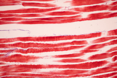
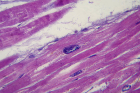
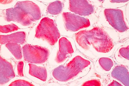
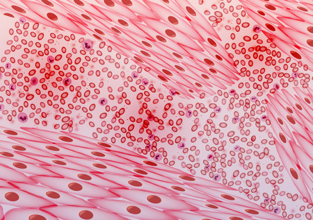
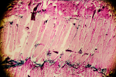
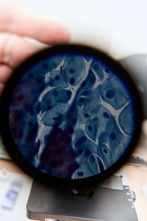
Striated muscle human under the microscope for education.
Коллекция по умолчанию
Коллекция по умолчанию
Создать новую

Trichinella spiralis larvae in muscle tissue under the microscope. Trichinella spiralis is a nematode parasite responsible for trichosis and affecting mammals.
Коллекция по умолчанию
Коллекция по умолчанию
Создать новую

Cardiac muscle inflammation Diseased tissue 100x
Коллекция по умолчанию
Коллекция по умолчанию
Создать новую

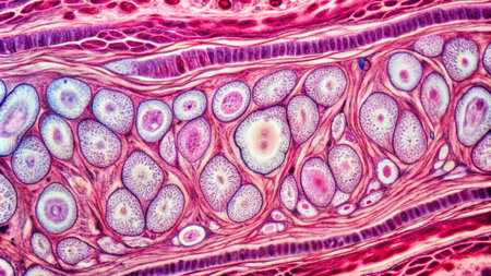
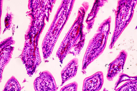
Human tongue section showing papillae and stratified epithelium
Коллекция по умолчанию
Коллекция по умолчанию
Создать новую
Portrait of a mature man reading a document on the street.
Коллекция по умолчанию
Коллекция по умолчанию
Создать новую
Photomicrograph of cardiac hypertrophy showing enlarged and thickened cardiac muscle fibers under the microscope.
Коллекция по умолчанию
Коллекция по умолчанию
Создать новую
Striated muscle human under the microscope for education.
Коллекция по умолчанию
Коллекция по умолчанию
Создать новую
Cardiac muscle fibers in longitudinal section, showing elongated nuclei and clear striations. High-resolution histology image.
Коллекция по умолчанию
Коллекция по умолчанию
Создать новую
Brightfield microscopy image capturing the bat membrane in a tissue culture model where it is artificially d for study or regenerative purposes
Коллекция по умолчанию
Коллекция по умолчанию
Создать новую
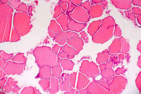
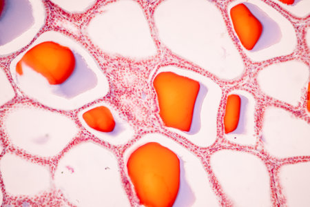
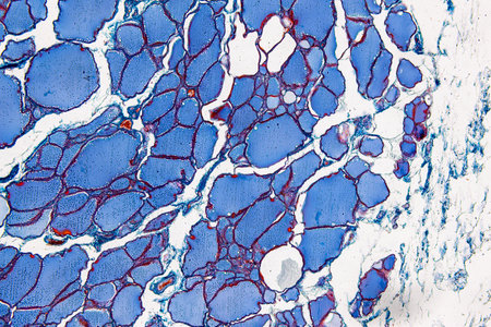
Goiter thyroid tissue with colloid 100x
Коллекция по умолчанию
Коллекция по умолчанию
Создать новую
A closeup examination of a cultured cell on a nanoengineered substrate with bright staining revealing cellular morphology and interaction with the surface at the nanoscale
Коллекция по умолчанию
Коллекция по умолчанию
Создать новую
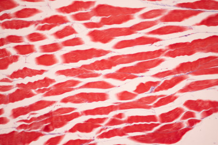
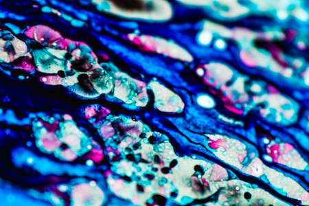
This detailed view highlights the contrasting red and white muscle fibers, illustrating their unique textures and anatomical features.
Коллекция по умолчанию
Коллекция по умолчанию
Создать новую
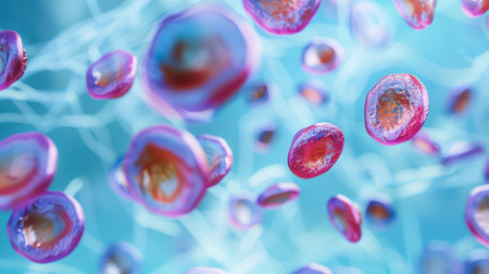
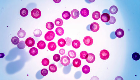
A microscopic image shows a cluster of pink and orange cells, with light refracting off their surfaces. The cells are suspended in a blue liquid, with a fine network of pale blue lines extending throughout the background.
Коллекция по умолчанию
Коллекция по умолчанию
Создать новую
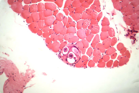
Trichinella spiralis larvae in muscle tissue, photomicrograph showing encysted parasites. Causes trichinosis, a parasitic infection from undercooked meat.
Коллекция по умолчанию
Коллекция по умолчанию
Создать новую
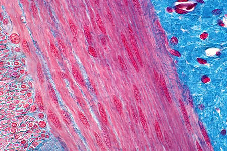
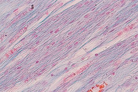
Histology of human smooth muscle under light microscope view. Haematoxylin and eosin staining technique for histology.
Коллекция по умолчанию
Коллекция по умолчанию
Создать новую
Thyroid cancer, light micrograph, photo under microscope
Коллекция по умолчанию
Коллекция по умолчанию
Создать новую
Anatomy and Histological Bone, Elastic cartilage human and Joint of human foetus under the microscope for education.
Коллекция по умолчанию
Коллекция по умолчанию
Создать новую
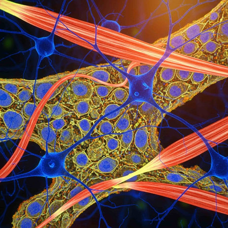
3D illustration of human nerve cell anatomy with neurons and nervous system
Коллекция по умолчанию
Коллекция по умолчанию
Создать новую
Pancreas cancer cells under microscope view for medical education.
Коллекция по умолчанию
Коллекция по умолчанию
Создать новую
Bladder cat- cell nature background. Abstract- photo macro sections with high magnification with light microscope
Коллекция по умолчанию
Коллекция по умолчанию
Создать новую
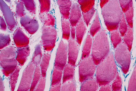
Education anatomy and Histological sample Striated (Skeletal) muscle of mammal Tissue under the microscope.
Коллекция по умолчанию
Коллекция по умолчанию
Создать новую
Backgrounds of Characteristics Tissue of Stomach Human, Small intestine Human, Pancreas Human and Large intestine Human under the microscope in Lab.
Коллекция по умолчанию
Коллекция по умолчанию
Создать новую
Explore the intricate world of vibrant cellular structures portrayed with detailed swirling patterns and rich textures, showcasing the beauty of microscopic life.
Коллекция по умолчанию
Коллекция по умолчанию
Создать новую
Heart muscle fibers contracting, highlighting the intricate structure and function of the muscle at a macro level with clear lighting.
Коллекция по умолчанию
Коллекция по умолчанию
Создать новую
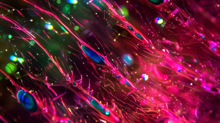
Intricate muscle fibers are displayed in a macro view, revealing a blend of textures and luminous details that highlight their complexity and structure.
Коллекция по умолчанию
Коллекция по умолчанию
Создать новую
Abstract human internal organs, Anatomy, Medical education concept, Generative AI
Коллекция по умолчанию
Коллекция по умолчанию
Создать новую
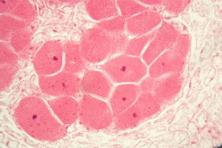
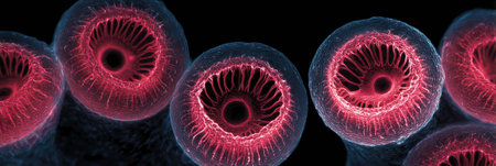
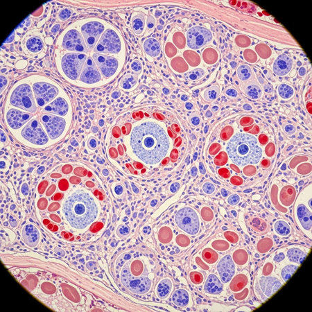
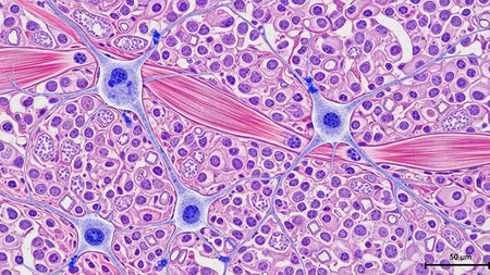
Light micrograph of a seminiferous tubule in the testis.A seminiferous tubule is a long, coiled tube in the testes where sperm is produced. The tubules are lined with Sertoli cells, which support and nourish the developing sperm cells. The tubules are also surrounded by Leydig cells, which produce testosterone.
Коллекция по умолчанию
Коллекция по умолчанию
Создать новую
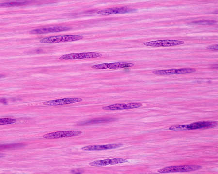
Nuclei of smooth muscle cells. These cells show a very elongated fusiform nucleus which contains small nucleoli. The small incisures in the nucleus surface are due to a contraction of the smooth muscle fiber.
Коллекция по умолчанию
Коллекция по умолчанию
Создать новую
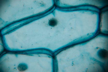
Study of protozoa and plant cells under the microscope for education.
Коллекция по умолчанию
Коллекция по умолчанию
Создать новую
Root bacteria nodules in a bean root under the microscope.
Коллекция по умолчанию
Коллекция по умолчанию
Создать новую
Cellular structures are magnified to reveal their complex shapes and vibrant colors, emphasizing the beauty of biology at a microscopic level.
Коллекция по умолчанию
Коллекция по умолчанию
Создать новую

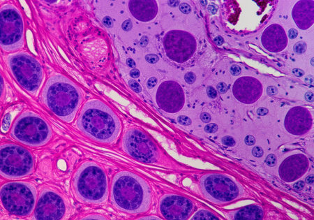
Education anatomy and Histological sample of Human under the microscope.
Коллекция по умолчанию
Коллекция по умолчанию
Создать новую
This captivating abstract image features delicate cellular structures against a vibrant gradient backdrop, perfect for scientific or artistic themes, emphasizing intricate details.
Коллекция по умолчанию
Коллекция по умолчанию
Создать новую
This stunning microscopic image captures the beauty of colorful microorganisms in an aqua background, showcasing intricate details and soft glowing light effects.
Коллекция по умолчанию
Коллекция по умолчанию
Создать новую

Abstract background of a mix white blue paints, like a fantastic flowers
Коллекция по умолчанию
Коллекция по умолчанию
Создать новую

Abstract creative marbling pattern templat for fabric, design background texture
Коллекция по умолчанию
Коллекция по умолчанию
Создать новую
Anatomy and Histological Bone, Elastic cartilage human and Joint of human foetus under the microscope for education.
Коллекция по умолчанию
Коллекция по умолчанию
Создать новую
Histopathology of the human skin. Human skin under microscope.
Коллекция по умолчанию
Коллекция по умолчанию
Создать новую
Education anatomy and Histological sample of Human under the microscope.
Коллекция по умолчанию
Коллекция по умолчанию
Создать новую
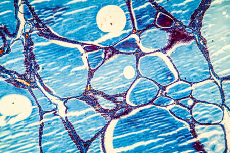
Pathology and Histology Tissue of Mammals under microscope.
Коллекция по умолчанию
Коллекция по умолчанию
Создать новую
Cancer cell, carcinoma, disease, dangerous illness, recovery from chemotherapy, joy in life, morale boosting, harmful cell destruction treatments, dna, uncontrolled nuclear cell division
Коллекция по умолчанию
Коллекция по умолчанию
Создать новую
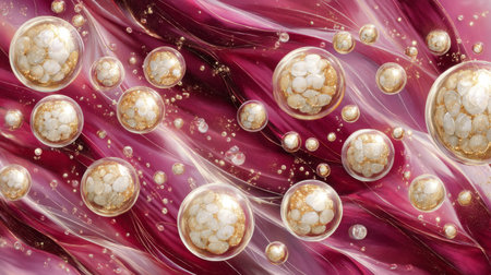
A mesmerizing abstract image featuring floating golden bubbles against a swirling colorful background, creating a sense of elegance and fluid movement.
Коллекция по умолчанию
Коллекция по умолчанию
Создать новую
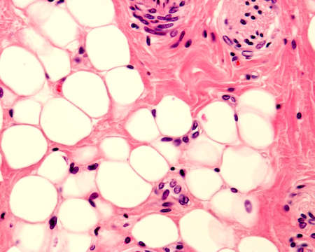
Small adipocyte lobule located in a connective tissue. A small nerve is located in the upper right corner. Light micrograph. H&E stain.
Коллекция по умолчанию
Коллекция по умолчанию
Создать новую
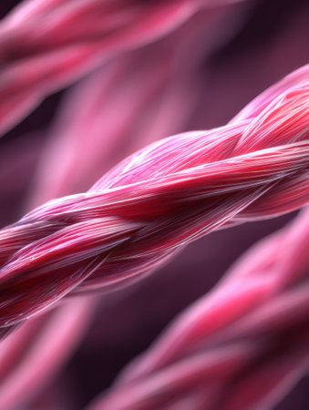
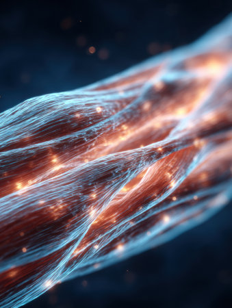
Macro view of muscle fibers rendered in 3D, highlighting their complex structure and texture with a soft blurred background for depth.
Коллекция по умолчанию
Коллекция по умолчанию
Создать новую

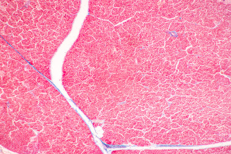
Stomach tissue under the microscope 100x
Коллекция по умолчанию
Коллекция по умолчанию
Создать новую
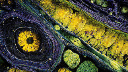
This stunning image captures the intricate structures of plant cells, showcasing vibrant yellow and purple colors through microscopy, revealing nature's complexity.
Коллекция по умолчанию
Коллекция по умолчанию
Создать новую
Thyroid cancer, light micrograph, photo under microscope
Коллекция по умолчанию
Коллекция по умолчанию
Создать новую
Blood Vessels, Veins and Arteries - Vector Illustration
Коллекция по умолчанию
Коллекция по умолчанию
Создать новую
Education anatomy and Histological sample of Human under the microscope.
Коллекция по умолчанию
Коллекция по умолчанию
Создать новую
Vibrant cross-section of a developing seed under UV light, highlighting the embryo and endosperm in bright colors
Коллекция по умолчанию
Коллекция по умолчанию
Создать новую

Human tongue section showing papillae and stratified epithelium
Коллекция по умолчанию
Коллекция по умолчанию
Создать новую
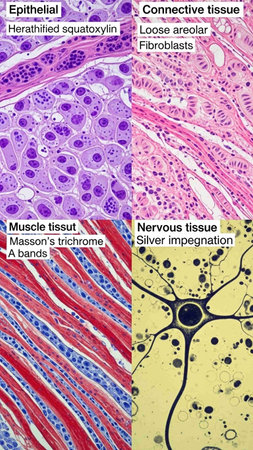
Microscopic macro close-up shot scientific research epithelial tissue biological anatomical capture of human animal plant cells muscles osmosis. Detailed microscope view nature organism body structure
Коллекция по умолчанию
Коллекция по умолчанию
Создать новую
Human tongue section showing papillae and stratified epithelium
Коллекция по умолчанию
Коллекция по умолчанию
Создать новую
Beautiful colorful 3d backgrounds, chromatic, hexagon, fascinated, illusion, mysterious, abstrack mood backgrounds,
Коллекция по умолчанию
Коллекция по умолчанию
Создать новую
beautiful electronic microscopy of bacteria fungi fantasy microbiology in blue tones microscopic life generative ai
Коллекция по умолчанию
Коллекция по умолчанию
Создать новую
Confocal microscopy image of a presynaptic terminal undergoing active recycling of synaptic vesicles after a burst of neurotransmitter release
Коллекция по умолчанию
Коллекция по умолчанию
Создать новую
Histopathology of alveoli, light micrograph, photo under microscope
Коллекция по умолчанию
Коллекция по умолчанию
Создать новую

Uterine cancer, light micrograph, photo under microscope
Коллекция по умолчанию
Коллекция по умолчанию
Создать новую

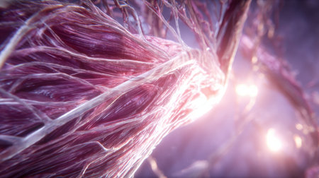
The macro 3D render showcases intricate muscle fibers with a side blurred effect, highlighting texture and structure in an expressive way.
Коллекция по умолчанию
Коллекция по умолчанию
Создать новую

Tooth development from human under microscope view for education.
Коллекция по умолчанию
Коллекция по умолчанию
Создать новую
Close-up view of a textured surface featuring intricate patterns and vibrant blue tones, highlighting the beauty of natural materials and their unique formations
Коллекция по умолчанию
Коллекция по умолчанию
Создать новую
Abstract scientific background with translucent cells and glowing elements, AI Generated
Коллекция по умолчанию
Коллекция по умолчанию
Создать новую

Striated muscle human under the microscope for education.
Коллекция по умолчанию
Коллекция по умолчанию
Создать новую
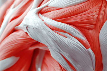
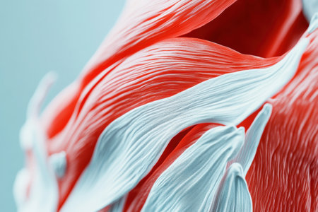
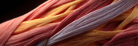
This detailed close-up reveals the intricate texture and structure of red and white muscle fibers, highlighting their complexity and design.
Коллекция по умолчанию
Коллекция по умолчанию
Создать новую
Education anatomy and Histological sample Touch corpuscles in skin Tissue under the microscope.
Коллекция по умолчанию
Коллекция по умолчанию
Создать новую
This detailed microscopic image showcases various cellular structures, highlighted in striking purple tones. The intricate patterns and textures reveal the complexity of biological tissues, making it a valuable resource for educational and scientific purposes
Коллекция по умолчанию
Коллекция по умолчанию
Создать новую
Showing Light micrograph of the Thyroid gland and Thymus gland human Child under the microscope for education in the laboratory.
Коллекция по умолчанию
Коллекция по умолчанию
Создать новую
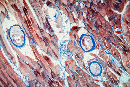
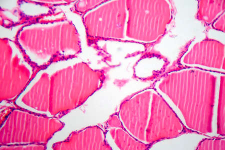
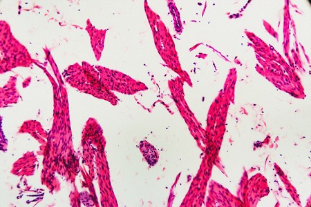
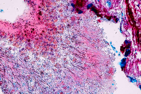
Trichinic parasites in muscle tissue 100x
Коллекция по умолчанию
Коллекция по умолчанию
Создать новую
Human blood cells. 3D illustration showing the cells of the human blood cells
Коллекция по умолчанию
Коллекция по умолчанию
Создать новую
Histological Uterus human, Uterine tube human, Placenta human and Umbilical cord Human under the microscope for education.
Коллекция по умолчанию
Коллекция по умолчанию
Создать новую
A close up of a pink and blue cell with a tree inside of it. The cell is surrounded by a pink and blue background
Коллекция по умолчанию
Коллекция по умолчанию
Создать новую
Achievements of modern biological science. A 24-well cell culture plate against an abstract biological background.
Коллекция по умолчанию
Коллекция по умолчанию
Создать новую
Human lung tissue under microscope view. Lungs are the primary organs of the respiratory system in humans and many other animals
Коллекция по умолчанию
Коллекция по умолчанию
Создать новую
Medical illustration of cholesterol lipids flowing through blood.
Коллекция по умолчанию
Коллекция по умолчанию
Создать новую
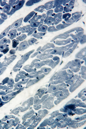
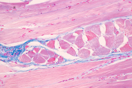
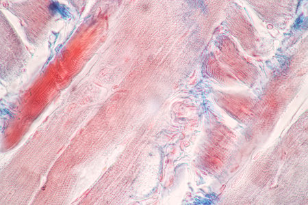
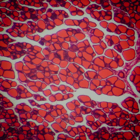
Cardiac muscle view through the microscope.
Коллекция по умолчанию
Коллекция по умолчанию
Создать новую

Histological Spermatic cord human, Seminal vesicle human, Prostate human and Human chromosomes under the microscope for education.
Коллекция по умолчанию
Коллекция по умолчанию
Создать новую
3D illustration mockup of the human organ systems, circulatory, digestive, red and white bloodcells wtih blurred backgroun. Medical education concept, Generative AI illustration
Коллекция по умолчанию
Коллекция по умолчанию
Создать новую
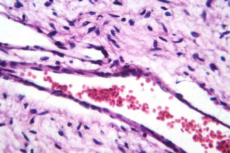
A small blood vessel with red blood cells in neurofibroma tissue sample, light photomicrograph.
Коллекция по умолчанию
Коллекция по умолчанию
Создать новую
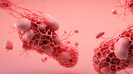
Revealing showing macro view of a repeating pattern of translucent spherical cells with glowing orange and purple internal structures and a dark background...
Коллекция по умолчанию
Коллекция по умолчанию
Создать новую
science medical anthropotomy physiology microscopic section of human thyroid gland background
Коллекция по умолчанию
Коллекция по умолчанию
Создать новую
An intricate digital representation of bacteria displaying vibrant colors and glowing elements, highlighting the fascinating world of microscopic life in a stunning abstract setting.
Коллекция по умолчанию
Коллекция по умолчанию
Создать новую
Beautiful image in microscope of microorganisms in the lab
Коллекция по умолчанию
Коллекция по умолчанию
Создать новую
Blue and pink refill ink spilled onto the white washbasin and the ink mixed into abstract blobs and patterns.
Коллекция по умолчанию
Коллекция по умолчанию
Создать новую
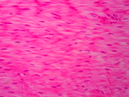
Histology of human smooth muscle under microscope view
Коллекция по умолчанию
Коллекция по умолчанию
Создать новую

HeLa cervical cancer cells, stained with Coomassie blue, under microscope.
Коллекция по умолчанию
Коллекция по умолчанию
Создать новую

Bladder cat- cell nature background. Abstract- photo macro sections with high magnification with light microscope
Коллекция по умолчанию
Коллекция по умолчанию
Создать новую
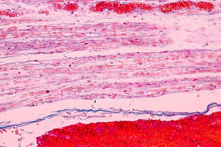
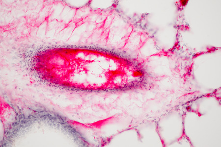
Cross section of blood vessels under microscope view for education in laboratory.
Коллекция по умолчанию
Коллекция по умолчанию
Создать новую
Characteristics of Lichen, hyphae and Symbiotic algae under the microscope for education.
Коллекция по умолчанию
Коллекция по умолчанию
Создать новую
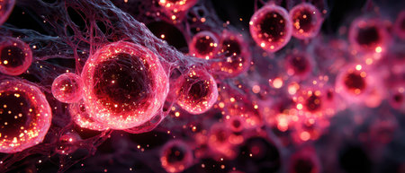
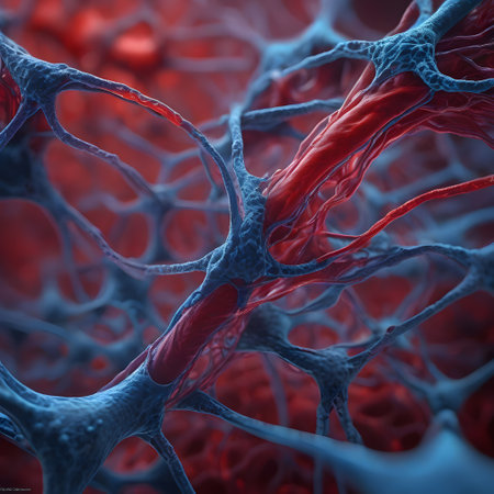
Glowing red cells connected by fine filaments create a mesmerizing view showcasing intricate biological structures in a dark environment.
Коллекция по умолчанию
Коллекция по умолчанию
Создать новую
Pathology and Histology Tissue of Mammals under microscope.
Коллекция по умолчанию
Коллекция по умолчанию
Создать новую

Proglottid (body unit) of tapeworm Taenia saginata, 3D illustration. A flatworm parasitizing animal and human intestine. Proglottid contains uterus with 12-30 primary lateral branches filled with eggs
Коллекция по умолчанию
Коллекция по умолчанию
Создать новую
Animation Skin Cell Medical Background Cosmetics Beauty Skin Care Concept. 3d Skin Collagen through Wrinkle Layer Reduce Up Saggy Skin of Body Molecule Element. Serum and Vitamin 3d Graphic
Коллекция по умолчанию
Коллекция по умолчанию
Создать новую
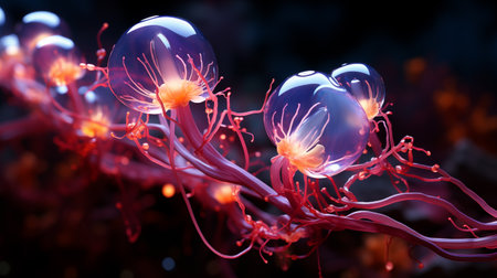
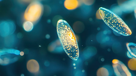
little jellyfishes.
Коллекция по умолчанию
Коллекция по умолчанию
Создать новую
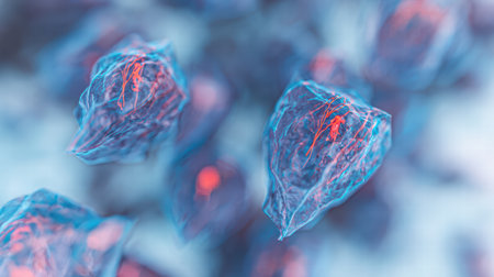
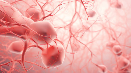
Blood vessel system and organs in the human body
Коллекция по умолчанию
Коллекция по умолчанию
Создать новую
Tooth development from human under microscope view for education.
Коллекция по умолчанию
Коллекция по умолчанию
Создать новую
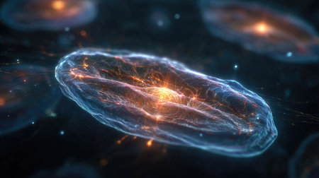
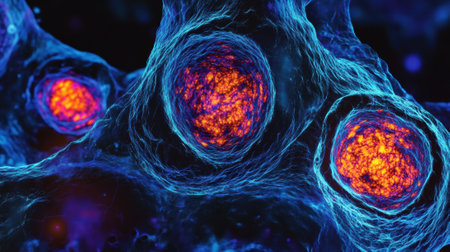
An artistic rendering presents a luminous cell-like structure featuring a radiant core. Its translucent form is highlighted by a blend of blues and oranges, suggesting an internal energy source. This composition utilizes a digital style, with a shallow depth of field, suitable for scientific or educational material, or for use in technology-related concepts.
Коллекция по умолчанию
Коллекция по умолчанию
Создать новую
This diagram illustrates the complex layering of skin and muscle, highlighting various textures and colors to enhance understanding of human anatomy.
Коллекция по умолчанию
Коллекция по умолчанию
Создать новую
Blood cells in human body under microscope view for education in laboratory.
Коллекция по умолчанию
Коллекция по умолчанию
Создать новую
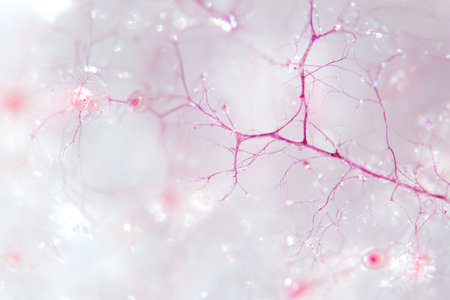
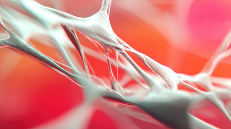
A close-up of the delicate network within an animal's brain, highlighting tiny, pinky-sized neurons in various stages of activity against a soft, white background. The focus is on intricate details and the subtle interplay between light and shadow that highlights these small connections. Soft lighting enhances the ethereal quality of this high-resolution macro photograph. --ar 3:2 --v 6.1 Job ID: a689d88f-4821-4583-9651-98e6d9d09fa5
Коллекция по умолчанию
Коллекция по умолчанию
Создать новую

Showing Light micrograph of the Thyroid gland and Thymus gland human Child under the microscope for education in the laboratory.
Коллекция по умолчанию
Коллекция по умолчанию
Создать новую
Legion-Media
Создайте свои проекты на основе качественных стоковых фотографий и видео.


Copyright © Legion-Media.