
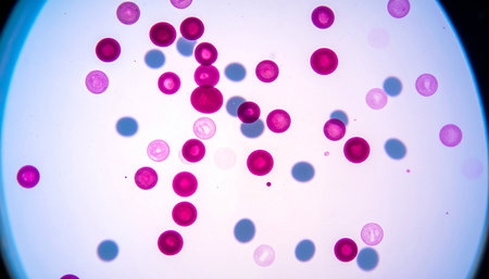
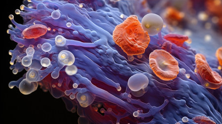
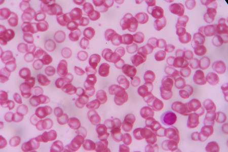
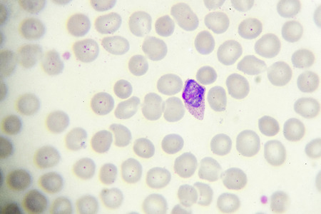
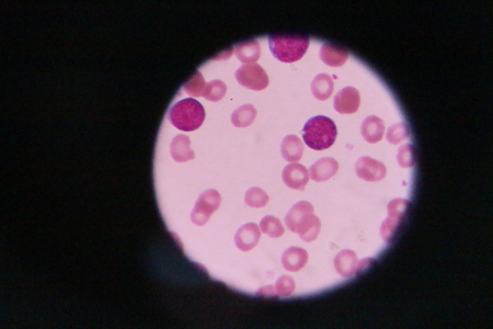
Malaria blood parasite infected red blood cells laboratory background.
Коллекция по умолчанию
Коллекция по умолчанию
Создать новую

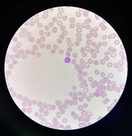
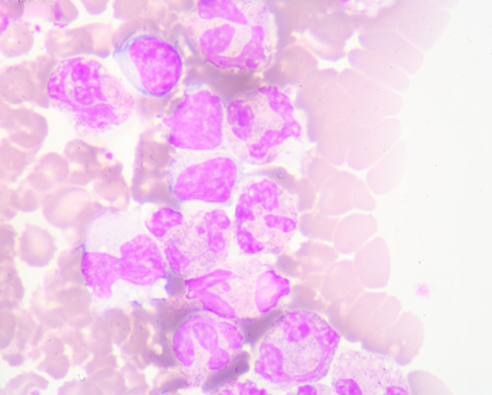
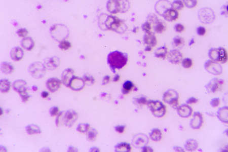
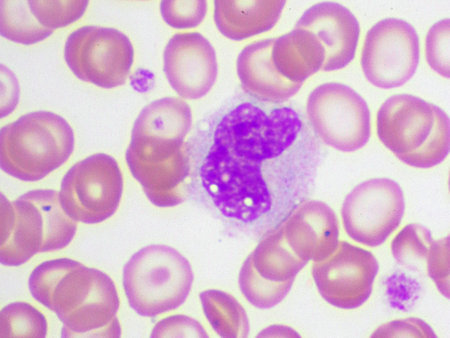
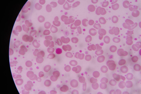
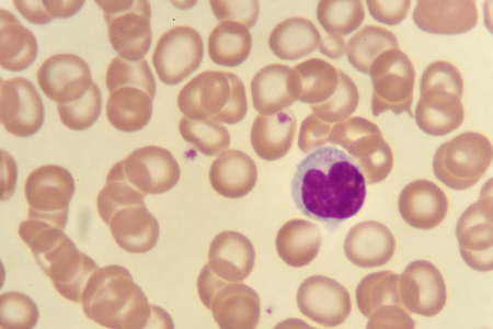
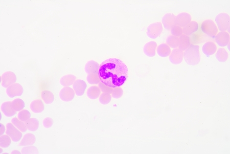
neutrophils. blood smear is often used as a follow-up test to abnormal results on a complete blood count (CBC) to evaluate the different types of blood cells.
Коллекция по умолчанию
Коллекция по умолчанию
Создать новую
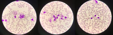
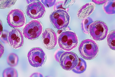
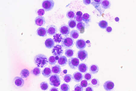
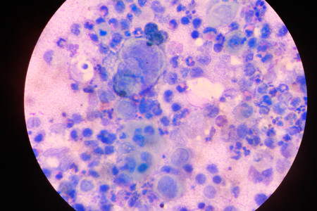
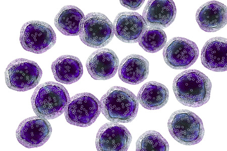
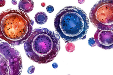
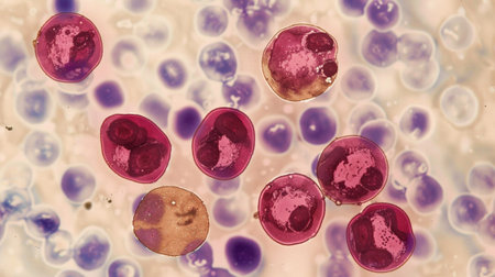
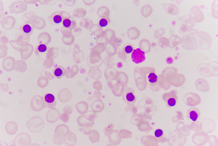
Immature white blood cells in leukemia.Science concept.
Коллекция по умолчанию
Коллекция по умолчанию
Создать новую
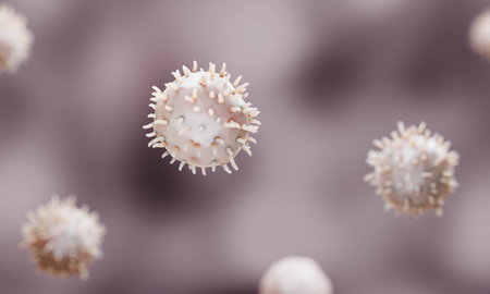
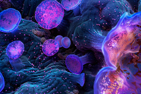
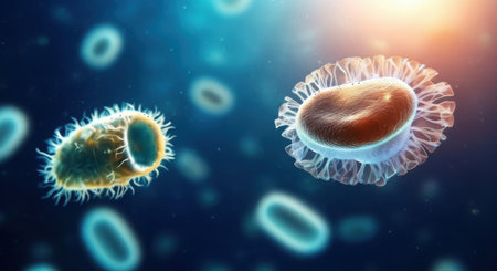
Microscopic view of cells, bacteria and viruses. Pathogens and microscopic organisms. Vivid biomedical backdrop. Banner. Concept of microbiology, immunology, health research, infection
Коллекция по умолчанию
Коллекция по умолчанию
Создать новую
Microscopic View Rendered Image of Abnormal, Diseased Cells in Biology and Medicine Illustration
Коллекция по умолчанию
Коллекция по умолчанию
Создать новую

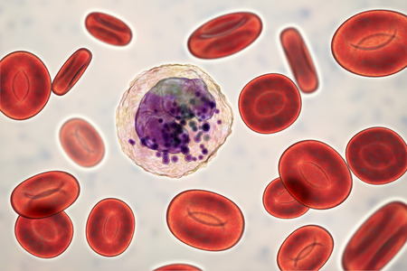
Neutrophil cell (white blood cell) in peripheral blood smear
Коллекция по умолчанию
Коллекция по умолчанию
Создать новую
Neutrophil show in blood smear CBC test find with microscope.
Коллекция по умолчанию
Коллекция по умолчанию
Создать новую

Chronic myeloid leukemia cells or CML, analyze by microscope, original magnification 1000x
Коллекция по умолчанию
Коллекция по умолчанию
Создать новую
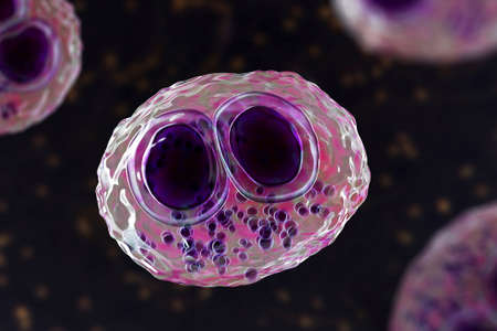
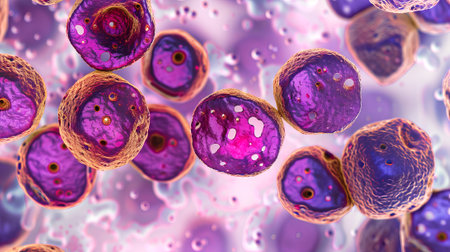
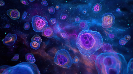
Cytomegalovirus CMV in a human cell, owl's eye inclusion in nucleus, multinucleated cell, 3D illustration. It is herpes virus, causes diseases in fetus, organ transplant patients, HIV infected people
Коллекция по умолчанию
Коллекция по умолчанию
Создать новую
Immature cells in myeloid serie myelocyte metamyelocyte.
Коллекция по умолчанию
Коллекция по умолчанию
Создать новую
Promyelocye
Коллекция по умолчанию
Коллекция по умолчанию
Создать новую
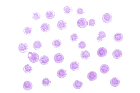
white blood cells
Коллекция по умолчанию
Коллекция по умолчанию
Создать новую
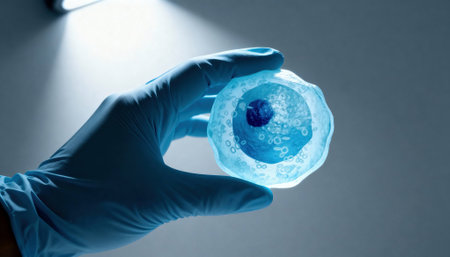
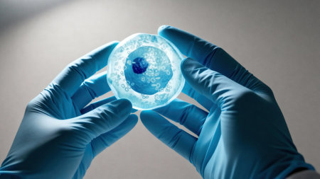
Gloved hand holds a translucent cell model under focused laboratory light with visible nucleus and organelle structures, suggesting scientific research and educational demonstration, with empty background space available for text
Коллекция по умолчанию
Коллекция по умолчанию
Создать новую
Nanoparticles Functionalization Therapeutics, Nanoparticles application in bioiechnology illustration
Коллекция по умолчанию
Коллекция по умолчанию
Создать новую
Blood smear with red blood cells in human body, medical background.
Коллекция по умолчанию
Коллекция по умолчанию
Создать новую
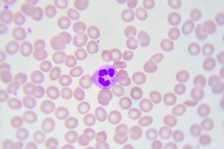
Neutrophil cell
Коллекция по умолчанию
Коллекция по умолчанию
Создать новую

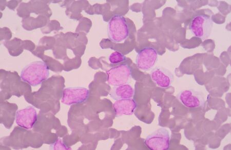
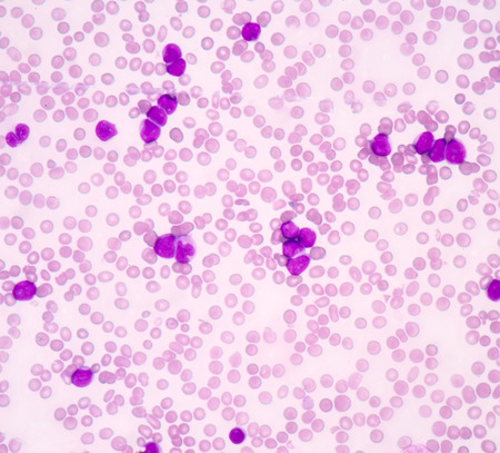
Picture of acute lymphocytic leukemia or ALL cells in blood smear, analyze by microscope, 400x
Коллекция по умолчанию
Коллекция по умолчанию
Создать новую
White blood cells of a human, photomicrograph panorama as seen under the microscope
Коллекция по умолчанию
Коллекция по умолчанию
Создать новую
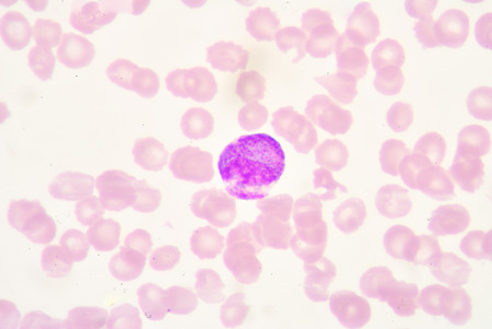
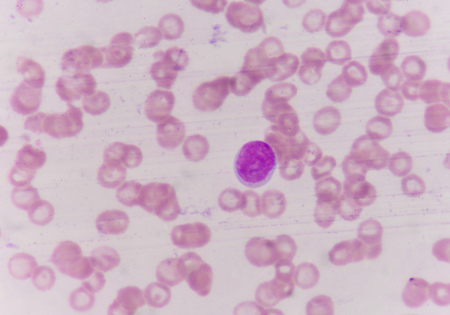
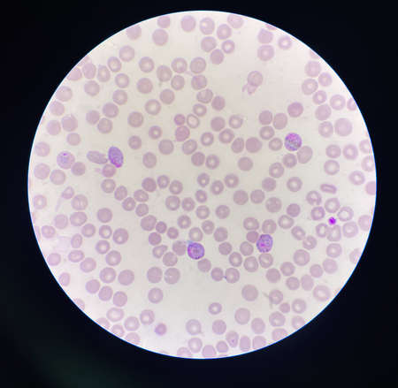
blood smear is often used as a follow-up test to abnormal results on a complete blood count (CBC) to evaluate the different types of blood cells.Atypical lymphocyte.
Коллекция по умолчанию
Коллекция по умолчанию
Создать новую
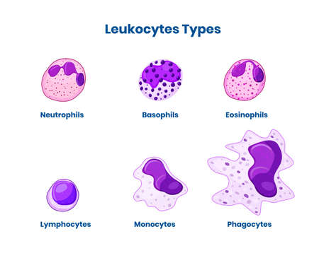
Types of the white blood cells. Leucocyte isolated on white vector illustration
Коллекция по умолчанию
Коллекция по умолчанию
Создать новую
a close up of a colorful structure
Коллекция по умолчанию
Коллекция по умолчанию
Создать новую

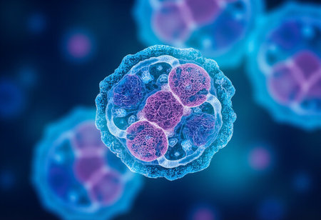
3d rendered medically accurate illustration of cells
Коллекция по умолчанию
Коллекция по умолчанию
Создать новую
Microscopic close-up of vibrant stained human cells on a blue backdrop
Коллекция по умолчанию
Коллекция по умолчанию
Создать новую
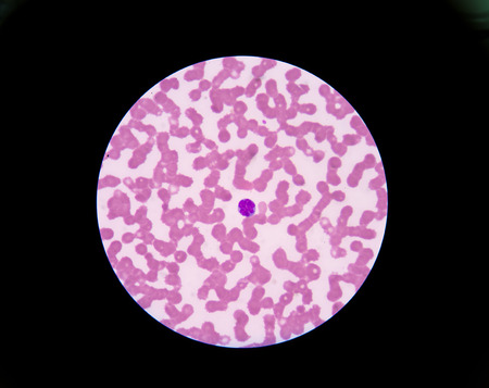
nucleated red cell
Коллекция по умолчанию
Коллекция по умолчанию
Создать новую
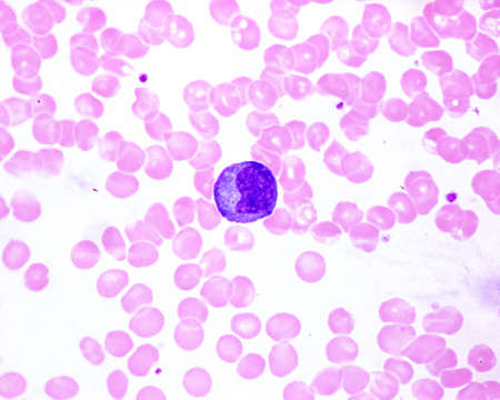
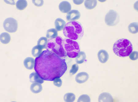
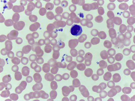
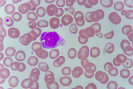
Human blood smear showing a monocyte with a basophilic cytoplasm in an infectious mononucleosis. It is the largest leukocyte (compare with red blood cell size).
Коллекция по умолчанию
Коллекция по умолчанию
Создать новую
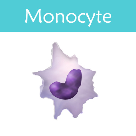
Leukocytes. Monocyte. White blood cell. Vector medical illustration
Коллекция по умолчанию
Коллекция по умолчанию
Создать новую

Leukemia cells
Коллекция по умолчанию
Коллекция по умолчанию
Создать новую

Comparison white blood cell Eosinophil and Neutrophil laboratory science concept.
Коллекция по умолчанию
Коллекция по умолчанию
Создать новую
Blood smear showing white and red blood cells
Коллекция по умолчанию
Коллекция по умолчанию
Создать новую
Leukemia cells
Коллекция по умолчанию
Коллекция по умолчанию
Создать новую
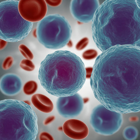
This image showcases a vibrant blue cell delicately surrounded by red blood cells in a dark, captivating space, illustrating fundamental biological concepts.
Коллекция по умолчанию
Коллекция по умолчанию
Создать новую
complete blood count
Коллекция по умолчанию
Коллекция по умолчанию
Создать новую
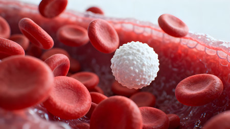
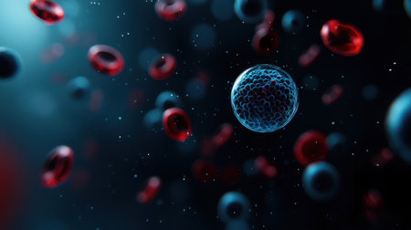
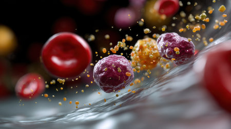
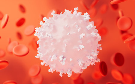
Ultra-detailed 3D render of a white blood cell floating among red blood cells in bloodstream, medical and scientific concept.
Коллекция по умолчанию
Коллекция по умолчанию
Создать новую
Blood cells in human body under microscope view for education in laboratory.
Коллекция по умолчанию
Коллекция по умолчанию
Создать новую
Picture of acute lymphocytic leukemia or ALL cells in blood smear, analyze by microscope, 400x
Коллекция по умолчанию
Коллекция по умолчанию
Создать новую
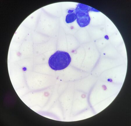
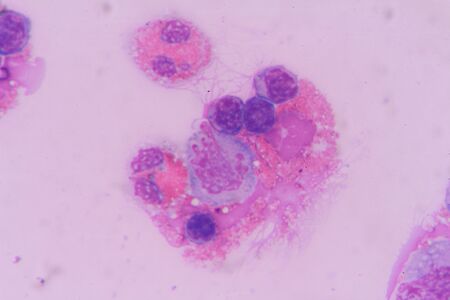
Multinucleated cell in Tzanck test finding with microscope in laboratory.
Коллекция по умолчанию
Коллекция по умолчанию
Создать новую
Virus cells, 3D illustration. Viruses and bacteria in human body. Viruses in infected organism.
Коллекция по умолчанию
Коллекция по умолчанию
Создать новую

Photomicrograph of canine eosinphil
Коллекция по умолчанию
Коллекция по умолчанию
Создать новую
A blood smear is often used as a follow-up test to abnormal results on a complete blood count (CBC) to evaluate the different types of blood cells.
Коллекция по умолчанию
Коллекция по умолчанию
Создать новую
White blood cells of a human, Eosinophil photomicrograph panorama as seen under the microscope
Коллекция по умолчанию
Коллекция по умолчанию
Создать новую

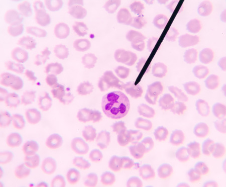
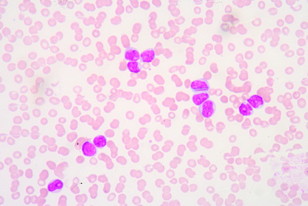
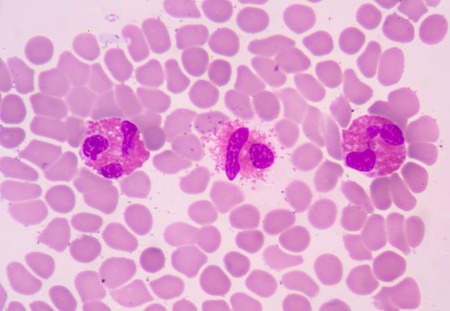
Blood smear showing, in the center, three neutrophil with hypersegmented nucleus. These cells appear in pathological situations such as megaloblastic anemias. Wright stain.
Коллекция по умолчанию
Коллекция по умолчанию
Создать новую
Blood smear with white blood cells and red blood cells. Medical background.
Коллекция по умолчанию
Коллекция по умолчанию
Создать новую

Blast cells in leukemia.blood smear is often used as a follow-up test to abnormal results on a complete blood count (CBC) to evaluate the different types of blood cells.
Коллекция по умолчанию
Коллекция по умолчанию
Создать новую
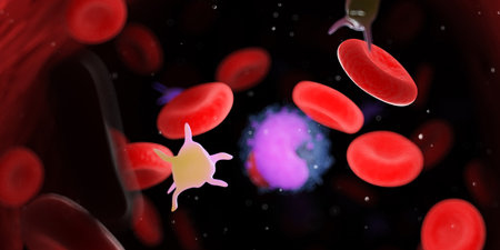
A captivating close-up view of red blood cells and immune cells interacting within a microscopic environment, showcasing biological processes in action.
Коллекция по умолчанию
Коллекция по умолчанию
Создать новую
blood smear is often used as a follow-up test to abnormal results on a complete blood count (CBC) to evaluate the different types of blood cells.Medical science background showing blast cells(AML)
Коллекция по умолчанию
Коллекция по умолчанию
Создать новую
Blast cells in blood smear specimen Leukemia petient.
Коллекция по умолчанию
Коллекция по умолчанию
Создать новую
Chromosomes Human under the microscope for education.
Коллекция по умолчанию
Коллекция по умолчанию
Создать новую
Numerous tiny white bubbles float gracefully on the surface of the water. The bubbles appear to be delicate and light, moving with the gentle current in a mesmerizing manner.
Коллекция по умолчанию
Коллекция по умолчанию
Создать новую

Monocyte cell in blood smear
Коллекция по умолчанию
Коллекция по умолчанию
Создать новую
A microscopic image shows a cluster of pink and orange cells, with light refracting off their surfaces. The cells are suspended in a blue liquid, with a fine network of pale blue lines extending throughout the background.
Коллекция по умолчанию
Коллекция по умолчанию
Создать новую
Colorful cell structures under microscopic view
Коллекция по умолчанию
Коллекция по умолчанию
Создать новую
multinucleated giant
Коллекция по умолчанию
Коллекция по умолчанию
Создать новую
Viruses that cause infection in the human body. The study of cells under a microscope.
Коллекция по умолчанию
Коллекция по умолчанию
Создать новую
Exploring the microscopic world of cells and tissues, AI generated
Коллекция по умолчанию
Коллекция по умолчанию
Создать новую
abstract acrylic watercolor paint brush stroke texture isolated on white background for logo and banner. design, creative, and illustration.
Коллекция по умолчанию
Коллекция по умолчанию
Создать новую

Lymphocyte (left) and monocyte (right) surrounded by red blood cells, 3D illustration
Коллекция по умолчанию
Коллекция по умолчанию
Создать новую
Delicious glazed donuts covered in colorful sprinkles ready to delight everyone
Коллекция по умолчанию
Коллекция по умолчанию
Создать новую
complete blood count
Коллекция по умолчанию
Коллекция по умолчанию
Создать новую

One infected cell among healthy
Коллекция по умолчанию
Коллекция по умолчанию
Создать новую
Malaria blood parasite infected red blood cells laboratory background.
Коллекция по умолчанию
Коллекция по умолчанию
Создать новую

Leukemia cells in blood smear
Коллекция по умолчанию
Коллекция по умолчанию
Создать новую
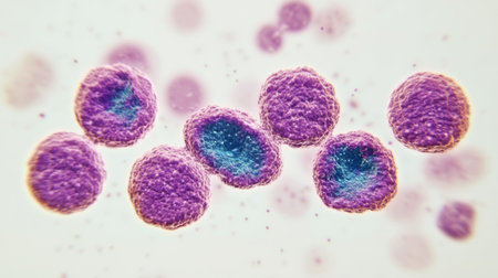
Burkitts lymphoma cells, a cancer of the lymphatic system, monoclonal B-cell tumor, 3D illustration
Коллекция по умолчанию
Коллекция по умолчанию
Создать новую
3d rendered medically accurate illustration of cells
Коллекция по умолчанию
Коллекция по умолчанию
Создать новую
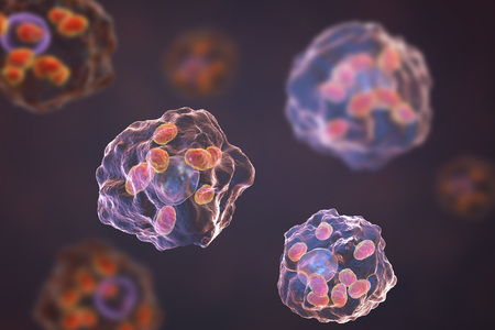
Macrophages infected by Leishmania amastigotes, 3D illustration
Коллекция по умолчанию
Коллекция по умолчанию
Создать новую
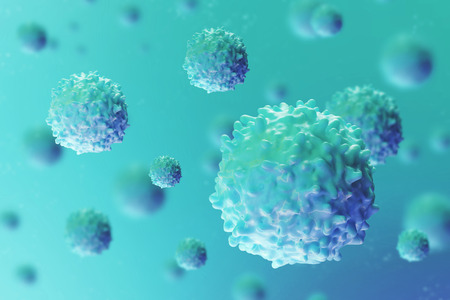
Lymphocytes and biological immune system, 3d rendering. 3D illustration.
Коллекция по умолчанию
Коллекция по умолчанию
Создать новую
Gloved hands hold a translucent cell culture sample with visible cellular structures and bubbles under focused clinical light, indicating laboratory research and microscopy, with available space for text on a neutral background
Коллекция по умолчанию
Коллекция по умолчанию
Создать новую
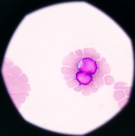
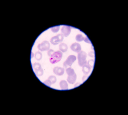
Malaria parasite in blood smear, gemetocyte stage
Коллекция по умолчанию
Коллекция по умолчанию
Создать новую
Vibrant and colorful cells are depicted in a dynamic 3D environment, showcasing the richness of microscopic life against a dark and abstract backdrop.
Коллекция по умолчанию
Коллекция по умолчанию
Создать новую
blood films for Malaria parasite
Коллекция по умолчанию
Коллекция по умолчанию
Создать новую
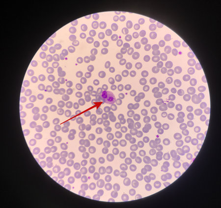
Red arrow showing neutrophil with toxic granule active PMN.
Коллекция по умолчанию
Коллекция по умолчанию
Создать новую
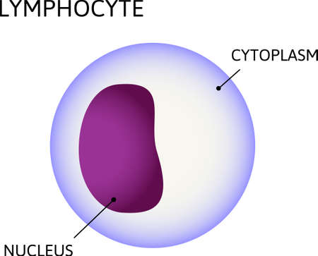
lymphocyte, variety of white blood cells. Consist of cytoplasm and nuclei. Vector medical illustration
Коллекция по умолчанию
Коллекция по умолчанию
Создать новую

3d rendered medically accurate illustration of the human blood cells and lymphocytes
Коллекция по умолчанию
Коллекция по умолчанию
Создать новую
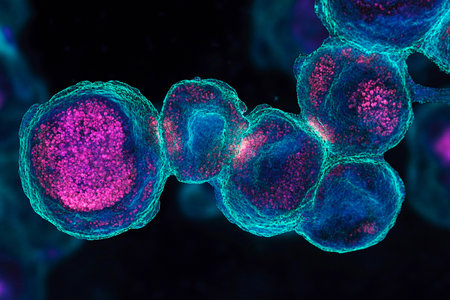
Colorful cells are showcased under a microscope, revealing their unique shapes and structures. The vibrant blues and purples illustrate complex biological interactions at a microscopic level.
Коллекция по умолчанию
Коллекция по умолчанию
Создать новую

Bacterial infection in the blood. Viruses attack the human body. Microorganisms, germs and microbes. 3D colorful illustration on microbiology
Коллекция по умолчанию
Коллекция по умолчанию
Создать новую

Acute promyelocytic leukemia cells or APL, analyze by microscope, original magnification 1000x
Коллекция по умолчанию
Коллекция по умолчанию
Создать новую

Meningococcal meningitis, cerebrospinal fluid smear containing neutrophils with and without bacteria Neisseria meningitidis
Коллекция по умолчанию
Коллекция по умолчанию
Создать новую
Microscopic view of human cells under microscope.
Коллекция по умолчанию
Коллекция по умолчанию
Создать новую
Eosinophil
Коллекция по умолчанию
Коллекция по умолчанию
Создать новую
A detailed microscopic view showing red cells and blue spherical cells floating in a fluid environment, highlighting their textures and surfaces.
Коллекция по умолчанию
Коллекция по умолчанию
Создать новую
Lymphocyte cells in blood smear
Коллекция по умолчанию
Коллекция по умолчанию
Создать новую
Stem cell, the building blocks of life, versatile and potent, medical breakthroughs, regeneration, and personalized therapies in the realm of modern medicine and biotechnology.
Коллекция по умолчанию
Коллекция по умолчанию
Создать новую
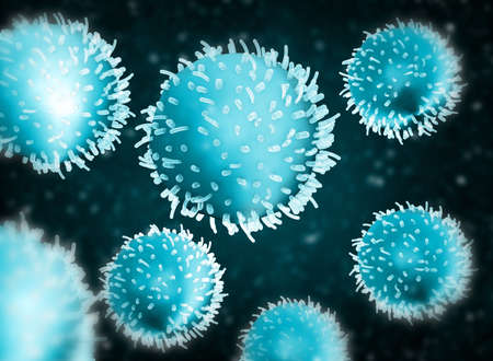
Close-up view of glowing bacteria and viruses with spiky exteriors, floating in a dark blue, luminous, and abstract background.
Коллекция по умолчанию
Коллекция по умолчанию
Создать новую
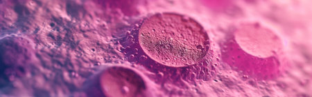
Abstract pink cancer cell organism background 3d render digital illustration
Коллекция по умолчанию
Коллекция по умолчанию
Создать новую
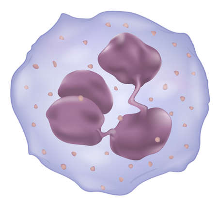
Illustration showing a white blood cell
Коллекция по умолчанию
Коллекция по умолчанию
Создать новую
Virus cells. Viral disease outbreak. 3d illustration
Коллекция по умолчанию
Коллекция по умолчанию
Создать новую
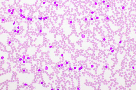
Blood smear show acute myeloblastic leukemia(AML).The smear shows large number of cancer leukemia cells (large blue cells) with the smaller red to pink normal red blood cells or erythrocytes.
Коллекция по умолчанию
Коллекция по умолчанию
Создать новую
A detailed artwork showcases a single cell featuring a softly glowing blue nucleus and shimmering green and purple cytoplasm. The background gradient enhances its vivid colors and intricate details.
Коллекция по умолчанию
Коллекция по умолчанию
Создать новую
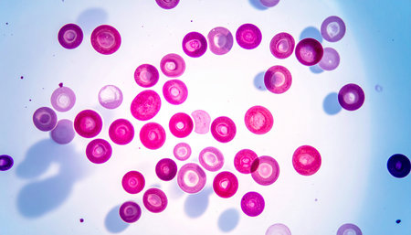
This image shows a detailed microscopic view of blood cells with vibrant staining, highlighting the structure and variety of cellular components for educational and research purposes.
Коллекция по умолчанию
Коллекция по умолчанию
Создать новую
Abnormal red blood cells in Blood smear Thalassemia patient.
Коллекция по умолчанию
Коллекция по умолчанию
Создать новую
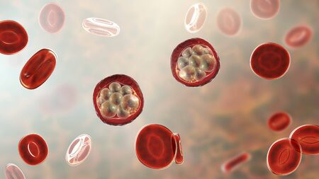
Red blood cells infected with malaria parasite, 3D illustration showing Plasmodium parasites inside red blood cells in the stage of schizont
Коллекция по умолчанию
Коллекция по умолчанию
Создать новую
Neutrophils are a type of phagocyte and are normally found in the bloodstream.
Коллекция по умолчанию
Коллекция по умолчанию
Создать новую
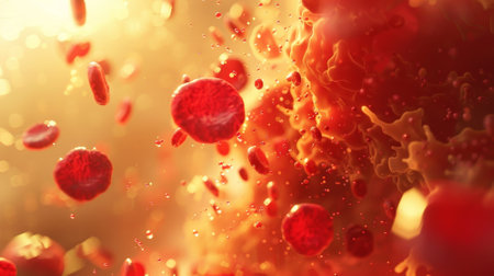
A close up of red blood cells in motion. Concept of urgency and chaos as the red blood cells are seen flying through the air
Коллекция по умолчанию
Коллекция по умолчанию
Создать новую

Acanthocytes, abnormal red blood cells with thorn-like projections, 3D illustration. They appear in severe liver disease, vitamin E defficiency, splenectomy, malabsorption, hypothyroidism
Коллекция по умолчанию
Коллекция по умолчанию
Создать новую
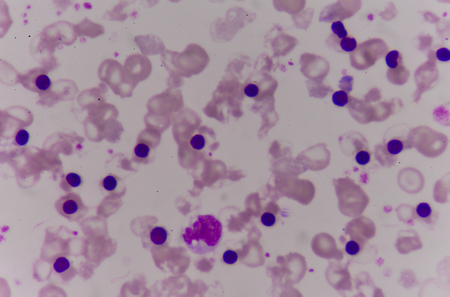
Abnormal red blood cells with moderate Nucleated red blood cells
Коллекция по умолчанию
Коллекция по умолчанию
Создать новую
A mesmerizing abstract illustration of cells floating in an enchanting cosmic space, featuring vivid colors and intricate details that inspire imagination.
Коллекция по умолчанию
Коллекция по умолчанию
Создать новую
blood smear
Коллекция по умолчанию
Коллекция по умолчанию
Создать новую
Close-up of a set of multicolored jelly candies
Коллекция по умолчанию
Коллекция по умолчанию
Создать новую
Basophil, a white blood cell, 3D illustration. Basophils are granulocytes taking part in inflammatory reactions and allergic diseases
Коллекция по умолчанию
Коллекция по умолчанию
Создать новую
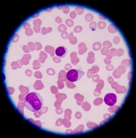
White blood cells in blood smear, analyze by microscope
Коллекция по умолчанию
Коллекция по умолчанию
Создать новую

Acute lymphoblastic leukemia, bone marrow smear, 3D illustration
Коллекция по умолчанию
Коллекция по умолчанию
Создать новую
Legion-Media
Создайте свои проекты на основе качественных стоковых фотографий и видео.


Copyright © Legion-Media.