

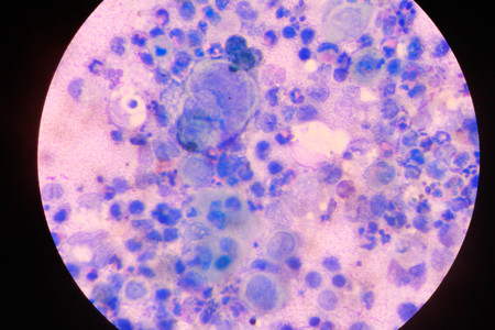
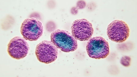
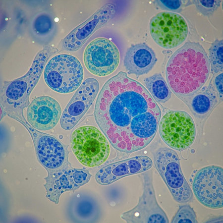
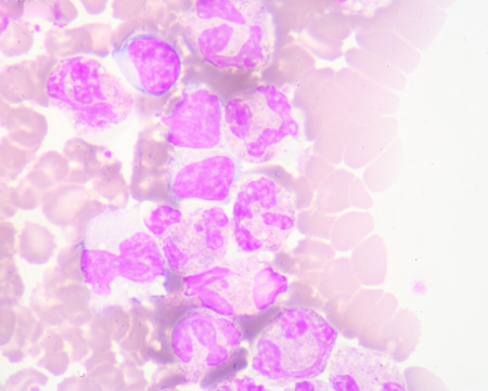
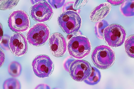
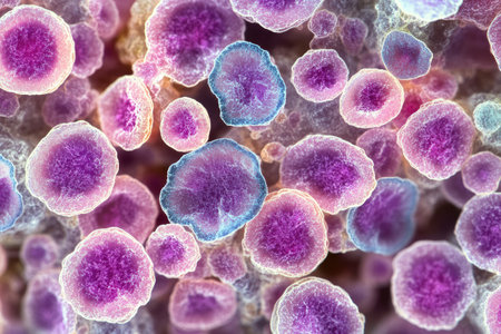
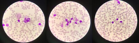
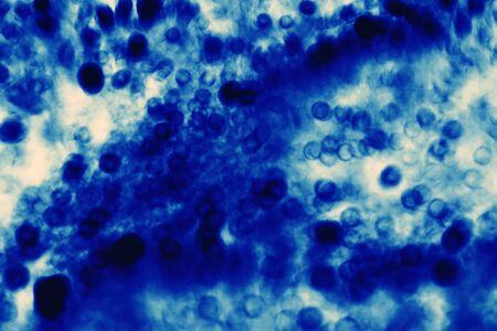
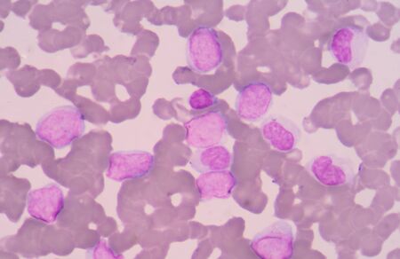
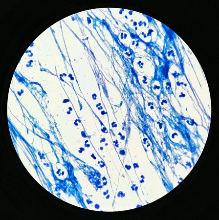
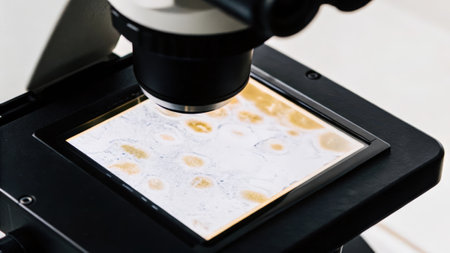
Chronic myeloid leukemia cells or CML, analyze by microscope, original magnification 1000x
Коллекция по умолчанию
Коллекция по умолчанию
Создать новую
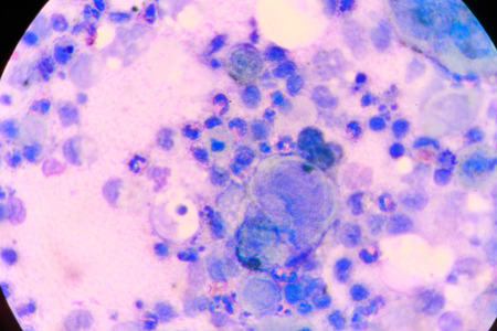
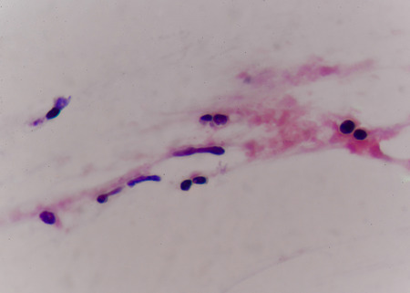
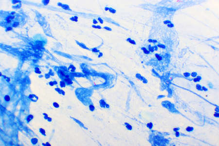
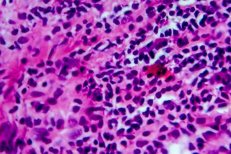
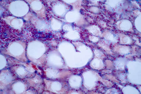
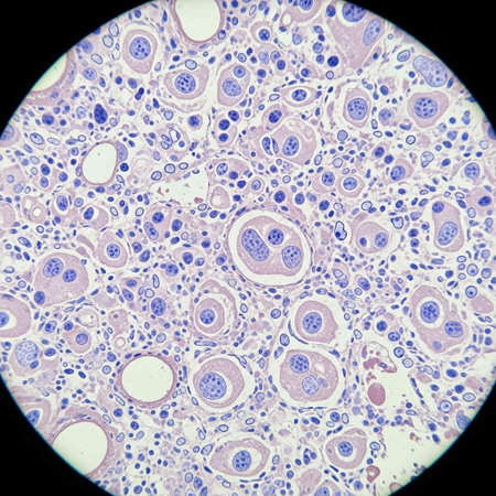
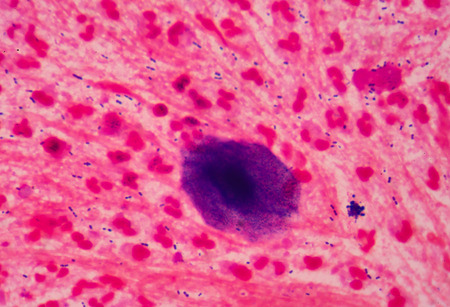
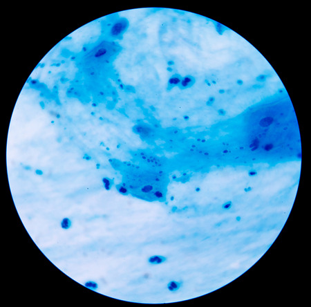
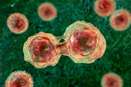
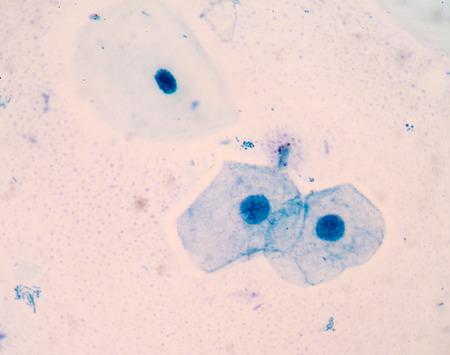
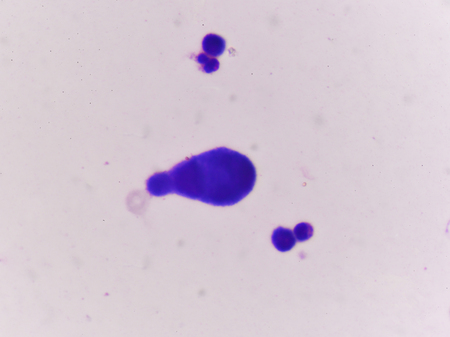
multinucleated giant
Коллекция по умолчанию
Коллекция по умолчанию
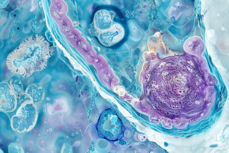
Создать новую
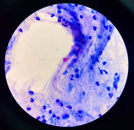
multinucleated giant
Коллекция по умолчанию
Коллекция по умолчанию
Создать новую
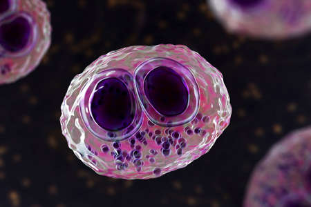
Cytomegalovirus CMV in a human cell, owl's eye inclusion in nucleus, multinucleated cell, 3D illustration. It is herpes virus, causes diseases in fetus, organ transplant patients, HIV infected people
Коллекция по умолчанию
Коллекция по умолчанию
Создать новую
Chaos ink texture background, ink in water pattern frost. Crystal winter design
Коллекция по умолчанию
Коллекция по умолчанию
Создать новую
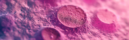
Microscopic View Rendered Image of Abnormal, Diseased Cells in Biology and Medicine Illustration
Коллекция по умолчанию
Коллекция по умолчанию
Создать новую

Abstract macro image of particles looking like bacteria, macro shot, microbiology theme
Коллекция по умолчанию
Коллекция по умолчанию
Создать новую

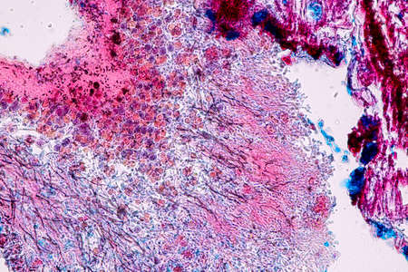
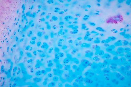
Human hyaline cartilage bone under microscope view for education pathology. Human tissue.
Коллекция по умолчанию
Коллекция по умолчанию
Создать новую
Chromosomes Human under the microscope for education.
Коллекция по умолчанию
Коллекция по умолчанию
Создать новую
Anatomy and Histological Ovary, Testis and Sperm human cells under microscope.
Коллекция по умолчанию
Коллекция по умолчанию
Создать новую

Picture of acute lymphocytic leukemia or ALL cells in blood smear, analyze by microscope, 400x
Коллекция по умолчанию
Коллекция по умолчанию
Создать новую
Ink colors are amazingly bright, luminous, translucent, free-flowing, and dry quickly. Abstract artwork. Trendy wallpaper. Natural pattern, luxury. Art for your design project. Transparent creativity.
Коллекция по умолчанию
Коллекция по умолчанию
Создать новую
Anatomy and Histological Ovary, Testis and Sperm human cells under microscope.
Коллекция по умолчанию
Коллекция по умолчанию
Создать новую
Chromosomes Human under the microscope for education.
Коллекция по умолчанию
Коллекция по умолчанию
Создать новую

Thyroid follicular carcinoma, light micrograph, photo under microscope
Коллекция по умолчанию
Коллекция по умолчанию
Создать новую

Cells in reproductive female cytology and histology education concept.
Коллекция по умолчанию
Коллекция по умолчанию
Создать новую
budding yeast cell with epithelial in gram stain method.
Коллекция по умолчанию
Коллекция по умолчанию
Создать новую
Eosinophil
Коллекция по умолчанию
Коллекция по умолчанию
Создать новую
Cell division under microscope view. Microscopic view of cell.
Коллекция по умолчанию
Коллекция по умолчанию
Создать новую
Blue and pink refill ink spilled onto the white washbasin and the ink mixed into abstract blobs and patterns.
Коллекция по умолчанию
Коллекция по умолчанию
Создать новую
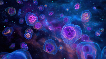
A mesmerizing abstract illustration of cells floating in an enchanting cosmic space, featuring vivid colors and intricate details that inspire imagination.
Коллекция по умолчанию
Коллекция по умолчанию
Создать новую

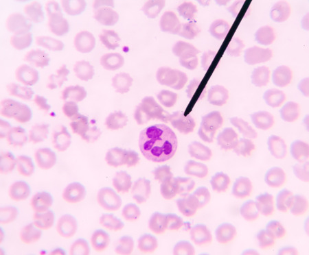
Blood smear showing, in the center, three neutrophil with hypersegmented nucleus. These cells appear in pathological situations such as megaloblastic anemias. Wright stain.
Коллекция по умолчанию
Коллекция по умолчанию
Создать новую
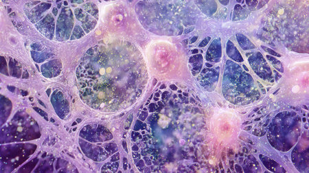
Colorful cells are showcased under a microscope, revealing their unique shapes and structures. The vibrant blues and purples illustrate complex biological interactions at a microscopic level.
Коллекция по умолчанию
Коллекция по умолчанию
Создать новую
Mycobacterium tuberculosis positive (small red rod) in sputum smear, acid-fast stain, analyze by microscope 1000x
Коллекция по умолчанию
Коллекция по умолчанию
Создать новую
Moderate Red cell on center Mycobacterium Tuberculosis bacteria.
Коллекция по умолчанию
Коллекция по умолчанию
Создать новую
Lung adenocarcinoma, light micrograph, photo under microscope
Коллекция по умолчанию
Коллекция по умолчанию
Создать новую
Wax droplets in water. Abstract blue background
Коллекция по умолчанию
Коллекция по умолчанию
Создать новую
Hodgkin's lymphoma, light micrograph, photo under microscope. High magnification
Коллекция по умолчанию
Коллекция по умолчанию
Создать новую
Yeast cells with epithelial tissue in Gram stain method.
Коллекция по умолчанию
Коллекция по умолчанию
Создать новую
Testicular seminoma, light micrograph, photo under microscope. A most common germ cell tumor of the testis
Коллекция по умолчанию
Коллекция по умолчанию
Создать новую
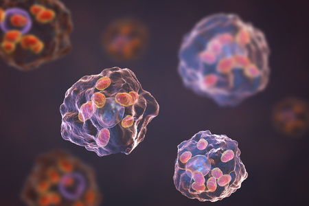
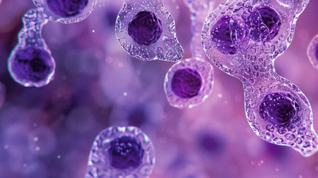
Macrophages infected by Leishmania amastigotes, 3D illustration
Коллекция по умолчанию
Коллекция по умолчанию
Создать новую
Human lung pathology under light microscope, The lungs is organs of the respiratory system in humans. Human pathology education. Haematoxylin and eosin staining technique slide.
Коллекция по умолчанию
Коллекция по умолчанию
Создать новую
A colorful image of a cell with a purple and blue blob in the center. The image is abstract and has a mood of curiosity and wonder
Коллекция по умолчанию
Коллекция по умолчанию
Создать новую

Characteristics of Lichen, hyphae and Symbiotic algae under the microscope for education.
Коллекция по умолчанию
Коллекция по умолчанию
Создать новую
Microscopic view of cells, bacteria and viruses. Pathogens and microscopic organisms. Vivid biomedical backdrop. Banner. Concept of microbiology, immunology, health research, infection
Коллекция по умолчанию
Коллекция по умолчанию
Создать новую

Hodgkins lymphoma, light micrograph, photo under microscope. High magnification
Коллекция по умолчанию
Коллекция по умолчанию
Создать новую
Hodgkin's lymphoma, light micrograph, photo under microscope. High magnification
Коллекция по умолчанию
Коллекция по умолчанию
Создать новую
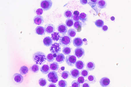
white blood cells
Коллекция по умолчанию
Коллекция по умолчанию
Создать новую
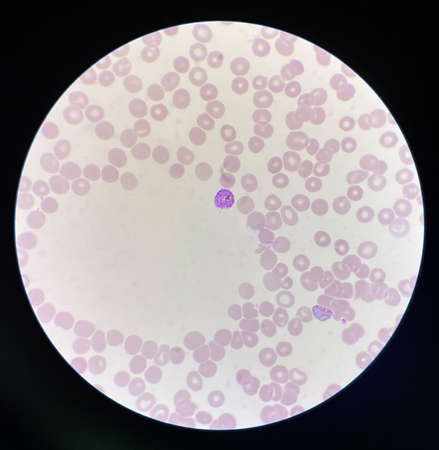
Neutrophil show in blood smear CBC test find with microscope.
Коллекция по умолчанию
Коллекция по умолчанию
Создать новую
Hodgkins lymphoma, light micrograph, photo under microscope. High magnification
Коллекция по умолчанию
Коллекция по умолчанию
Создать новую
Malaria blood parasite infected red blood cells laboratory background.
Коллекция по умолчанию
Коллекция по умолчанию
Создать новую
Histopathology of human liver under microscope view for medical education.
Коллекция по умолчанию
Коллекция по умолчанию
Создать новую
microscope lens, viewing Trypanosoma cruzi parasitic protozoan, causer of Chagas disease
Коллекция по умолчанию
Коллекция по умолчанию
Создать новую
Moderate red white blood cells with gram negative diplococci intracellular Gram-negative coffee bean-shaped diplococci bacteria responsible for the sexually transmitted infection gonorrhea
Коллекция по умолчанию
Коллекция по умолчанию
Создать новую
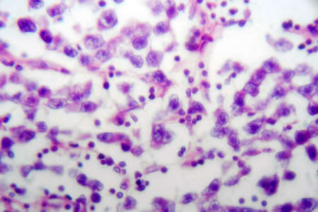
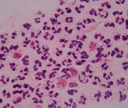
Abnormal neutrophil in pleural fluid smear.
Коллекция по умолчанию
Коллекция по умолчанию
Создать новую
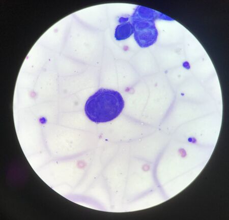
Microscopic view of human cells under microscope.
Коллекция по умолчанию
Коллекция по умолчанию
Создать новую
Chromosomes Human under the microscope for education.
Коллекция по умолчанию
Коллекция по умолчанию
Создать новую
nucleated red cell
Коллекция по умолчанию
Коллекция по умолчанию
Создать новую
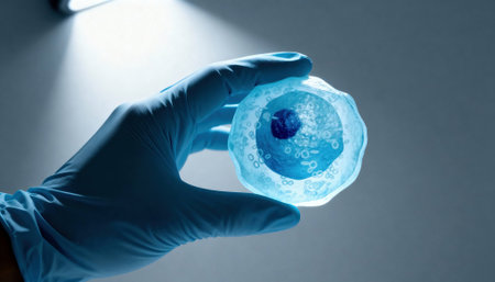
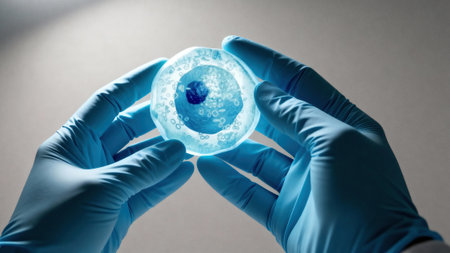
Gloved hand holds a translucent cell model under focused laboratory light with visible nucleus and organelle structures, suggesting scientific research and educational demonstration, with empty background space available for text
Коллекция по умолчанию
Коллекция по умолчанию
Создать новую
Microscopic close-up of vibrant stained human cells on a blue backdrop
Коллекция по умолчанию
Коллекция по умолчанию
Создать новую
Marbled black ink patterns create a striking effect against a white background
Коллекция по умолчанию
Коллекция по умолчанию
Создать новую
Characteristics of Lichen, hyphae and Symbiotic algae under the microscope for education.
Коллекция по умолчанию
Коллекция по умолчанию
Создать новую

Bacteria gram staining
Коллекция по умолчанию
Коллекция по умолчанию
Создать новую
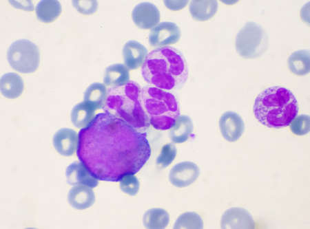
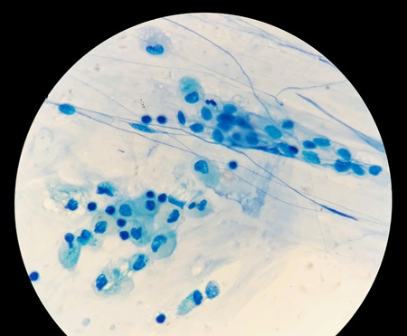
Immature cells in myeloid serie myelocyte metamyelocyte.
Коллекция по умолчанию
Коллекция по умолчанию
Создать новую

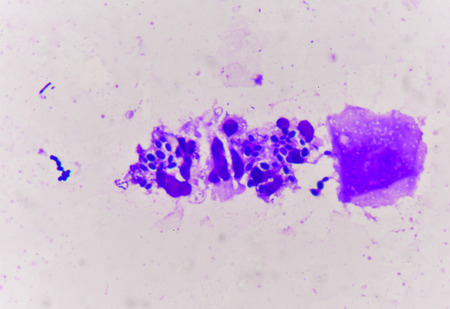
mesothelial cell in pleural fluid
Коллекция по умолчанию
Коллекция по умолчанию
Создать новую
Close-up of a Cluster of Purple and Blue Cells
Коллекция по умолчанию
Коллекция по умолчанию
Создать новую
bacterium medical science background.
Коллекция по умолчанию
Коллекция по умолчанию
Создать новую
Cell division process, micro
Коллекция по умолчанию
Коллекция по умолчанию
Создать новую

Ascaris lumbricoides, a large roundworm, fertilized egg, 3D illustration
Коллекция по умолчанию
Коллекция по умолчанию
Создать новую

Wax droplets in water. Abstract blue background
Коллекция по умолчанию
Коллекция по умолчанию
Создать новую
abstract acrylic watercolor paint brush stroke texture isolated on white background for logo and banner. design, creative, and illustration.
Коллекция по умолчанию
Коллекция по умолчанию
Создать новую
Immature white blood cells in leukemia.Science concept.
Коллекция по умолчанию
Коллекция по умолчанию
Создать новую

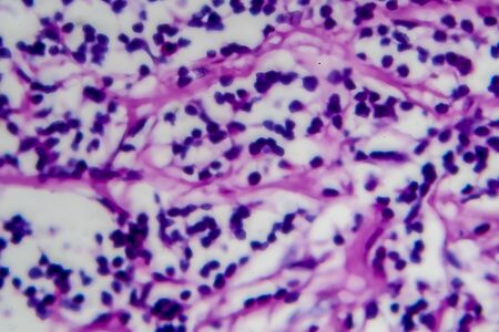
Histopathology of interstitial nephritis, light micrograph, photo under microscope. High magnification
Коллекция по умолчанию
Коллекция по умолчанию
Создать новую
Gloved hands hold a translucent cell culture sample with visible cellular structures and bubbles under focused clinical light, indicating laboratory research and microscopy, with available space for text on a neutral background
Коллекция по умолчанию
Коллекция по умолчанию
Создать новую
Branching budding yeast cells with pseudohyphae in sputum gram stain fine with microscope.
Коллекция по умолчанию
Коллекция по умолчанию
Создать новую
A closeup examination of a cultured cell on a nanoengineered substrate with bright staining revealing cellular morphology and interaction with the surface at the nanoscale
Коллекция по умолчанию
Коллекция по умолчанию
Создать новую
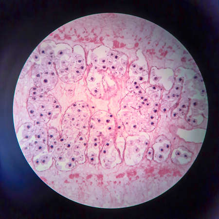
Hyaline cartilage, Elastic cartilage and Bone Human under the microscope in Lab.
Коллекция по умолчанию
Коллекция по умолчанию
Создать новую
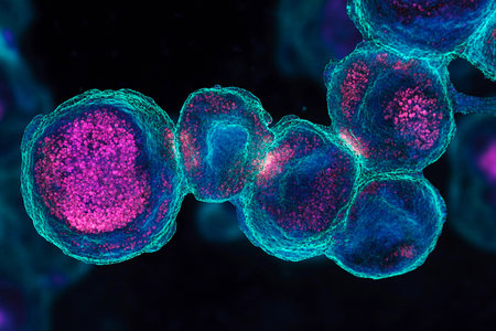
The activation of TLRs leading to the production of proinflammatory cytokines as seen in a micrograph of immune cells
Коллекция по умолчанию
Коллекция по умолчанию
Создать новую
Numerous tiny white bubbles float gracefully on the surface of the water. The bubbles appear to be delicate and light, moving with the gentle current in a mesmerizing manner.
Коллекция по умолчанию
Коллекция по умолчанию
Создать новую

Tooth development from human under microscope view for education.
Коллекция по умолчанию
Коллекция по умолчанию
Создать новую
abstract of blur blue for background used
Коллекция по умолчанию
Коллекция по умолчанию
Создать новую
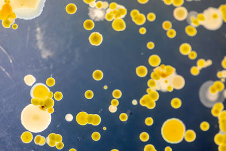
Backgrounds of Characteristics and Different shaped Colony of Bacteria and Mold growing on agar plates from Soil samples for education in Microbiology laboratory.
Коллекция по умолчанию
Коллекция по умолчанию
Создать новую
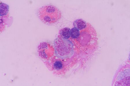
Multinucleated cell in Tzanck test finding with microscope in laboratory.
Коллекция по умолчанию
Коллекция по умолчанию
Создать новую
Smear of Acid-Fast bacilli AFB stained from sputum specimen, under 100X light microscope.
Коллекция по умолчанию
Коллекция по умолчанию
Создать новую
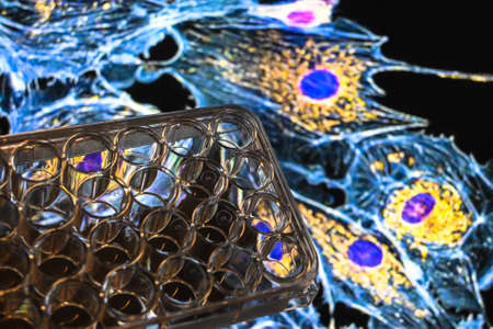
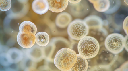
Achievements of modern biological science. A 24-well cell culture plate against an abstract biological background.
Коллекция по умолчанию
Коллекция по умолчанию
Создать новую
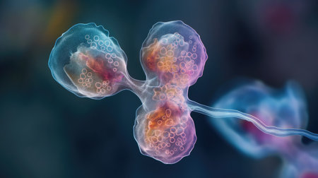
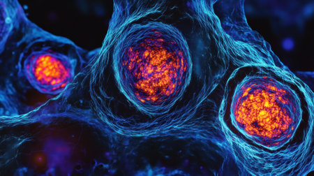
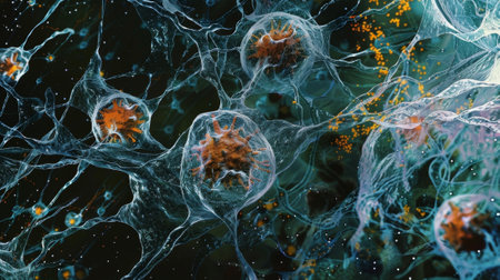
Dividing stem cells, 3D illustration. Research and scientific background
Коллекция по умолчанию
Коллекция по умолчанию
Создать новую

Watercolor texture. Abstract red watercolor paint pattern isolated on water color background. Splash ink stain for brush design
Коллекция по умолчанию
Коллекция по умолчанию
Создать новую
Cancer Cell in human showing abnormal cells.
Коллекция по умолчанию
Коллекция по умолчанию
Создать новую

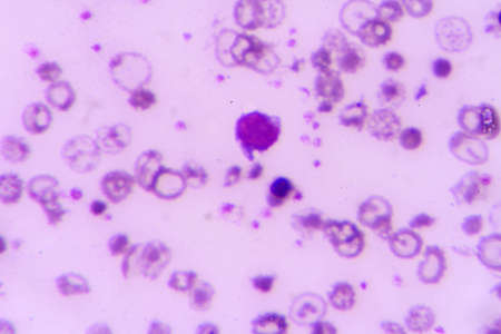
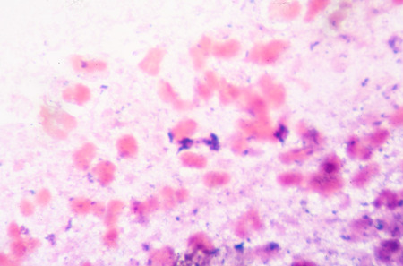
Blast cells in leukemia.blood smear is often used as a follow-up test to abnormal results on a complete blood count (CBC) to evaluate the different types of blood cells.
Коллекция по умолчанию
Коллекция по умолчанию
Создать новую
close-up of vibrant bacteria colonies on a petri dish, created with generative ai
Коллекция по умолчанию
Коллекция по умолчанию
Создать новую

Abstract creative marbling pattern templat for fabric, design background texture
Коллекция по умолчанию
Коллекция по умолчанию
Создать новую
Microscopic view of cells dividing and multiplying, forming complex colorful patterns in an organic environment, showing the beauty and complexity of biological processes
Коллекция по умолчанию
Коллекция по умолчанию
Создать новую
Abstract background of acrylic paint in pink and blue tones
Коллекция по умолчанию
Коллекция по умолчанию
Создать новую
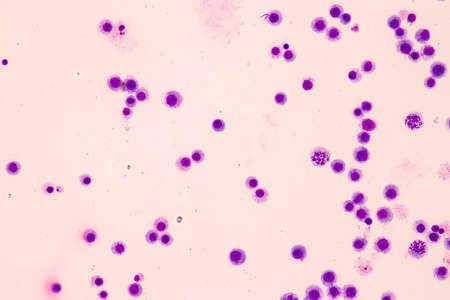
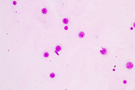
Leukemia cells
Коллекция по умолчанию
Коллекция по умолчанию
Создать новую
Abstract hand drawn pink floral pattern, watercolor vivid background
Коллекция по умолчанию
Коллекция по умолчанию
Создать новую
Pork tapeworm Taenia solium, section through the body, light micrograph
Коллекция по умолчанию
Коллекция по умолчанию
Создать новую
squamous cell
Коллекция по умолчанию
Коллекция по умолчанию
Создать новую
Characteristics of Lichen, hyphae and Symbiotic algae under the microscope for education.
Коллекция по умолчанию
Коллекция по умолчанию
Создать новую
Coccidiosis, coccidia in liver, light micrograph. Micrograph shows bile duct hyperplasia and fibrosis with periductal inflammation, groups of coccidia, large violet cells
Коллекция по умолчанию
Коллекция по умолчанию
Создать новую
abstract soft cloud shapes floating gently, symbolizing mental relaxation and serenity --chaos 30 --ar 16:9 --v 6.1 Job ID: a7aa97f5-d63f-4de4-8c88-c2a1381e6422
Коллекция по умолчанию
Коллекция по умолчанию
Создать новую
Smear of Acid-Fast bacilli AFB stained with WBC and mucous, under 100X light microscope.
Коллекция по умолчанию
Коллекция по умолчанию
Создать новую
Abnomal cells look like malignant cells from specimen plural fluid.
Коллекция по умолчанию
Коллекция по умолчанию
Создать новую

PC-3 human prostate cancer cells, stained with Coomassie blue, under differencial interference contrast microscope.
Коллекция по умолчанию
Коллекция по умолчанию
Создать новую
microscope slide with magnified view of human blood cells, created with generative ai
Коллекция по умолчанию
Коллекция по умолчанию
Создать новую
Meiosis In Grasshopper Testis under the light microscope view.
Коллекция по умолчанию
Коллекция по умолчанию
Создать новую
A microscope eyepiece capturing the view of edited cells with a display showing realtime imaging where genetic changes can be seen highlighted in bright colors..
Коллекция по умолчанию
Коллекция по умолчанию
Создать новую
Magnified view reveals networks detailed of cells showing unique shapes and textures, highlighting the complexity of biological forms and processes.
Коллекция по умолчанию
Коллекция по умолчанию
Создать новую
blood smear is often used as a follow-up test to abnormal results on a complete blood count (CBC) to evaluate the different types of blood cells.
Коллекция по умолчанию
Коллекция по умолчанию
Создать новую

malaria parasite plasmodium falciparum on a thick blood smear reading under a microscope
Коллекция по умолчанию
Коллекция по умолчанию
Создать новую
Microscopic view of cells in vibrant purple
Коллекция по умолчанию
Коллекция по умолчанию
Создать новую
Legion-Media
Создайте свои проекты на основе качественных стоковых фотографий и видео.


Copyright © Legion-Media.