
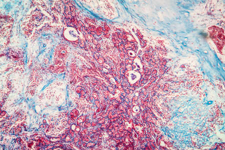
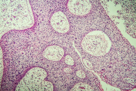
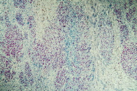
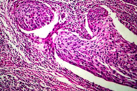
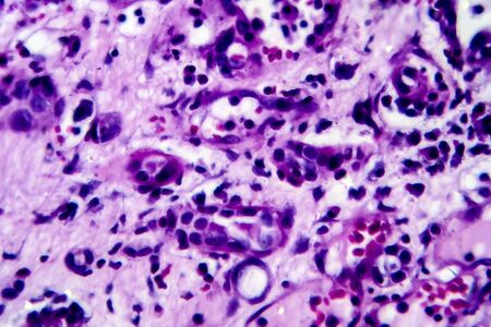
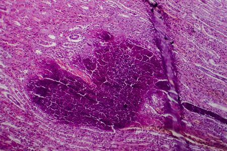
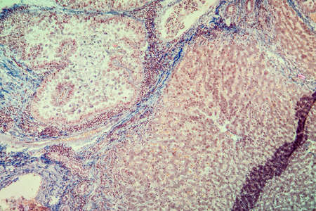
Basal cell cancer Diseased tissue 100x
Коллекция по умолчанию
Коллекция по умолчанию
Создать новую
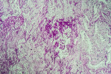
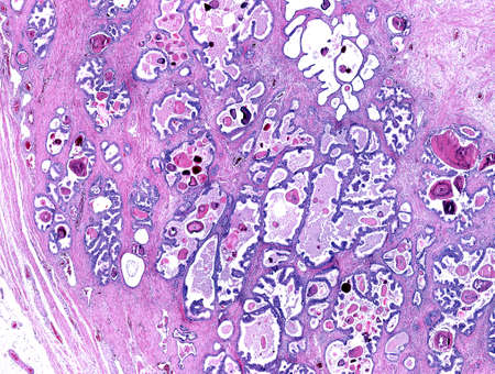
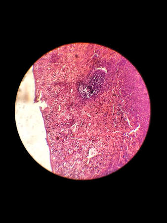
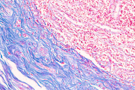
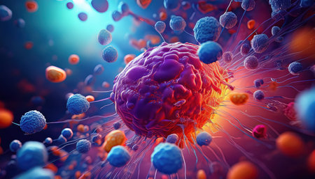
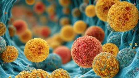
Breast cancer of the woman diseased tissue 100x
Коллекция по умолчанию
Коллекция по умолчанию
Создать новую
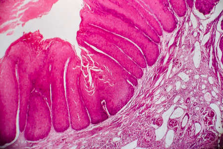
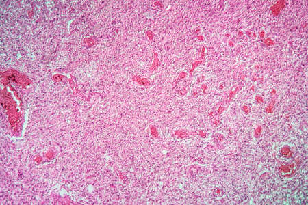
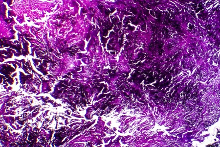
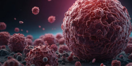
Squamous cell carcinoma diseased tissue under the microscope 100x
Коллекция по умолчанию
Коллекция по умолчанию
Создать новую
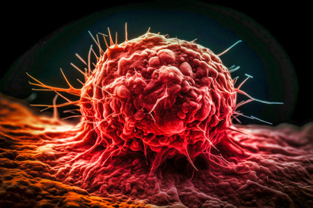
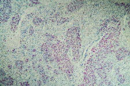
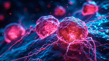
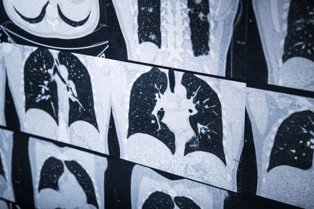
Colon inflammation in Crohn's disease 100x
Коллекция по умолчанию
Коллекция по умолчанию
Создать новую
fish caviar as a background. macro
Коллекция по умолчанию
Коллекция по умолчанию
Создать новую
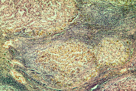
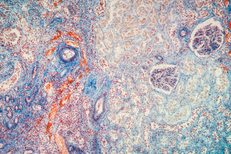
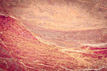
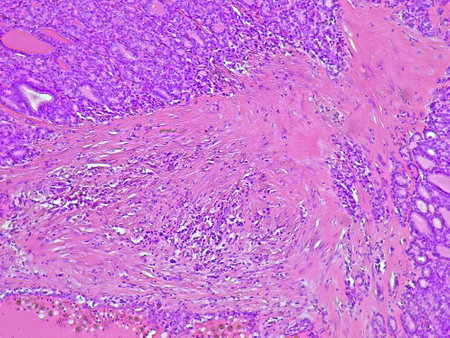
Liver cirrhosis tissue affected 100x after alcohol abuse
Коллекция по умолчанию
Коллекция по умолчанию
Создать новую
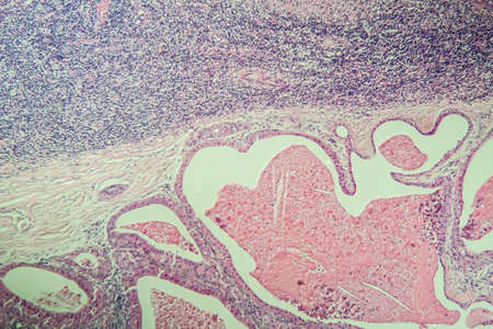
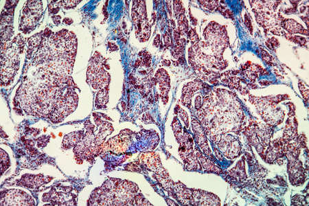
Salivary gland swollen diseased tissue under the microscope 100x
Коллекция по умолчанию
Коллекция по умолчанию
Создать новую

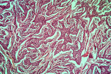
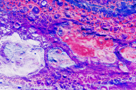
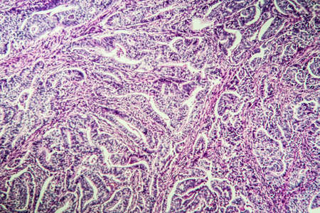
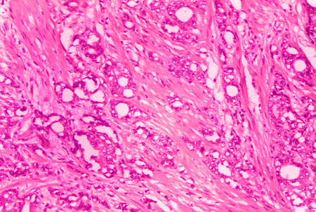
Ovarian cancer, light micrograph, photo under microscope. Photograph shows a fragment of a cancerous tumor in the female ovary. Selective focus
Коллекция по умолчанию
Коллекция по умолчанию
Создать новую
Painting acrylic paint- abstract drawing. Texture background
Коллекция по умолчанию
Коллекция по умолчанию
Создать новую
Atrophy kidney tissue under the microscope 100x
Коллекция по умолчанию
Коллекция по умолчанию
Создать новую
Gastric carcinoma in tissue section 100x
Коллекция по умолчанию
Коллекция по умолчанию
Создать новую
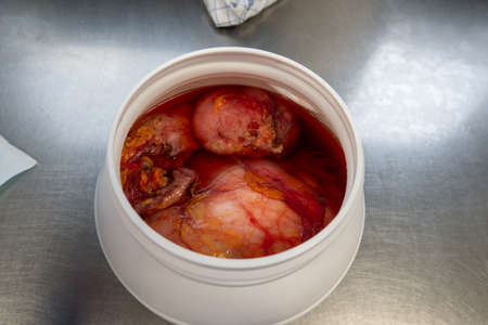
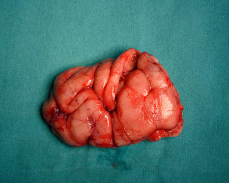
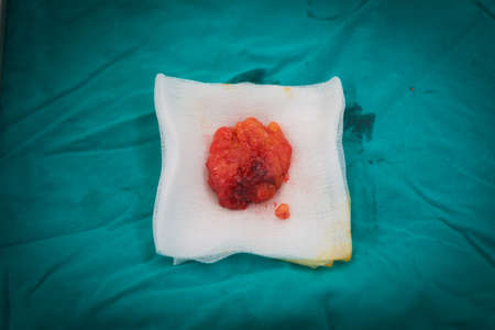
a large tumor, which was removed, lies pickled in formalin
Коллекция по умолчанию
Коллекция по умолчанию
Создать новую

Doctor incising specimen of bacterial brain abscess on sterile gauze for pathological examination.
Коллекция по умолчанию
Коллекция по умолчанию
Создать новую
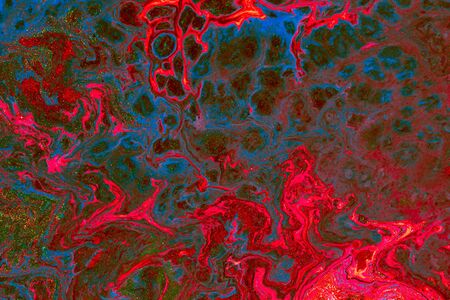
Liquid colored acrylic paints. Abstract colorful background.
Коллекция по умолчанию
Коллекция по умолчанию
Создать новую
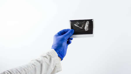
Doctor demonstrates an X-ray of the male prostate gland, to diagnose the X-ray picture of the prostate, on a white background.
Коллекция по умолчанию
Коллекция по умолчанию
Создать новую
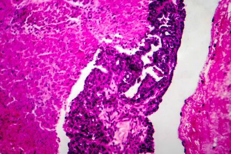
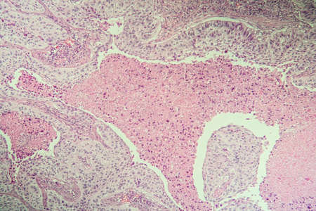
Papillary serous ovarian adenocarcinoma, cancer of ovary, light micrograph, photo under microscope
Коллекция по умолчанию
Коллекция по умолчанию
Создать новую
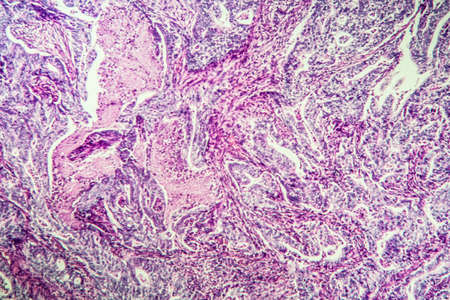
Gastric carcinoma in tissue section 100x
Коллекция по умолчанию
Коллекция по умолчанию
Создать новую
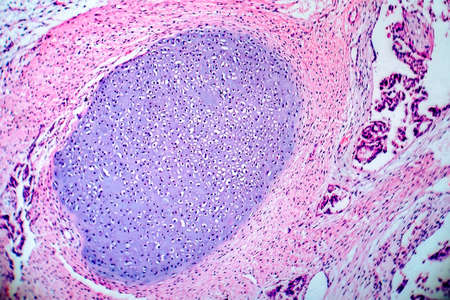
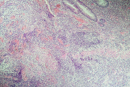
Light micrograph of teratoma, a tumor made up of several different types of tissue, such as hair, teeth, muscle, or bone. Teratoma is typically found in the ovary, testicle, or coccyx
Коллекция по умолчанию
Коллекция по умолчанию
Создать новую
Fibroepithelium Diseased tissue 100x
Коллекция по умолчанию
Коллекция по умолчанию
Создать новую
lungs Infection with Candida and Aspergillus in AIDS patients 200x
Коллекция по умолчанию
Коллекция по умолчанию
Создать новую
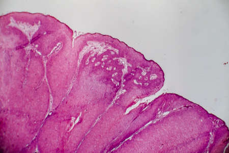
Condyloma acuminatum, also known as genital warts. Light micrograph, photo under microscope
Коллекция по умолчанию
Коллекция по умолчанию
Создать новую
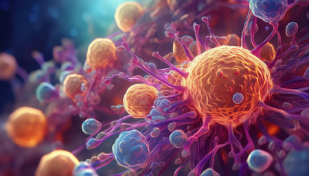
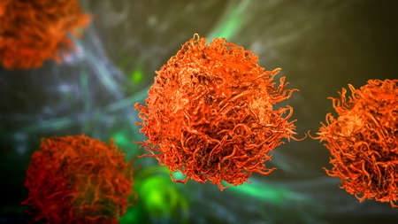
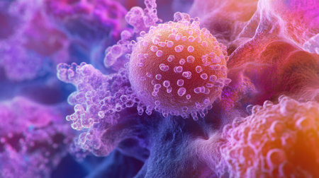
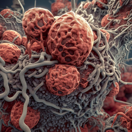
cancer tumor close-up. Generative AI
Коллекция по умолчанию
Коллекция по умолчанию
Создать новую
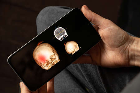
3D computed tomography of the brain with a fracture of the frontal part of the skull after injury on the screens of phones in the hands, mobile application
Коллекция по умолчанию
Коллекция по умолчанию
Создать новую

Cells of a human spleen with chronic myelogenous leukemia, under the microscope.
Коллекция по умолчанию
Коллекция по умолчанию
Создать новую
Condyloma acuminatum, also known as genital warts. Light micrograph, photo under microscope
Коллекция по умолчанию
Коллекция по умолчанию
Создать новую
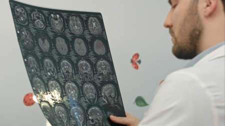
Senior man doctor examines MRI image in hospital
Коллекция по умолчанию
Коллекция по умолчанию
Создать новую

Histopathology of interstitial nephritis, light micrograph, photo under microscope
Коллекция по умолчанию
Коллекция по умолчанию
Создать новую
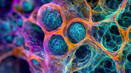
Vivid portrayal of cellular structures showcasing detailed biological research.
Коллекция по умолчанию
Коллекция по умолчанию
Создать новую

ovary taken during surgery after castration of the cat in a veterinary clinic
Коллекция по умолчанию
Коллекция по умолчанию
Создать новую
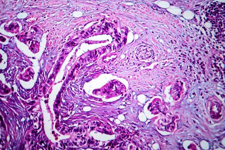
Histopathology of adenocarcinoma of the prostate
Коллекция по умолчанию
Коллекция по умолчанию
Создать новую
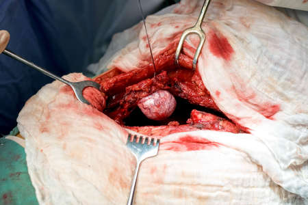
A large tumor in the chest of the patient before removing
Коллекция по умолчанию
Коллекция по умолчанию
Создать новую
Close-up view of abnormal brain specimen causing epilepsy after surgical excision on green cloth.
Коллекция по умолчанию
Коллекция по умолчанию
Создать новую
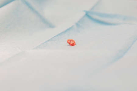
a piece of tissue taken during a surgery in a veterinary clinic
Коллекция по умолчанию
Коллекция по умолчанию
Создать новую
Bowen's Disease Tumor under the microscope 100x
Коллекция по умолчанию
Коллекция по умолчанию
Создать новую
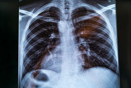
Chest X-ray image for physician's examination
Коллекция по умолчанию
Коллекция по умолчанию
Создать новую
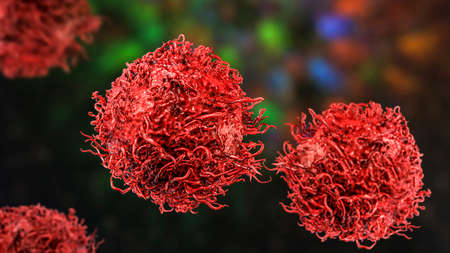
Stomach cancer cells, 3D illustration showing morphology of cancerous cells
Коллекция по умолчанию
Коллекция по умолчанию
Создать новую
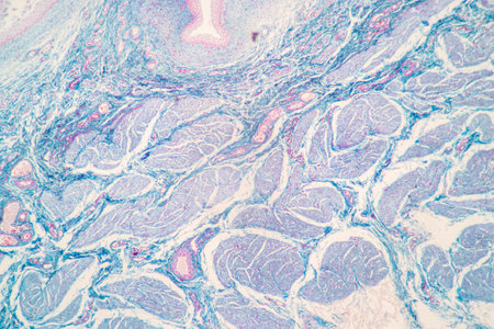
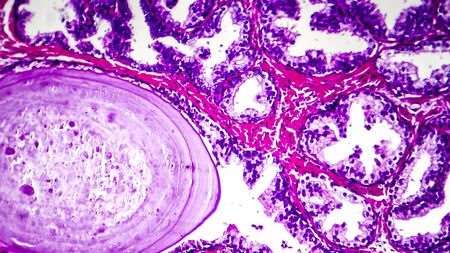
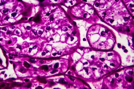
Low magnification of a human prostate gland in a 70-year-old man. The prostate gland appears with dilated alveoli, which contains many corpora amylacea (prostatic concretions) in their lumen. Light microscope micrograph. Hematoxylin & eosin stain.
Коллекция по умолчанию
Коллекция по умолчанию
Создать новую

Bowen's Disease Tumor under the microscope 100x
Коллекция по умолчанию
Коллекция по умолчанию
Создать новую
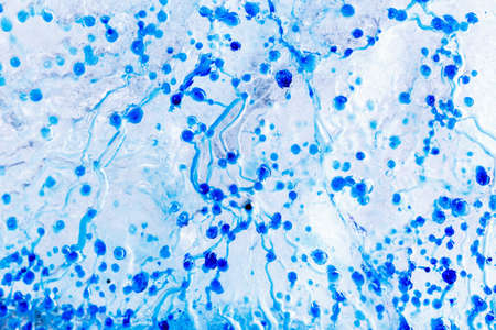
AIDS with fungi 100x infected tissue
Коллекция по умолчанию
Коллекция по умолчанию
Создать новую
Biological histological fixed colored preparation of the spleen - a secondary organ of the immune system
Коллекция по умолчанию
Коллекция по умолчанию
Создать новую
Glioma tumor with diseased tissue 100x
Коллекция по умолчанию
Коллекция по умолчанию
Создать новую
AIDS with fungi 100x infected tissue
Коллекция по умолчанию
Коллекция по умолчанию
Создать новую
macro of human bloods. microscope view for education physiology .3d microbiology rendering
Коллекция по умолчанию
Коллекция по умолчанию
Создать новую
Squamous cell carcinoma of the uterus, light micrograph, photo under microscope
Коллекция по умолчанию
Коллекция по умолчанию
Создать новую
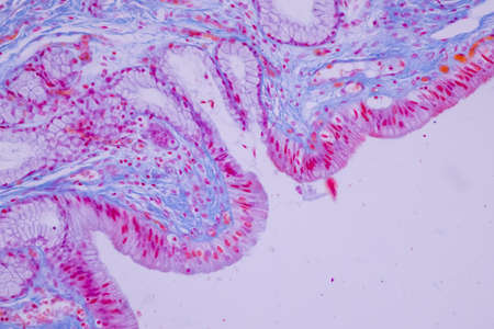
Tissue of Small intestine (Duodenum) and Vermiform appendix Human under the microscope in Lab.
Коллекция по умолчанию
Коллекция по умолчанию
Создать новую
Microscopic View of Cancer Cells, Depicting Growth, Division, and Spread, for Scientific Use
Коллекция по умолчанию
Коллекция по умолчанию
Создать новую
Histopathology of interstitial nephritis, light micrograph, photo under microscope. High magnification
Коллекция по умолчанию
Коллекция по умолчанию
Создать новую
Vivid portrayal of diverse cells interacting in a lab, highlighting science in action.
Коллекция по умолчанию
Коллекция по умолчанию
Создать новую
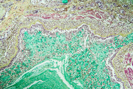
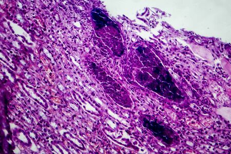
Histopathology of silicosis, the most prevalent chronic occupational disease. Light micrograph, photo under microscope
Коллекция по умолчанию
Коллекция по умолчанию
Создать новую
Cancer cell. Oncology research structure mutation, somatic cell of body. genetic predisposition. Neoplasms, cancerous disease, malignant tumor, Danger fear the unknown, biology medicine dna immune
Коллекция по умолчанию
Коллекция по умолчанию
Создать новую
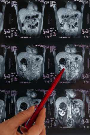
Magnetic resonance imaging, MRI, computed tomography, x-ray image. Area of ​​the pelvis with kidneys infected with tumors
Коллекция по умолчанию
Коллекция по умолчанию
Создать новую
Showing Light micrograph of the Adrenal gland and Urinary bladder human under the microscope for education in the laboratory.
Коллекция по умолчанию
Коллекция по умолчанию
Создать новую
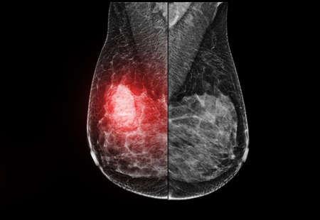
X-ray Digital Mammogram or mammography of the breast MLO view for diagnonsis Breast cancer in women isolated on black background.
Коллекция по умолчанию
Коллекция по умолчанию
Создать новую
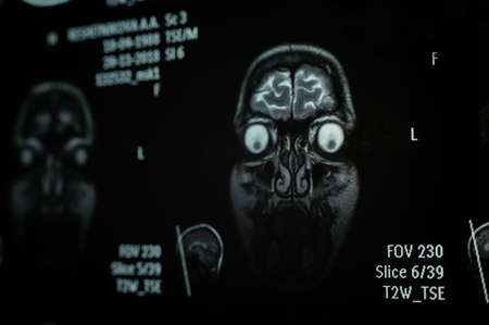
Close-up of magnetic resonance imaging of the human brain
Коллекция по умолчанию
Коллекция по умолчанию
Создать новую
Macro view of a cancerous cell under the microscope, highlighting its abnormal structure and the threat it poses to human health.
Коллекция по умолчанию
Коллекция по умолчанию
Создать новую

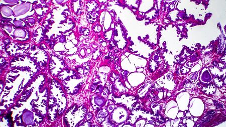
Endemic goiter, light micrograph, abnormal enlargement of the thyroid gland due to dietary iodine deficiency. Photomicrograph shows follicles of varying size, abundant colloid, lymphocytic infiltrate
Коллекция по умолчанию
Коллекция по умолчанию
Создать новую
Acute pyelonephritis, light micrograph, photo under microscope
Коллекция по умолчанию
Коллекция по умолчанию
Создать новую
Signet ring cell carcinoma of the stomach, light micrograph, photo under microscope
Коллекция по умолчанию
Коллекция по умолчанию
Создать новую

Ultrasonography of thyroid gland for diagnostics of the thyroid gland.
Коллекция по умолчанию
Коллекция по умолчанию
Создать новую
Cancer cell growth uncontrollably over tissue, Tumor infection cells and spreading, Invasive inflammation metastasis cancerous. reproduce by duplicating, cells expanding, Melanoma
Коллекция по умолчанию
Коллекция по умолчанию
Создать новую
Coccidiosis of liver tissue under the microscope 100x
Коллекция по умолчанию
Коллекция по умолчанию
Создать новую
Dynamic interplay of cells and microorganisms captured in a vivid environment, showing their distinctive colors and structures under a microscope in a laboratory setting.
Коллекция по умолчанию
Коллекция по умолчанию
Создать новую
AI Generated. National Cancer Control Month Background Illustration
Коллекция по умолчанию
Коллекция по умолчанию
Создать новую
X-ray image of a patient with pneumonia. Coronavirus pandemic. Lung damage. Closeup photo background.
Коллекция по умолчанию
Коллекция по умолчанию
Создать новую

thousands of tiny bacteria cells, forming into a beautiful organic shape, generative ai
Коллекция по умолчанию
Коллекция по умолчанию
Создать новую
A Glimpse into Cancer Prevention
Коллекция по умолчанию
Коллекция по умолчанию
Создать новую
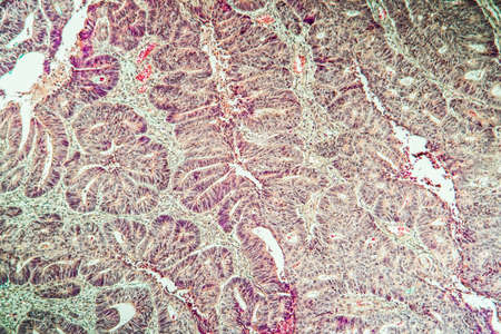
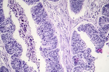
Colon carcinoma arising from adenoma, 100x
Коллекция по умолчанию
Коллекция по умолчанию
Создать новую
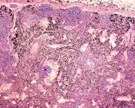
Anthracotic lymph node. Accumulation of carbon is most commonly found in intrapulmonary lymph nodes, due to coal dust, smoke or pollution. H & E stain
Коллекция по умолчанию
Коллекция по умолчанию
Создать новую
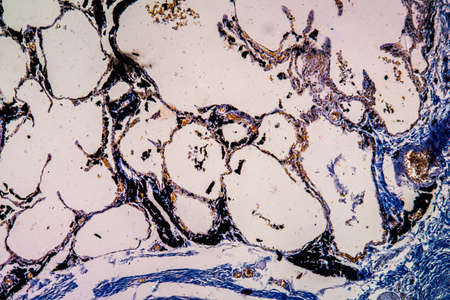
Charcoal dust lung tissue under the microscope 100x
Коллекция по умолчанию
Коллекция по умолчанию
Создать новую
Discover a vibrant abstract microstructure showcasing cells and biological elements, illuminated with stunning colors and intricate patterns that evoke creativity.
Коллекция по умолчанию
Коллекция по умолчанию
Создать новую
Stomach cancer cells, 3D illustration showing morphology of cancerous cells
Коллекция по умолчанию
Коллекция по умолчанию
Создать новую

Chronic pyelonephritis, light micrograph, photo under microscope. High magnification
Коллекция по умолчанию
Коллекция по умолчанию
Создать новую
Columnar epithelium of human gall bladder under the microscope in Lab.
Коллекция по умолчанию
Коллекция по умолчанию
Создать новую
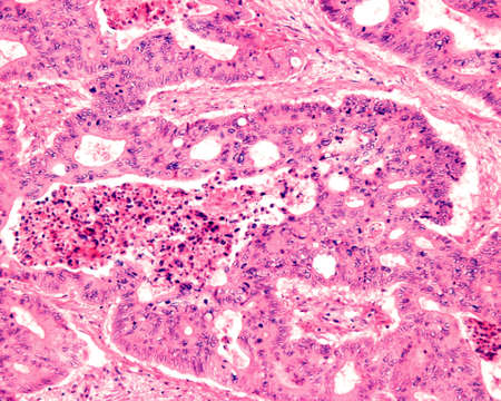
Large-bowel adenocarcinoma. Cancer cells arranged in cords or strands with empty central spaces remembering the normal crypts of the colon mucosa.
Коллекция по умолчанию
Коллекция по умолчанию
Создать новую
Chronic nephritis, light micrograph, photo under microscope
Коллекция по умолчанию
Коллекция по умолчанию
Создать новую
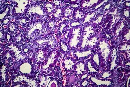
Histopathology of prostate gland hyperplasia, light micrograph, photo under microscope
Коллекция по умолчанию
Коллекция по умолчанию
Создать новую

Histology of human tissue, show nephritis as seen under the microscope
Коллекция по умолчанию
Коллекция по умолчанию
Создать новую

Lung adenocarcinoma, light micrograph, photo under microscope
Коллекция по умолчанию
Коллекция по умолчанию
Создать новую
Breast cancer, light micrograph, photo under microscope
Коллекция по умолчанию
Коллекция по умолчанию
Создать новую
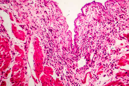
Asthma of the lungs diseased tissue under the microscope 100x
Коллекция по умолчанию
Коллекция по умолчанию
Создать новую
Macrophage cells, immune cells at high magnification, AI generative illustration,
Коллекция по умолчанию
Коллекция по умолчанию
Создать новую
abstract background
Коллекция по умолчанию
Коллекция по умолчанию
Создать новую
Esophageal squamous cell carcinoma, light micrograph, photo under microscope
Коллекция по умолчанию
Коллекция по умолчанию
Создать новую
lipoma
Коллекция по умолчанию
Коллекция по умолчанию
Создать новую

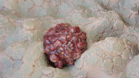
Colon polyp, one of the largest polyps
Коллекция по умолчанию
Коллекция по умолчанию
Создать новую
Colorful grunge stone texture background, creative abstract marble backdrop in red color.
Коллекция по умолчанию
Коллекция по умолчанию
Создать новую
Histopathology of prostate gland hyperplasia, light micrograph, photo under microscope
Коллекция по умолчанию
Коллекция по умолчанию
Создать новую

Thyroid follicular carcinoma, light micrograph, photo under microscope
Коллекция по умолчанию
Коллекция по умолчанию
Создать новую
Acute pyelonephritis, light micrograph, photo under microscope
Коллекция по умолчанию
Коллекция по умолчанию
Создать новую

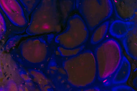
Abstract science background- pyloric division of the stomach of the dog. Cell biology
Коллекция по умолчанию
Коллекция по умолчанию
Создать новую
Handmade modern abstract painting. Made with fluid acrylic paints, by acrylic pouring with silicone and torching. Useable as a background or texture.
Коллекция по умолчанию
Коллекция по умолчанию
Создать новую
Photomicrograph showing histology of benign thyroid nodule under microscope with struma colloids cystica results in pathology laboratory
Коллекция по умолчанию
Коллекция по умолчанию
Создать новую
Ice texture background, ink in water pattern frost. Crystal winter design
Коллекция по умолчанию
Коллекция по умолчанию
Создать новую

cancer cell or tumor illustration in high detail for medical science or medical background
Коллекция по умолчанию
Коллекция по умолчанию
Создать новую
Kidney cancer, light micrograph, photo under microscope. High magnification
Коллекция по умолчанию
Коллекция по умолчанию
Создать новую
3d rendering of a colorful planet on a black background with smoke
Коллекция по умолчанию
Коллекция по умолчанию
Создать новую
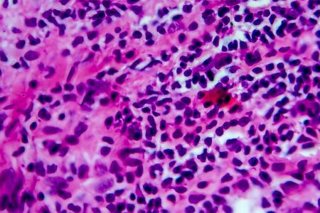
Hodgkin's lymphoma, light micrograph, photo under microscope. High magnification
Коллекция по умолчанию
Коллекция по умолчанию
Создать новую
Abstract creative marbling pattern templat for fabric, design background texture
Коллекция по умолчанию
Коллекция по умолчанию
Создать новую
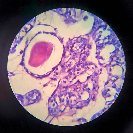
Thyroid follicular carcinoma, light micrograph, photo under microscope
Коллекция по умолчанию
Коллекция по умолчанию
Создать новую
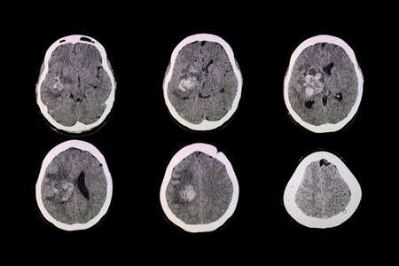
CT brian scan of the patient with hemorrhage and hematoma at right basal ganglia.
Коллекция по умолчанию
Коллекция по умолчанию
Создать новую
Legion-Media
Создайте свои проекты на основе качественных стоковых фотографий и видео.


Copyright © Legion-Media.