
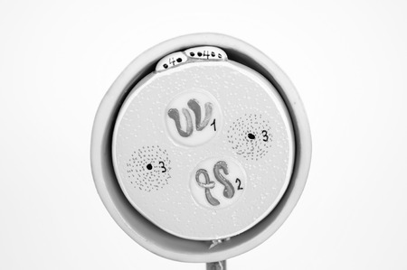
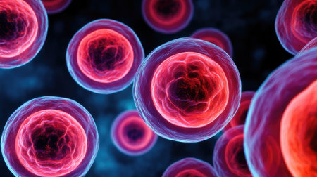
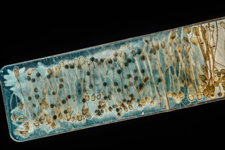
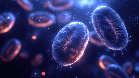
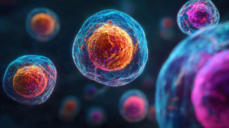
cell of fetus human model anatomy with black and white color concept
Коллекция по умолчанию
Коллекция по умолчанию
Создать новую
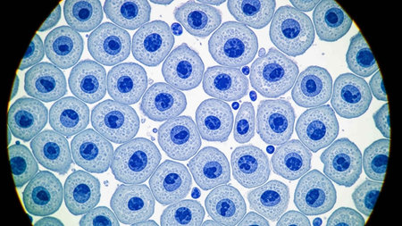
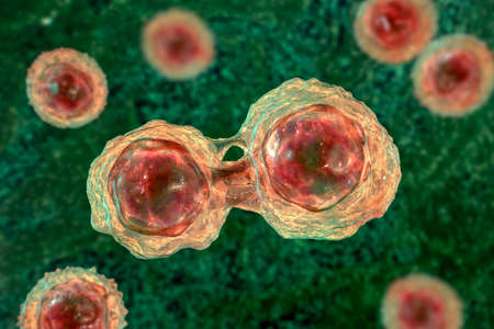
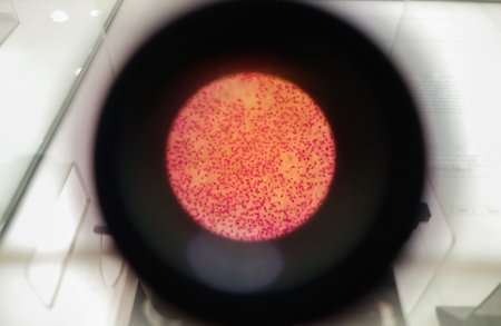
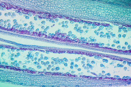
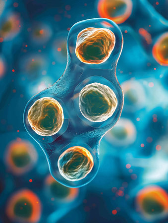
Pancreas cancer cells under microscope view for medical education.
Коллекция по умолчанию
Коллекция по умолчанию
Создать новую
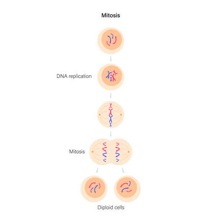
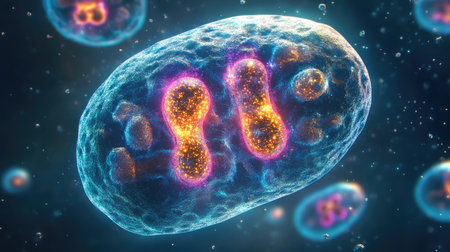
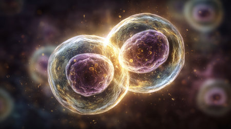
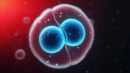
Mitosis cell division.
Коллекция по умолчанию
Коллекция по умолчанию
Создать новую
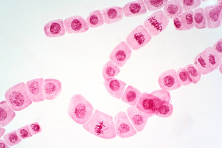
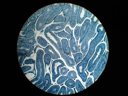
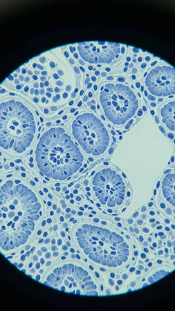
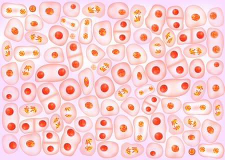
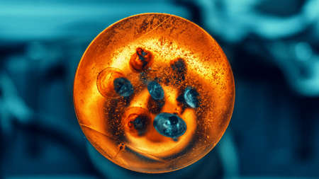
Mitosis cell of root tip of onion under the light microscope view.
Коллекция по умолчанию
Коллекция по умолчанию
Создать новую
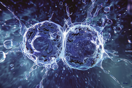
This close-up shot showcases water bubbles on a solid black background. The bubbles vary in size and shape, creating an interesting visual texture. The play of light on the water bubbles adds a dynamic element to the scene.
Коллекция по умолчанию
Коллекция по умолчанию
Создать новую
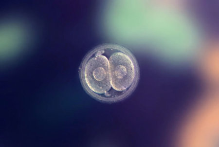
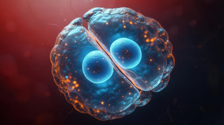
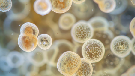
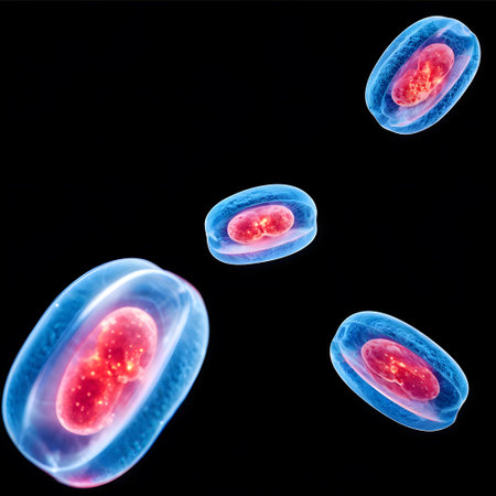
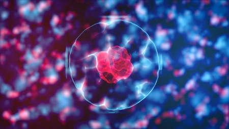
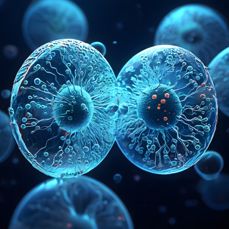
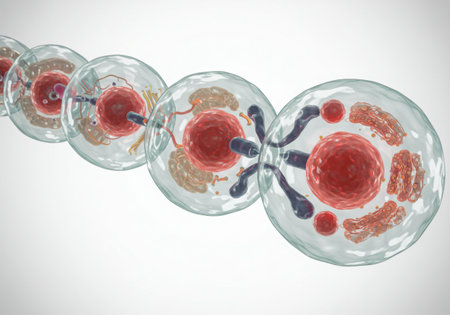
Human preimplantation embryo developing during in vitro fertilization, revealing the initial stages of cellular mitosis and supporting reproductive science
Коллекция по умолчанию
Коллекция по умолчанию
Создать новую
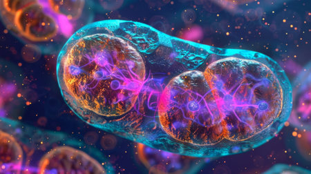
Depiction of the chemiosmotic theory illustrating the movement of protons across the inner mitochondrial membrane
Коллекция по умолчанию
Коллекция по умолчанию
Создать новую
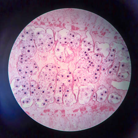
Pork tapeworm Taenia solium, section through the body, light micrograph
Коллекция по умолчанию
Коллекция по умолчанию
Создать новую
Dividing stem cells, 3D illustration. Research and scientific background
Коллекция по умолчанию
Коллекция по умолчанию
Создать новую
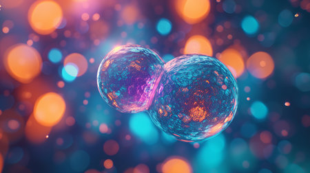
Vibrant 3D illustration showing the intricate process of cell division at the microscopic level
Коллекция по умолчанию
Коллекция по умолчанию
Создать новую
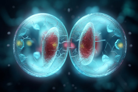
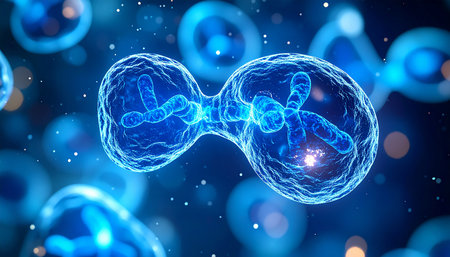
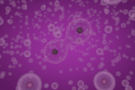
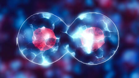
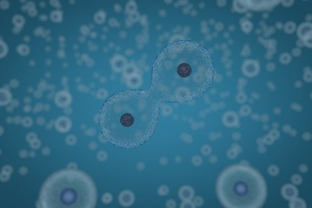
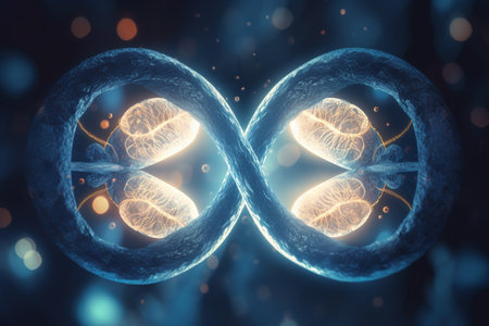
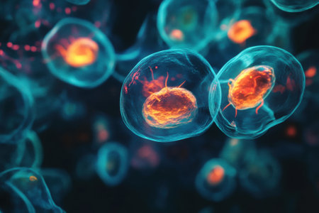
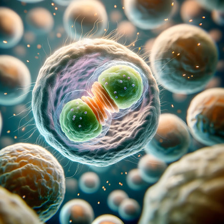
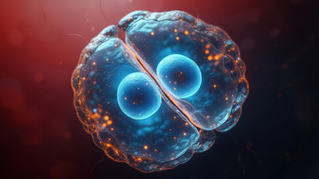
Detailed visualization of a cell in the process of mitosis, showing two distinct nuclei surrounded by a glowing membrane. Ideal for biology, science, and education themes.
Коллекция по умолчанию
Коллекция по умолчанию
Создать новую

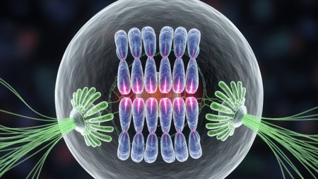
MITOSIS. Prophase, Metaphase, Anaphase, and Telophase. Cell division.
Коллекция по умолчанию
Коллекция по умолчанию
Создать новую
This vibrant illustration showcases a mitochondrion, highlighting its key features in stunning detail, perfect for educational materials and scientific presentations.
Коллекция по умолчанию
Коллекция по умолчанию
Создать новую
A detailed illustration of cells in various stages of mitosis and proliferation, showcasing cellular division and development.
Коллекция по умолчанию
Коллекция по умолчанию
Создать новую
Various steps of cellular division. 3D illustration.
Коллекция по умолчанию
Коллекция по умолчанию
Создать новую

Reproductive system concept
Коллекция по умолчанию
Коллекция по умолчанию
Создать новую
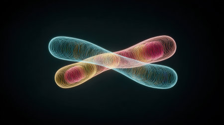
A colorful, abstract image of two DNA strands. The strands are in different colors and are twisted together. The image has a futuristic, otherworldly feel to it
Коллекция по умолчанию
Коллекция по умолчанию
Создать новую
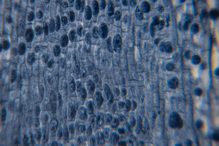
Close-up view of a textured surface featuring intricate patterns and vibrant blue tones, highlighting the beauty of natural materials and their unique formations
Коллекция по умолчанию
Коллекция по умолчанию
Создать новую

3D Illustration of Mitosis or cell division of biological stem cells.
Коллекция по умолчанию
Коллекция по умолчанию
Создать новую
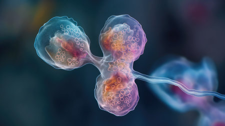
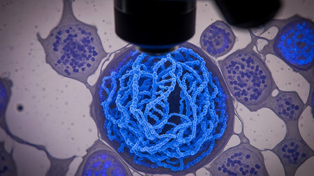
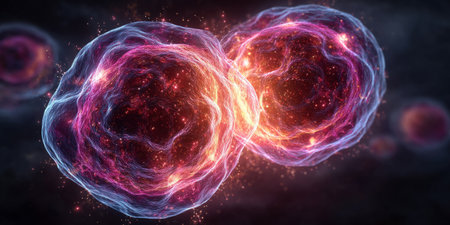
A stunning 3D rendering captures the intricate and beautiful moment of cell division, the fundamental process of life and growth. Glowing blue chromosomes separate as one cell becomes two, symbolizing creation, replication, and the future of biological science.
Коллекция по умолчанию
Коллекция по умолчанию
Создать новую

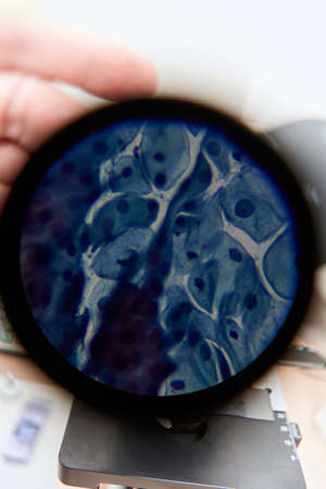
Root tip of Onion and Mitosis cell in the Root tip of Onion under a microscope.
Коллекция по умолчанию
Коллекция по умолчанию
Создать новую

Dividing cells on colorful background, 3D illustration
Коллекция по умолчанию
Коллекция по умолчанию
Создать новую

Proglottid (body unit) of tapeworm Taenia saginata, 3D illustration. A flatworm parasitizing animal and human intestine. Proglottid contains uterus with 12-30 primary lateral branches filled with eggs
Коллекция по умолчанию
Коллекция по умолчанию
Создать новую
Microscopic view of cell division under glowing light .Generative AI
Коллекция по умолчанию
Коллекция по умолчанию
Создать новую
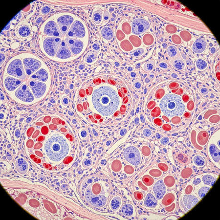
Cross section Human testis under microscope view. Shows spermatogonia, spermatocytes in meiosis, spermatids, and spermatozoa
Коллекция по умолчанию
Коллекция по умолчанию
Создать новую
Beautiful image in microscope of microorganisms in the lab
Коллекция по умолчанию
Коллекция по умолчанию
Создать новую
Cell Division
Коллекция по умолчанию
Коллекция по умолчанию
Создать новую

Chromosome with dark background, 3d rendering. Computer digital drawing.
Коллекция по умолчанию
Коллекция по умолчанию
Создать новую
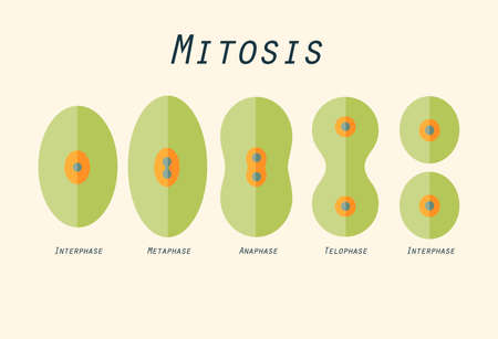
Illustration in flat design of cell division
Коллекция по умолчанию
Коллекция по умолчанию
Создать новую

meiosis. Cell division and Interphase. In the illustration labeled chiasma, Sister chromatids and homologous chromosomes. vector diagram
Коллекция по умолчанию
Коллекция по умолчанию
Создать новую
microscope lens, viewing Trypanosoma cruzi parasitic protozoan, causer of Chagas disease
Коллекция по умолчанию
Коллекция по умолчанию
Создать новую
Scientific illustration of cells dividing by osmosis, background with cells, 3D render illustration
Коллекция по умолчанию
Коллекция по умолчанию
Создать новую
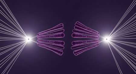
An abstract, symmetrical illustration of the anaphase stage of mitosis, where the chromosomes are pulled apart towards opposite poles by glowing spindle fibers. The scene is set against a dark purple background. The image represents concepts of biology, genetics, and cellular division.
Коллекция по умолчанию
Коллекция по умолчанию
Создать новую
Dividing cell with nucleus in mitosis and multiplication of cells for beauty and biology
Коллекция по умолчанию
Коллекция по умолчанию
Создать новую
Cross section of human cell under microscope view for education in laboratory.
Коллекция по умолчанию
Коллекция по умолчанию
Создать новую
Numerous tiny white bubbles float gracefully on the surface of the water. The bubbles appear to be delicate and light, moving with the gentle current in a mesmerizing manner.
Коллекция по умолчанию
Коллекция по умолчанию
Создать новую
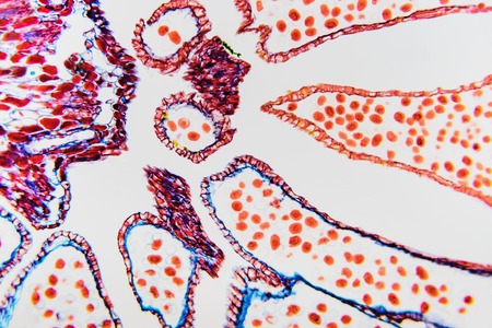
Abstract nature background- reproductive system flower. Ovary and ovule- plant cell.
Коллекция по умолчанию
Коллекция по умолчанию
Создать новую
A stunning 3D rendering captures the intricate process of cell division. Glowing blue chromosomes are visible within the nucleus, symbolizing the core of life and the future of genetic research and medical innovation. The surrounding cells create a sense of a larger biological system at work, perfect for concepts in science, technology, and healthcare.
Коллекция по умолчанию
Коллекция по умолчанию
Создать новую
Microscopic view of cell division under glowing light .Generative AI
Коллекция по умолчанию
Коллекция по умолчанию
Создать новую
Nestwurz orchid root cross 100x
Коллекция по умолчанию
Коллекция по умолчанию
Создать новую

Cell division during mitosis magnified under a microscope
Коллекция по умолчанию
Коллекция по умолчанию
Создать новую
3d illustration of Neisseria gonorrhoeae bacteria cells
Коллекция по умолчанию
Коллекция по умолчанию
Создать новую
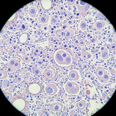
Histopathology of human liver under microscope view for medical education.
Коллекция по умолчанию
Коллекция по умолчанию
Создать новую
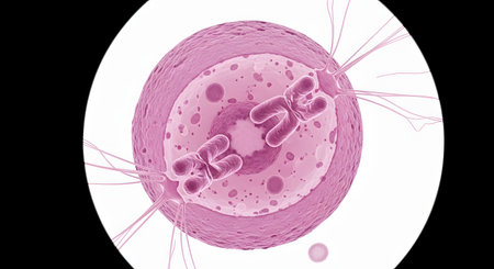
A highly detailed 3D render of a single cell in the process of mitosis or division, with chromosomes visible inside the nucleus. The pink and white image, seen as if through a microscope, illustrates a fundamental concept in biology and genetics.
Коллекция по умолчанию
Коллекция по умолчанию
Создать новую
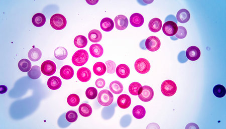
Blood cells in human body under microscope view for education in laboratory.
Коллекция по умолчанию
Коллекция по умолчанию
Создать новую
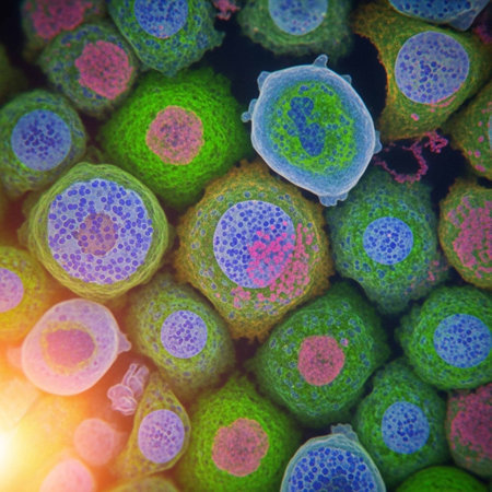
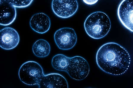
This image showcases a stunning microscopic view of vibrant cells, featuring intricate structures and a captivating dark background, perfect for educational and scientific projects.
Коллекция по умолчанию
Коллекция по умолчанию
Создать новую
Cell division background
Коллекция по умолчанию
Коллекция по умолчанию
Создать новую
viruses and anticlonal anti body under the microscope
Коллекция по умолчанию
Коллекция по умолчанию
Создать новую

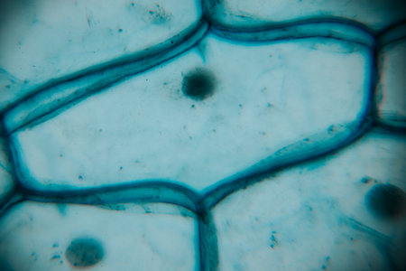
Close up Plant epidermis with stomata or Leaf Epidermis (Stomata) under microscope.
Коллекция по умолчанию
Коллекция по умолчанию
Создать новую
A detailed scientific illustration presents a cell in the process of division. Centered within a transparent sphere, a cluster of chromosomes are arranged. The image showcases detailed cellular components, highlighted by gradient colors and dynamic lighting. Suitable for educational materials, scientific publications, and conceptual visuals.
Коллекция по умолчанию
Коллекция по умолчанию
Создать новую
microscope slide with detailed view of plant stem, complete with cells and minutiae, created with generative ai
Коллекция по умолчанию
Коллекция по умолчанию
Создать новую
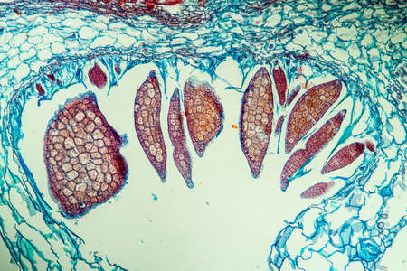
Flower with pollen along 100x
Коллекция по умолчанию
Коллекция по умолчанию
Создать новую
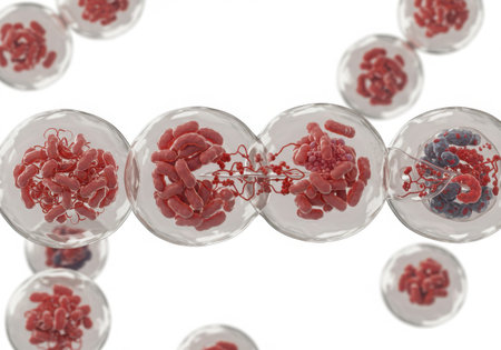
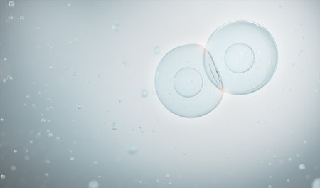
Stem cell division during mitosis, highlighting two blue nuclei within a transparent cell membrane. Ideal for scientific, educational, or medical content.
Коллекция по умолчанию
Коллекция по умолчанию
Создать новую
Glass transparent lamp night light with discs inside and air bubbles. Abstraction, background, selective soft focus
Коллекция по умолчанию
Коллекция по умолчанию
Создать новую
Scientific illustration of cells dividing by osmosis, background with cells, 3D render illustration
Коллекция по умолчанию
Коллекция по умолчанию
Создать новую
Scientific illustration of cells dividing by osmosis, cells multiplication, background with cells, 3D render illustration
Коллекция по умолчанию
Коллекция по умолчанию
Создать новую
Cell division process, micro
Коллекция по умолчанию
Коллекция по умолчанию
Создать новую
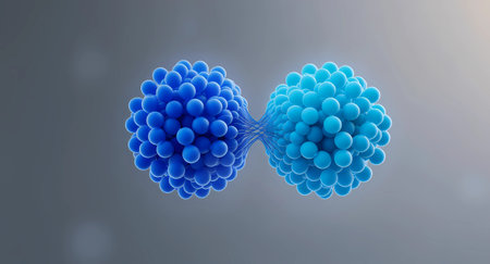
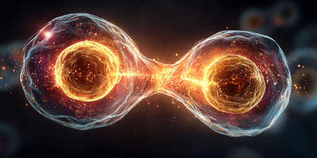
A minimalist and high-detail 3D rendering featuring two distinct clusters of blue spheres, symbolizing a cell division, molecular binding, or complex data transfer concept. The clusters are linked by a fine, nerve-like network or thread, suggesting connection, communication, or information exchange at a cellular or quantum level. This abstract visualization is perfect for themes related to biology, technology, networking, or scientific research.
Коллекция по умолчанию
Коллекция по умолчанию
Создать новую
Cosmetic hydrating product or ingredient concept. Glowing blue drops of transparent liquid on a black background. Drops gel or oil close up. Abstract dark backdrop.
Коллекция по умолчанию
Коллекция по умолчанию
Создать новую

Diagram of Mitosis. Prophase, Metaphase, Anaphase, and Telophase.
Коллекция по умолчанию
Коллекция по умолчанию
Создать новую
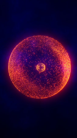
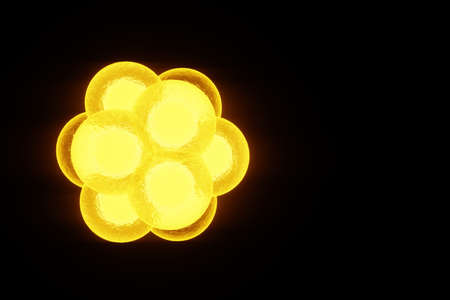
Fiery glowing sphere of orange and red particles floating in dark space, symbolizing cosmic energy or digital sun.
Коллекция по умолчанию
Коллекция по умолчанию
Создать новую
Vibrant 3D illustration showing the intricate process of cell division at the microscopic level
Коллекция по умолчанию
Коллекция по умолчанию
Создать новую
Bubbles in water on white background. 3d illustration.
Коллекция по умолчанию
Коллекция по умолчанию
Создать новую
An intricate digital representation of bacteria displaying vibrant colors and glowing elements, highlighting the fascinating world of microscopic life in a stunning abstract setting.
Коллекция по умолчанию
Коллекция по умолчанию
Создать новую

Vegetation cone of the water pest plant 100x
Коллекция по умолчанию
Коллекция по умолчанию
Создать новую
3D illustration of Neisseria gonorrhoeae bacteria
Коллекция по умолчанию
Коллекция по умолчанию
Создать новую
Chromosomes, gene mutation, genetic code. 3D rendering
Коллекция по умолчанию
Коллекция по умолчанию
Создать новую

This image features a detailed illustration of chromosomes, set against a blurred, blue backdrop. The visual style is suggestive of scientific or medical visualization, with a focus on biological structures. The composition uses depth of field and soft lighting to create a sense of three-dimensionality. It could be suitable for educational materials or science-related publications.
Коллекция по умолчанию
Коллекция по умолчанию
Создать новую
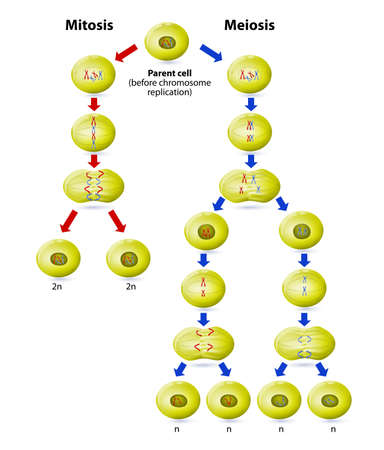
Meiosis and Mitosis. difference. Mitosis is the process of asexual reproduction. The process results in no new genetic combinations or no new genes. Meiosis is sexual reproduction with two pairs of genes. This is the method of reproduction favored by evol
Коллекция по умолчанию
Коллекция по умолчанию
Создать новую
3d rendering of Chromosome Abstract Scientific Background, 3d illustration.
Коллекция по умолчанию
Коллекция по умолчанию
Создать новую
Microscopic view of cells dividing and multiplying in 3d render
Коллекция по умолчанию
Коллекция по умолчанию
Создать новую
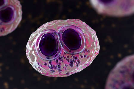
Cytomegalovirus CMV in a human cell, owl's eye inclusion in nucleus, multinucleated cell, 3D illustration. It is herpes virus, causes diseases in fetus, organ transplant patients, HIV infected people
Коллекция по умолчанию
Коллекция по умолчанию
Создать новую
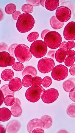
Red blood cells are visibly floating and interacting under a microscope showcasing their circular shape and vibrant red color in detail.
Коллекция по умолчанию
Коллекция по умолчанию
Создать новую
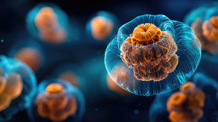
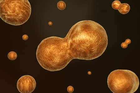
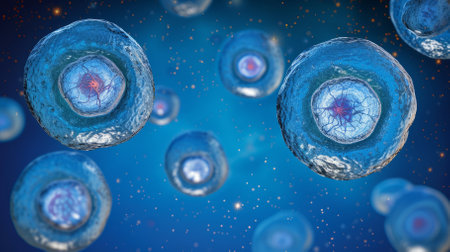
An artistic illustration showcases multiple biological cells, each featuring a layered orange core encapsulated within a translucent, blue-tinged membrane. The image utilizes a shallow depth of field, with soft focus on certain areas. Suitable for scientific publications, educational resources, or conceptual design projects.
Коллекция по умолчанию
Коллекция по умолчанию
Создать новую
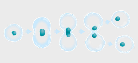
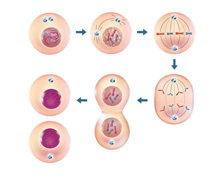
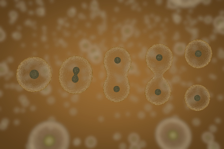
The graphic shows in 5 steps the division of a cell and duplication of the nucleus. vector images
Коллекция по умолчанию
Коллекция по умолчанию
Создать новую
Creative background image of the nucleus of the atom. The concept of nuclear fusion, nuclear fission, energy. 3D illustration, 3D rendering
Коллекция по умолчанию
Коллекция по умолчанию
Создать новую
3D illustration of a microscope and a human cell under microscope.
Коллекция по умолчанию
Коллекция по умолчанию
Создать новую
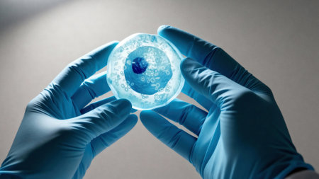
Gloved hands hold a translucent cell culture sample with visible cellular structures and bubbles under focused clinical light, indicating laboratory research and microscopy, with available space for text on a neutral background
Коллекция по умолчанию
Коллекция по умолчанию
Создать новую
the smallest unit that can live on its own and that makes up all living organisms and the tissues of the body.
Коллекция по умолчанию
Коллекция по умолчанию
Создать новую

A blue and orange strand of DNA is shown in a close up. The strand is twisted and has a cross shape. Concept of complexity and mystery, as DNA is the building block of life
Коллекция по умолчанию
Коллекция по умолчанию
Создать новую
A close-up, highly detailed macro photograph showcasing the process of cell division. The image captures the moment of mitosis, with one cell clearly visible in the process of splitting into two. The background is filled with a blur of cellular activity, emphasizing the singular focus on the cell undergoing division. Natural colors are used to accurately represent the cells structure, including the distinct chromosomes becoming visible as they separate, and the formation of the mitotic spindle. The overall lighting should highlight the intricate details of cellular components, creating a vivid and educational visual.
Коллекция по умолчанию
Коллекция по умолчанию
Создать новую

Gloved hands hold a translucent cell culture sample with visible cellular structures and bubbles under focused clinical light, indicating laboratory research and microscopy, with available space for text on a neutral background
Коллекция по умолчанию
Коллекция по умолчанию
Создать новую
Microscopic close up of male sperm fertilizing a female egg in reproductive process
Коллекция по умолчанию
Коллекция по умолчанию
Создать новую
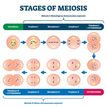
Stages of meiosis illustration. Labeled cell division process explanation scheme from genetic aspect. Interphase and interkinesis diagram with phases structural changes. Educational infographic
Коллекция по умолчанию
Коллекция по умолчанию
Создать новую
3D illustration of a group of stem cells
Коллекция по умолчанию
Коллекция по умолчанию
Создать новую
tiny cells during cell division and reproduction
Коллекция по умолчанию
Коллекция по умолчанию
Создать новую
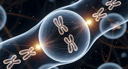
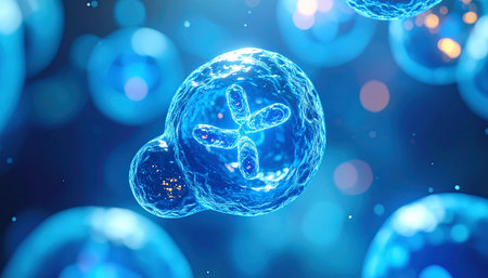
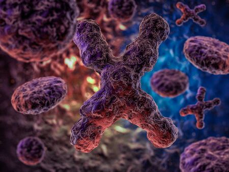
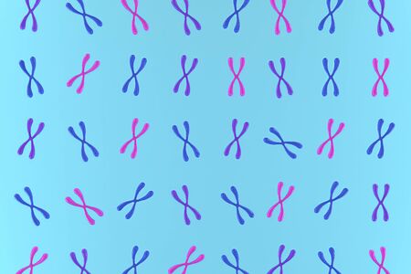
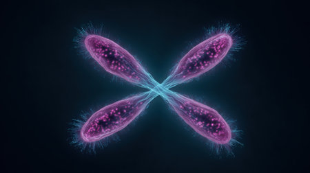
A 3D scientific illustration shows a detailed view of X-shaped human chromosomes inside a cell during the process of cell division (mitosis or meiosis). This visualization is a powerful educational tool for explaining concepts in genetics, heredity, and cellular biology.
Коллекция по умолчанию
Коллекция по умолчанию
Создать новую
Microscopic view of textured surface showing circular patterns and lines in gray and blue, highlighting cellular structures and organic shapes in detailed close-up
Коллекция по умолчанию
Коллекция по умолчанию
Создать новую

Composite image of chromosomes on blue background
Коллекция по умолчанию
Коллекция по умолчанию
Создать новую
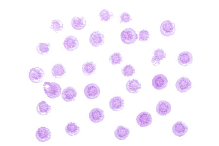
abstract acrylic watercolor paint brush stroke texture isolated on white background for logo and banner. design, creative, and illustration.
Коллекция по умолчанию
Коллекция по умолчанию
Создать новую
A microscopic view of cells undergoing mitosis, with chromosomes condensing and separating, leading to cell division.
Коллекция по умолчанию
Коллекция по умолчанию
Создать новую
A close up of a purple and blue cell with a pink center. The cell is surrounded by a blue and purple frame
Коллекция по умолчанию
Коллекция по умолчанию
Создать новую
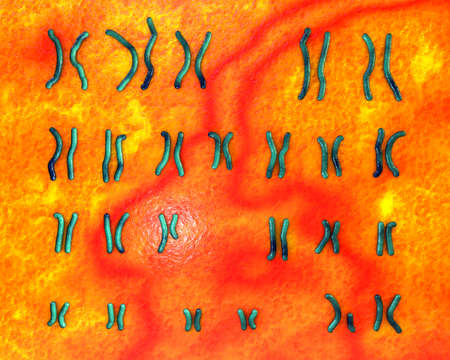
Karyotype of Prader-Willi syndrome
Коллекция по умолчанию
Коллекция по умолчанию
Создать новую
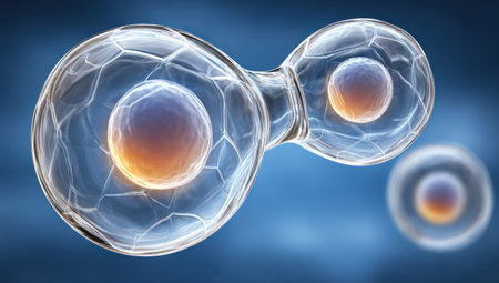
Illustration of cells dividing, showcasing nucleus within each, translucent spheres, blue backdrop
Коллекция по умолчанию
Коллекция по умолчанию
Создать новую
Detailed visualization of a cell in the process of mitosis, showing two distinct nuclei surrounded by a glowing membrane. Ideal for biology, science, and education themes.
Коллекция по умолчанию
Коллекция по умолчанию
Создать новую
Microscopic view of cell division under glowing light .Generative AI
Коллекция по умолчанию
Коллекция по умолчанию
Создать новую

Transparent cell with biotechnology and cosmetic concept, 3d rendering. 3D illustration.
Коллекция по умолчанию
Коллекция по умолчанию
Создать новую
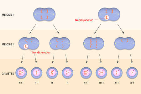
Meiotic nondisjunction. Abnormal chromosome number.
Коллекция по умолчанию
Коллекция по умолчанию
Создать новую
A group of colorful cells are shown in a close up. The cells are in various shades of blue, red, and purple. Concept of vibrancy and life, as the colors of the cells are bright
Коллекция по умолчанию
Коллекция по умолчанию
Создать новую
Histopathology of alveoli, light micrograph, photo under microscope
Коллекция по умолчанию
Коллекция по умолчанию
Создать новую
Legion-Media
Создайте свои проекты на основе качественных стоковых фотографий и видео.


Copyright © Legion-Media.