
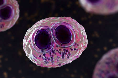
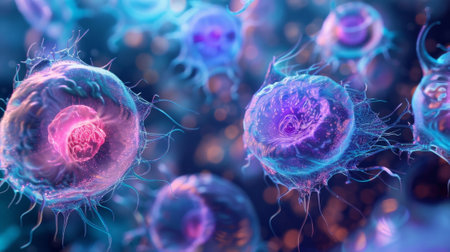
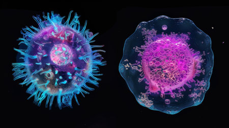
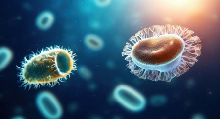
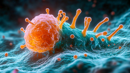
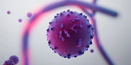
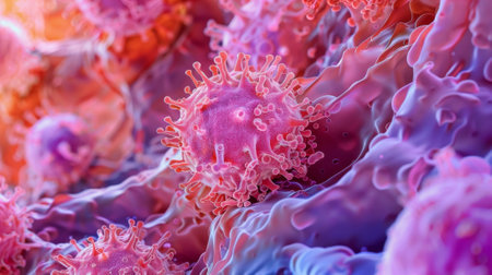
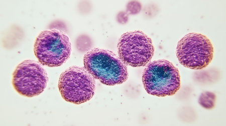
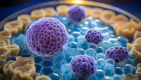
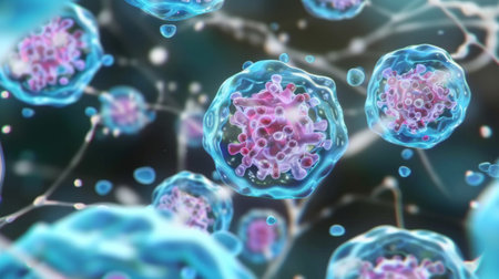
Cytomegalovirus CMV in a human cell, owl's eye inclusion in nucleus, multinucleated cell, 3D illustration. It is herpes virus, causes diseases in fetus, organ transplant patients, HIV infected people
Коллекция по умолчанию
Коллекция по умолчанию
Создать новую
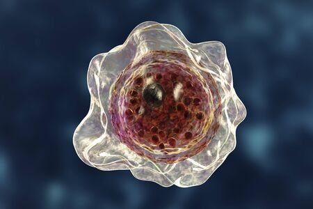
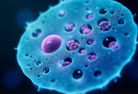
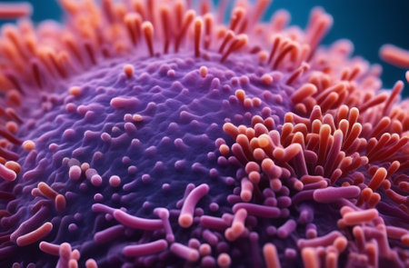
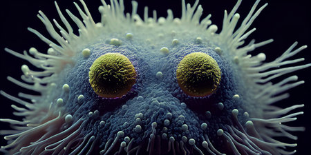
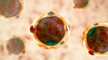
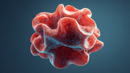
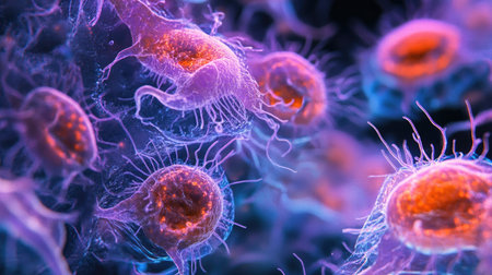
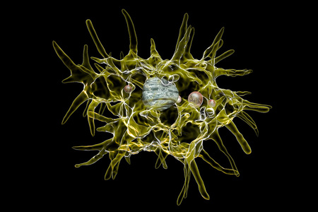
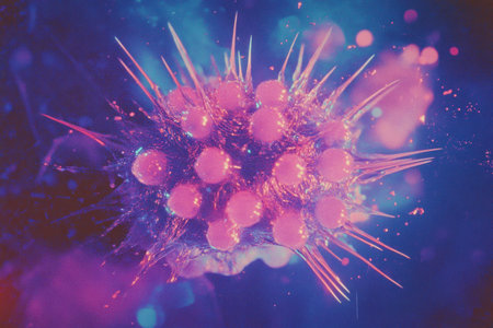
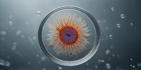
Cyst of Balamuthia mandrillaris amoeba, 3D illustration. A free-living protozoan in soil and water, can cause granulomatous amoebic encephalitis. Both cysts and trophozoites are infectious forms for humans
Коллекция по умолчанию
Коллекция по умолчанию
Создать новую
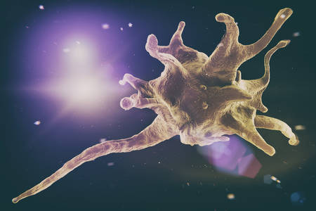
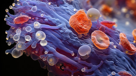
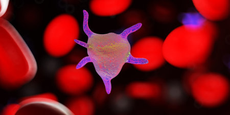
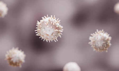
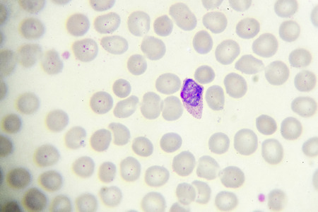
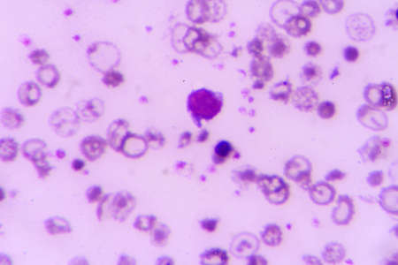
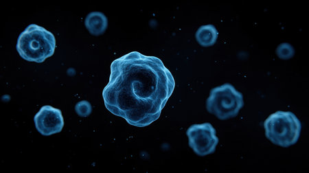
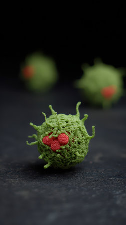
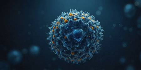
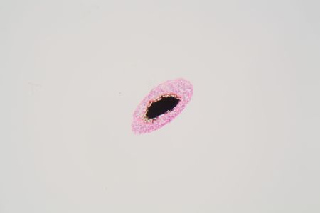
Activatet platelet cell, Thrombocyte are a component of blood whose function is to react to bleeding from blood vessel injury by clumping, thereby initiating a blood clot. 3d illustration
Коллекция по умолчанию
Коллекция по умолчанию
Создать новую
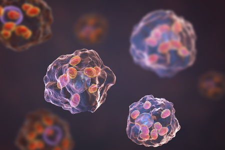
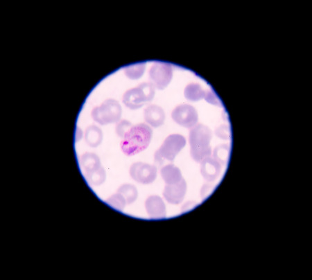
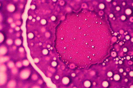
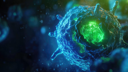
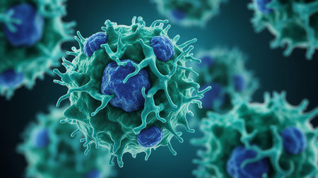
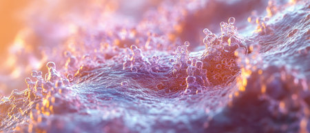
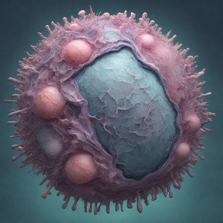
Macrophages infected by Leishmania amastigotes, 3D illustration
Коллекция по умолчанию
Коллекция по умолчанию
Создать новую
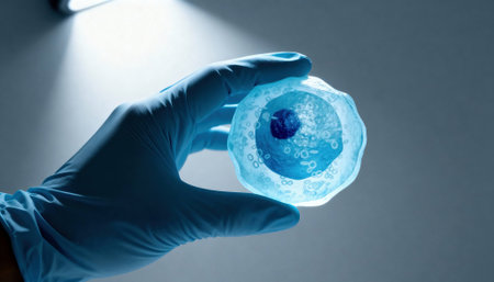
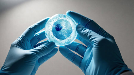
Gloved hand holds a translucent cell model under focused laboratory light with visible nucleus and organelle structures, suggesting scientific research and educational demonstration, with empty background space available for text
Коллекция по умолчанию
Коллекция по умолчанию
Создать новую
a close up of a colorful structure
Коллекция по умолчанию
Коллекция по умолчанию
Создать новую
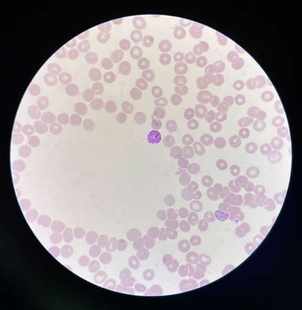
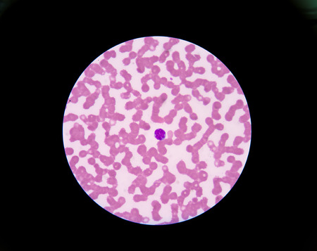
Malaria blood parasite infected red blood cells laboratory background.
Коллекция по умолчанию
Коллекция по умолчанию
Создать новую
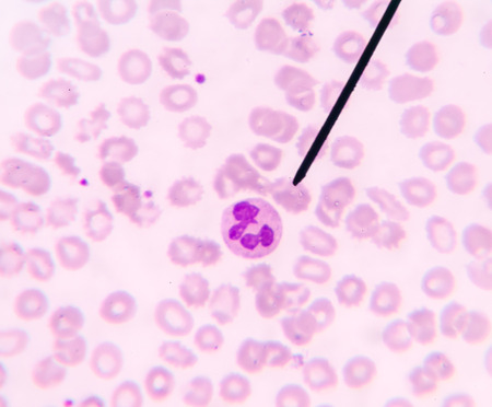
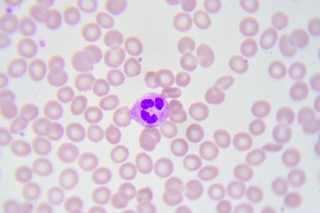
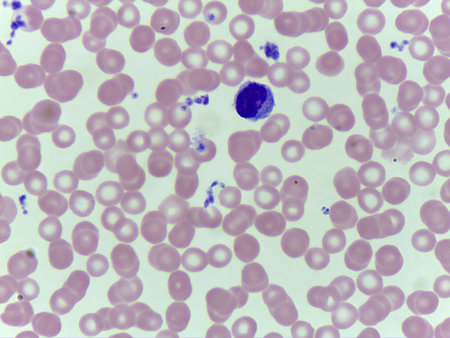
Neutrophil show in blood smear CBC test find with microscope.
Коллекция по умолчанию
Коллекция по умолчанию
Создать новую

Photomicrograph of canine eosinphil
Коллекция по умолчанию
Коллекция по умолчанию
Создать новую
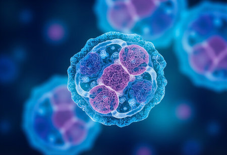
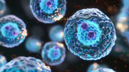
Microscopic view of human blood cell, 3D illustration.
Коллекция по умолчанию
Коллекция по умолчанию
Создать новую

neutrophils. blood smear is often used as a follow-up test to abnormal results on a complete blood count (CBC) to evaluate the different types of blood cells.
Коллекция по умолчанию
Коллекция по умолчанию
Создать новую
A microcosm of motion as cilia on different cells move in different directions yet work together to maintain healthy bodily functions
Коллекция по умолчанию
Коллекция по умолчанию
Создать новую
3d rendered medically accurate illustration of a platelet
Коллекция по умолчанию
Коллекция по умолчанию
Создать новую
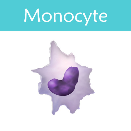
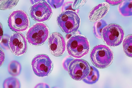
Leukocytes. Monocyte. White blood cell. Vector medical illustration
Коллекция по умолчанию
Коллекция по умолчанию
Создать новую
Nanoparticles Functionalization Therapeutics, Nanoparticles application in bioiechnology illustration
Коллекция по умолчанию
Коллекция по умолчанию
Создать новую

Gloved hands hold a translucent cell culture sample with visible cellular structures and bubbles under focused clinical light, indicating laboratory research and microscopy, with available space for text on a neutral background
Коллекция по умолчанию
Коллекция по умолчанию
Создать новую
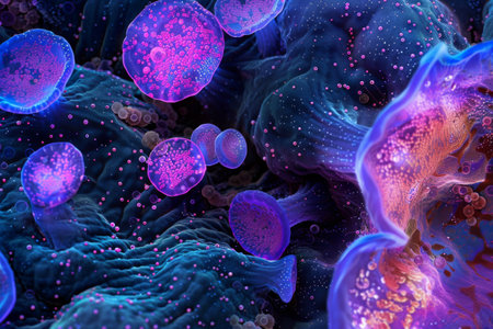
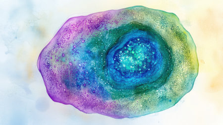
A colorful image of a cell with a purple and blue blob in the center. The image is abstract and has a mood of curiosity and wonder
Коллекция по умолчанию
Коллекция по умолчанию
Создать новую
Virus cells, 3D illustration. Viruses and bacteria in human body. Viruses in infected organism.
Коллекция по умолчанию
Коллекция по умолчанию
Создать новую
Exploring the microscopic world of cells and tissues, AI generated
Коллекция по умолчанию
Коллекция по умолчанию
Создать новую
A comparison of healthy and diseased immune cells with the latter displaying a significant decrease in phagolysosome formation hampering their ability to fight s
Коллекция по умолчанию
Коллекция по умолчанию
Создать новую
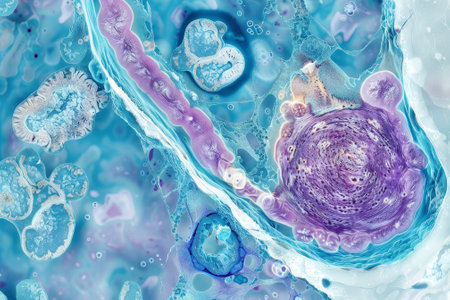
taste buds under the microscope, hairs inside the intestines, gastric mucosa, body microflora. High quality photo
Коллекция по умолчанию
Коллекция по умолчанию
Создать новую
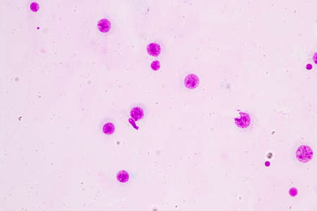
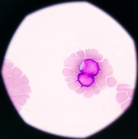
Malaria parasite in blood smear, gemetocyte stage
Коллекция по умолчанию
Коллекция по умолчанию
Создать новую
blood films for Malaria parasite
Коллекция по умолчанию
Коллекция по умолчанию
Создать новую

Ascaris lumbricoides, a large roundworm, fertilized egg, 3D illustration
Коллекция по умолчанию
Коллекция по умолчанию
Создать новую
Close-up view of glowing bacteria and viruses with spiky exteriors, floating in a dark blue, luminous, and abstract background.
Коллекция по умолчанию
Коллекция по умолчанию
Создать новую
A group of chaotically surreal bacteria with a face. cartoon picture
Коллекция по умолчанию
Коллекция по умолчанию
Создать новую
nucleated red cell
Коллекция по умолчанию
Коллекция по умолчанию
Создать новую

Neutrophil cell (white blood cell) in peripheral blood smear
Коллекция по умолчанию
Коллекция по умолчанию
Создать новую
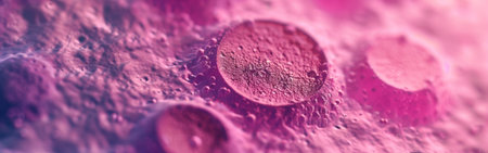
Abstract pink cancer cell organism background 3d render digital illustration
Коллекция по умолчанию
Коллекция по умолчанию
Создать новую
Microscopic view of human cells under microscope.
Коллекция по умолчанию
Коллекция по умолчанию
Создать новую
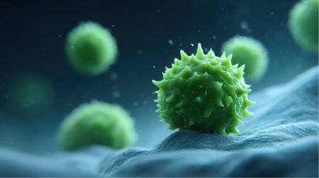
Several spiky green biological cells are resting on a light blue membrane. This appears to be a microscopic view of cells or viruses. Dark blue background.
Коллекция по умолчанию
Коллекция по умолчанию
Создать новую

The world of microorganisms
Коллекция по умолчанию
Коллекция по умолчанию
Создать новую

Meningococcal meningitis, cerebrospinal fluid smear containing neutrophils with and without bacteria Neisseria meningitidis
Коллекция по умолчанию
Коллекция по умолчанию
Создать новую

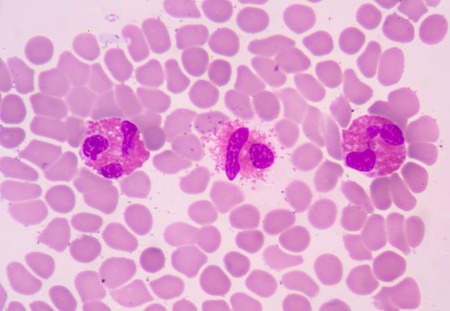
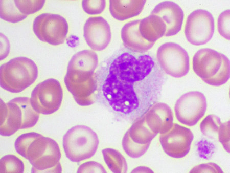
Blood smear showing, in the center, three neutrophil with hypersegmented nucleus. These cells appear in pathological situations such as megaloblastic anemias. Wright stain.
Коллекция по умолчанию
Коллекция по умолчанию
Создать новую
Gloved hands hold a translucent cell culture sample with visible cellular structures and bubbles under focused clinical light, indicating laboratory research and microscopy, with available space for text on a neutral background
Коллекция по умолчанию
Коллекция по умолчанию
Создать новую

3d rendered illustration of human cancer cells
Коллекция по умолчанию
Коллекция по умолчанию
Создать новую
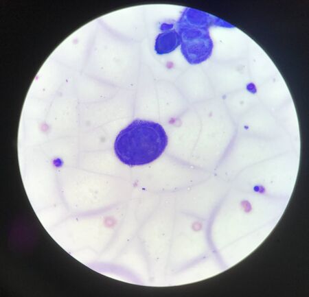
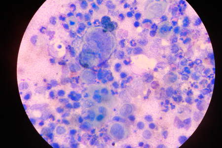
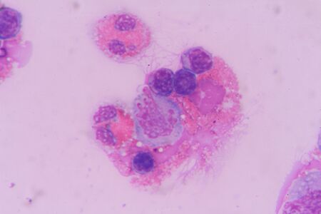
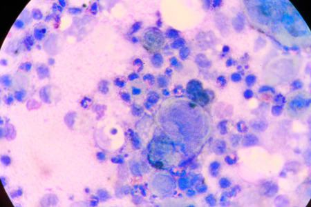
Multinucleated cell in Tzanck test finding with microscope in laboratory.
Коллекция по умолчанию
Коллекция по умолчанию
Создать новую
A glowing, glowing, glowing blob of light in the sky. It's a bit like a jellyfish, but it's not
Коллекция по умолчанию
Коллекция по умолчанию
Создать новую
Neutrophil cell
Коллекция по умолчанию
Коллекция по умолчанию
Создать новую
Microscopic close-up of vibrant stained human cells on a blue backdrop
Коллекция по умолчанию
Коллекция по умолчанию
Создать новую
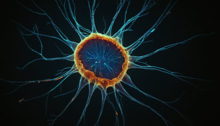
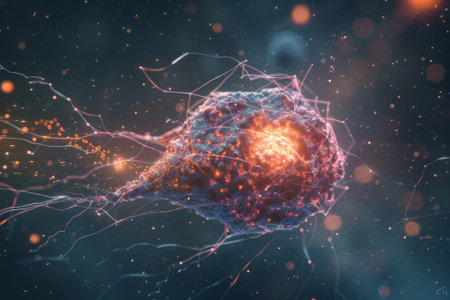
Radiant Neuron Structure
Коллекция по умолчанию
Коллекция по умолчанию
Создать новую
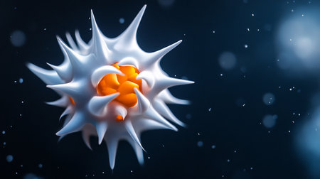
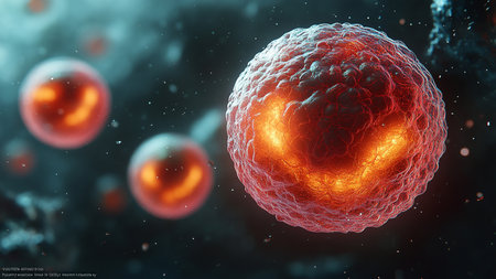
Abstract sphere with white, spiky exterior and orange interior, featuring a textured, slightly translucent surface and a complex, organic form.
Коллекция по умолчанию
Коллекция по умолчанию
Создать новую
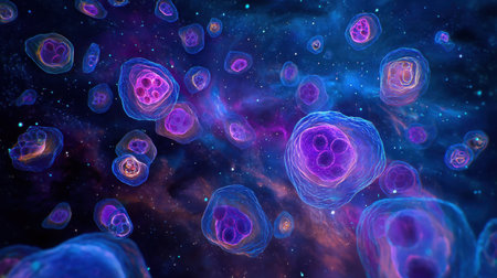
Blastocystis hominis parasite, 3D illustration. The causative agent of diarrheal infections in humans
Коллекция по умолчанию
Коллекция по умолчанию
Создать новую
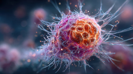
A detailed illustration showcases a colorful cell with filaments and textures, highlighting its complex structures and biological features in a laboratory environment.
Коллекция по умолчанию
Коллекция по умолчанию
Создать новую
White blood cells of a human, Eosinophil photomicrograph panorama as seen under the microscope
Коллекция по умолчанию
Коллекция по умолчанию
Создать новую
Captivating image of blue glowing cells on a dark background, ideal for illustrating concepts in biology, research, and microscopic life forms in scientific documents.
Коллекция по умолчанию
Коллекция по умолчанию
Создать новую
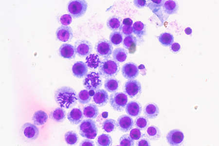
Immature white blood cells in leukemia.Science concept.
Коллекция по умолчанию
Коллекция по умолчанию
Создать новую
Blood smear with white blood cells and red blood cells. Medical background.
Коллекция по умолчанию
Коллекция по умолчанию
Создать новую
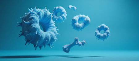
A probiotics-themed 3D illustration depicting Lactobacillus restoring gut flora in a blue setting
Коллекция по умолчанию
Коллекция по умолчанию
Создать новую

PC-3 human prostate cancer cells, stained with Coomassie blue, under differencial interference contrast microscope.
Коллекция по умолчанию
Коллекция по умолчанию
Создать новую
The cancer cell's aggressive and invasive nature is emphasized through its detailed, ominous appearance.
Коллекция по умолчанию
Коллекция по умолчанию
Создать новую
cancer cell or tumor illustration in high detail 3d render concept for medical science
Коллекция по умолчанию
Коллекция по умолчанию
Создать новую
A detailed representation of a green microorganism with red spots sits on a dark surface showcasing its unique shape and texture.
Коллекция по умолчанию
Коллекция по умолчанию
Создать новую
A futuristic glowing 3D model of a cell in vibrant blue and green, symbolizing life sciences.
Коллекция по умолчанию
Коллекция по умолчанию
Создать новую
multinucleated giant
Коллекция по умолчанию
Коллекция по умолчанию
Создать новую
AI Generated. Microscopic View of a Cell Under Observation
Коллекция по умолчанию
Коллекция по умолчанию
Создать новую

Chronic myeloid leukemia cells or CML, analyze by microscope, original magnification 1000x
Коллекция по умолчанию
Коллекция по умолчанию
Создать новую
Blood smear showing white and red blood cells
Коллекция по умолчанию
Коллекция по умолчанию
Создать новую
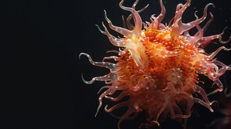
A detailed photograph of a vibrant orange sea anemone, showcasing its complex structure against a stark black backdrop.
Коллекция по умолчанию
Коллекция по умолчанию
Создать новую
Eosinophil
Коллекция по умолчанию
Коллекция по умолчанию
Создать новую
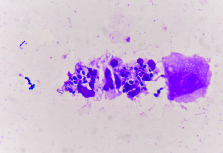
Yeast cells with epithelial tissue in Gram stain method.
Коллекция по умолчанию
Коллекция по умолчанию
Создать новую
Microbiology loop isolated on white background
Коллекция по умолчанию
Коллекция по умолчанию
Создать новую
Immature cells in myeloid serie myelocyte metamyelocyte.
Коллекция по умолчанию
Коллекция по умолчанию
Создать новую
Leukocytes squeezing in between epithelial cells to reach the lining of an organ
Коллекция по умолчанию
Коллекция по умолчанию
Создать новую
Stunning microscopic image of colorful cellular structures demonstrates the intricate details and vibrant colors found in the world of microbiology and life forms.
Коллекция по умолчанию
Коллекция по умолчанию
Создать новую
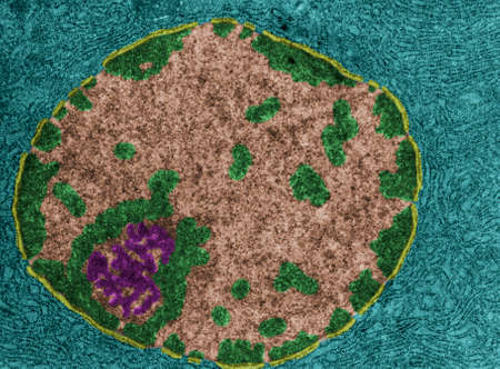
False colour transmission electron microscope (TEM) showing the nucleus of a protein-synthesizing cell. The nuclear envelope (yellow), chromatin (green), nucleoplasm (light brown) and nucleolus (magenta) can be seen. The cytoplasm (cian) is full of RER.
Коллекция по умолчанию
Коллекция по умолчанию
Создать новую
Digital illustration of blood cells in color background with alpha layer. 3D rendering
Коллекция по умолчанию
Коллекция по умолчанию
Создать новую

Electron micrograph of a macrophage
Коллекция по умолчанию
Коллекция по умолчанию
Создать новую

Plasma cell, a white blood cell, differenciated from B lymphocyte that secretes antibodies, 3D illustration
Коллекция по умолчанию
Коллекция по умолчанию
Создать новую

An abstract image of an orange splash with water droplets, set against a dark, blurred background.
Коллекция по умолчанию
Коллекция по умолчанию
Создать новую
multinucleated giant
Коллекция по умолчанию
Коллекция по умолчанию
Создать новую
Acanthamoeba castellanii amoeba, 3D illustration. Amoeba found in all aquatic habitats and soil, causes ceratitis especially amongst contact lens wearers
Коллекция по умолчанию
Коллекция по умолчанию
Создать новую
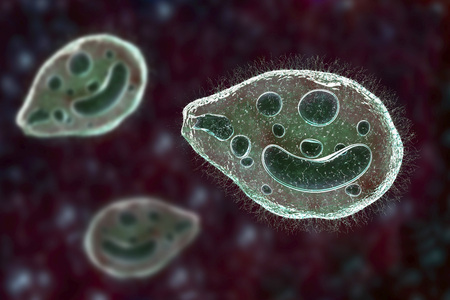
Balantidium coli protozoan, 3D illustration. Ciliated intestinal parasite that causes balantidiasis
Коллекция по умолчанию
Коллекция по умолчанию
Создать новую
A mesmerizing abstract illustration of cells floating in an enchanting cosmic space, featuring vivid colors and intricate details that inspire imagination.
Коллекция по умолчанию
Коллекция по умолчанию
Создать новую

Human hyaline cartilage bone under microscope view for education pathology. Human tissue.
Коллекция по умолчанию
Коллекция по умолчанию
Создать новую
Chromosomes Human under the microscope for education.
Коллекция по умолчанию
Коллекция по умолчанию
Создать новую
Coronavirus 2019-nCoV. SARS-CoV-2. Virus cells in infected organism, viral disease epidemic. 3D rendering
Коллекция по умолчанию
Коллекция по умолчанию
Создать новую
A detailed artwork showcases a single cell featuring a softly glowing blue nucleus and shimmering green and purple cytoplasm. The background gradient enhances its vivid colors and intricate details.
Коллекция по умолчанию
Коллекция по умолчанию
Создать новую
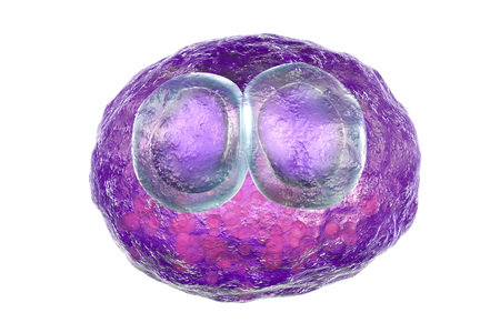
Cytomegalovirus CMV in human cell, owls eye inclusion in nucleus, multinucleated cell, 3D illustration. It is herpes virus, causes disease in fetus, organ transplant patients, HIV infected people
Коллекция по умолчанию
Коллекция по умолчанию
Создать новую
Microscopic View Rendered Image of Abnormal, Diseased Cells in Biology and Medicine Illustration
Коллекция по умолчанию
Коллекция по умолчанию
Создать новую
A grainy, blurry photograph of an abstract explosion in purple and blue colors. The photo is taken from a great distance, making the spines appear tiny but vibrant against their dark background. Up close, they look like spikes on fire with some glitch effects that give them a futuristic appearance. --ar 3:2 --v 6.1 Job ID: 70334657-f4da-4b0b-970e-2730ac7a063c
Коллекция по умолчанию
Коллекция по умолчанию
Создать новую
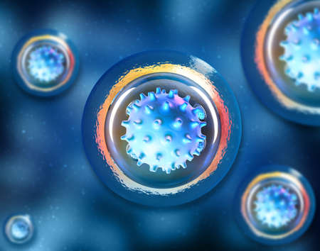
AI Generated. A close up of a cell with a blue nucleus
Коллекция по умолчанию
Коллекция по умолчанию
Создать новую
Sparkling water waves form vibrant bubbles during the morning light creating a captivating and tranquil atmosphere.
Коллекция по умолчанию
Коллекция по умолчанию
Создать новую
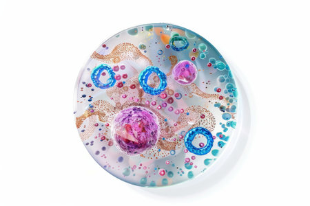
3d illustration of human papillomavirus cells in a plate
Коллекция по умолчанию
Коллекция по умолчанию
Создать новую
Abstract view of semiprecious resin surface with vivid grape-like purple clusters scattered across cloudy blue and white texture.
Коллекция по умолчанию
Коллекция по умолчанию
Создать новую
Numerous tiny white bubbles float gracefully on the surface of the water. The bubbles appear to be delicate and light, moving with the gentle current in a mesmerizing manner.
Коллекция по умолчанию
Коллекция по умолчанию
Создать новую
Virus cells. Viral disease outbreak. 3d illustration
Коллекция по умолчанию
Коллекция по умолчанию
Создать новую
Chromosomes Human under the microscope for education.
Коллекция по умолчанию
Коллекция по умолчанию
Создать новую
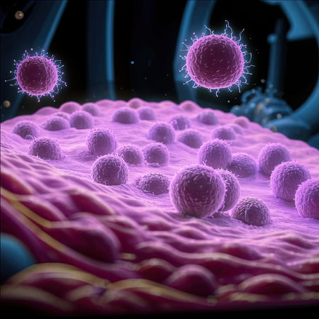
Illustration of a set of cells. Reproduction technologies. In vitro gametogenesis. This technique transforms skin cells into induced stem cells, which can then be turned into eggs and sperm.
Коллекция по умолчанию
Коллекция по умолчанию
Создать новую

Growing cancer cell concept image. 3d rendering
Коллекция по умолчанию
Коллекция по умолчанию
Создать новую
Human cell under microscope.
Коллекция по умолчанию
Коллекция по умолчанию
Создать новую
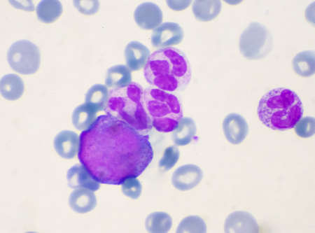
Promyelocye
Коллекция по умолчанию
Коллекция по умолчанию
Создать новую

malaria parasite plasmodium falciparum on a thick blood smear reading under a microscope
Коллекция по умолчанию
Коллекция по умолчанию
Создать новую
Human cell under microscope.
Коллекция по умолчанию
Коллекция по умолчанию
Создать новую
AI Generated. Close-up of a petri dish with bacterial growth
Коллекция по умолчанию
Коллекция по умолчанию
Создать новую

Characteristics of Squamous epithelial cell (Cell structure) of human under microscope view for education in laboratory.
Коллекция по умолчанию
Коллекция по умолчанию
Создать новую
Schistosoma mansoni under the microscope. Schistosoma mansoni is human parasite and causes schistosomiasis.
Коллекция по умолчанию
Коллекция по умолчанию
Создать новую
3D illustration of a microscopic view of a virus cell with cells.
Коллекция по умолчанию
Коллекция по умолчанию
Создать новую
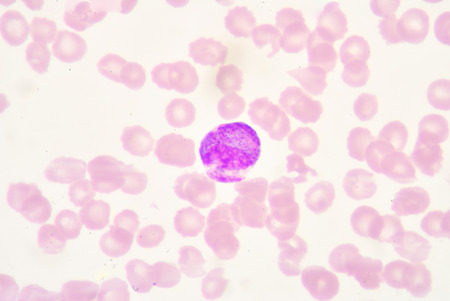
blood smear is often used as a follow-up test to abnormal results on a complete blood count (CBC) to evaluate the different types of blood cells.Atypical lymphocyte.
Коллекция по умолчанию
Коллекция по умолчанию
Создать новую
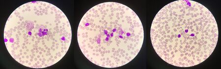
blood smear is often used as a follow-up test to abnormal results on a complete blood count (CBC) to evaluate the different types of blood cells.Medical science background showing blast cells(AML)
Коллекция по умолчанию
Коллекция по умолчанию
Создать новую
Legion-Media
Создайте свои проекты на основе качественных стоковых фотографий и видео.


Copyright © Legion-Media.