

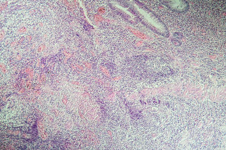
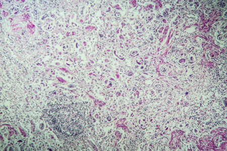
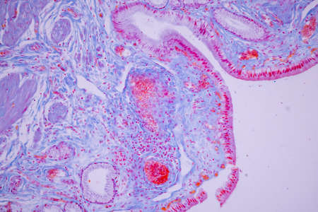
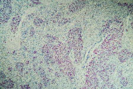
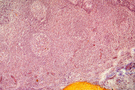
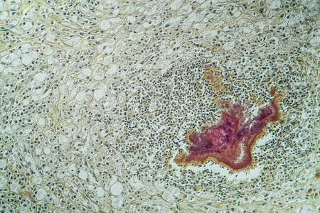
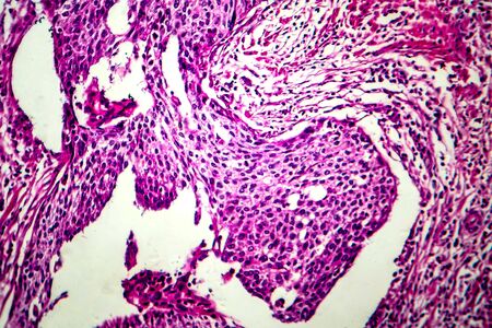
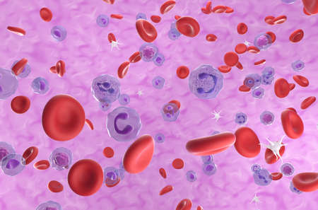
Bladder cancer, light micrograph, photo under microscope
Коллекция по умолчанию
Коллекция по умолчанию
Создать новую
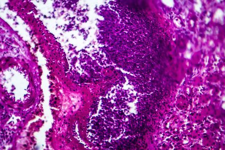
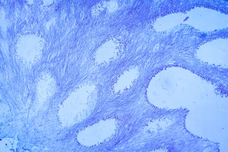
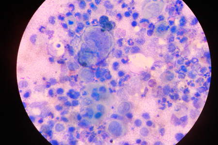
Colon inflammation in Crohn's disease 100x
Коллекция по умолчанию
Коллекция по умолчанию
Создать новую
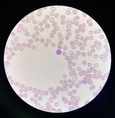
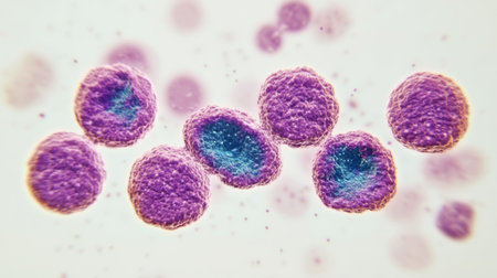
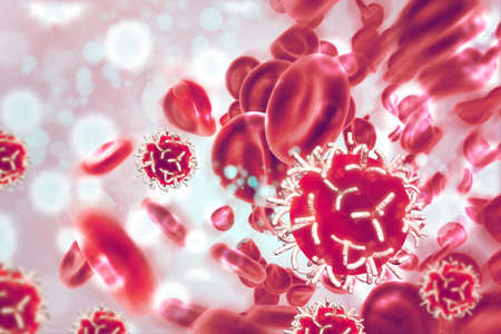
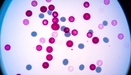
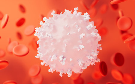
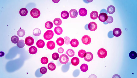
Malaria blood parasite infected red blood cells laboratory background.
Коллекция по умолчанию
Коллекция по умолчанию
Создать новую

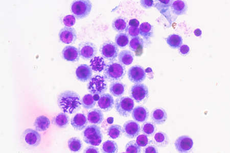
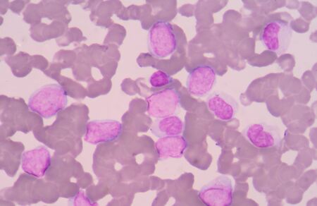
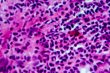
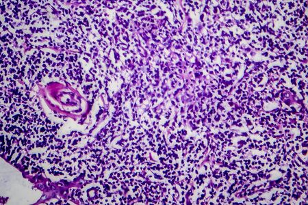
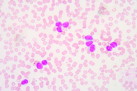
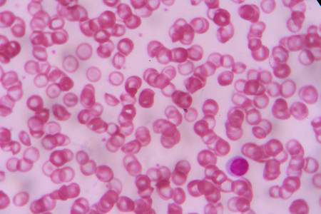
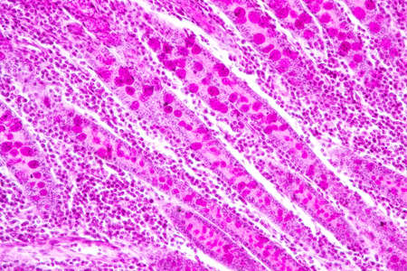
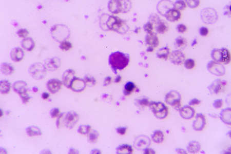
Picture of acute lymphocytic leukemia or ALL cells in blood smear, analyze by microscope, 400x
Коллекция по умолчанию
Коллекция по умолчанию
Создать новую
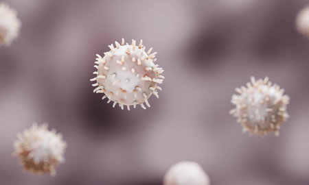
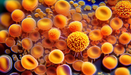
Nanoparticles Functionalization Therapeutics, Nanoparticles application in bioiechnology illustration
Коллекция по умолчанию
Коллекция по умолчанию
Создать новую

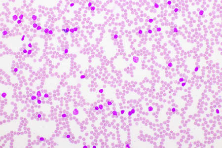
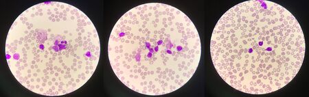
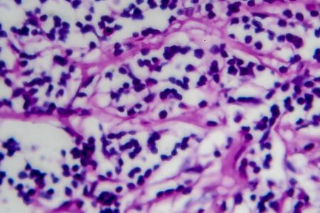
Chronic myeloid leukemia cells or CML, analyze by microscope, original magnification 1000x
Коллекция по умолчанию
Коллекция по умолчанию
Создать новую
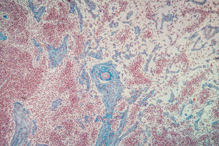
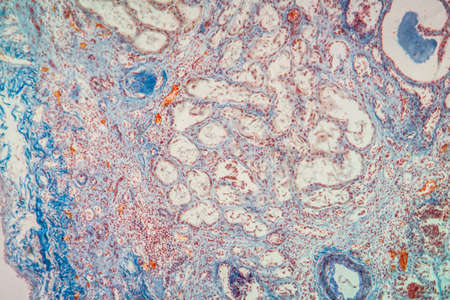
Histopathology of acute nephritis, light micrograph, photo under microscope
Коллекция по умолчанию
Коллекция по умолчанию
Создать новую

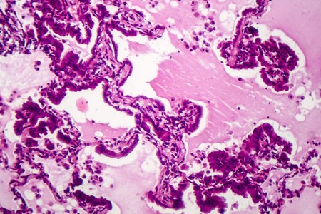
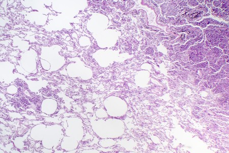
Histopathology of interstitial pneumonia, light micrograph, photo under microscope showing diffuse alveolar damage and fibrosis
Коллекция по умолчанию
Коллекция по умолчанию
Создать новую

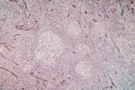
Ovarian cancer, light micrograph, photo under microscope. Photograph shows a fragment of a cancerous tumor in the female ovary. Selective focus
Коллекция по умолчанию
Коллекция по умолчанию
Создать новую
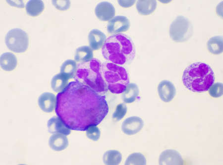
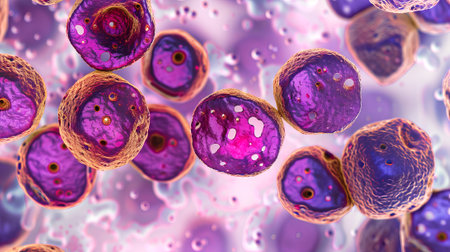
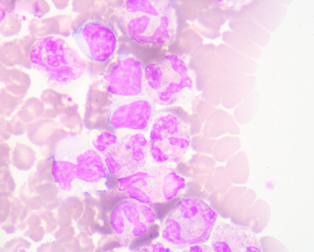
Immature cells in myeloid serie myelocyte metamyelocyte.
Коллекция по умолчанию
Коллекция по умолчанию
Создать новую
abstract acrylic watercolor paint brush stroke texture isolated on white background for logo and banner. design, creative, and illustration.
Коллекция по умолчанию
Коллекция по умолчанию
Создать новую
Picture of acute lymphocytic leukemia or ALL cells in blood smear, analyze by microscope, 400x
Коллекция по умолчанию
Коллекция по умолчанию
Создать новую
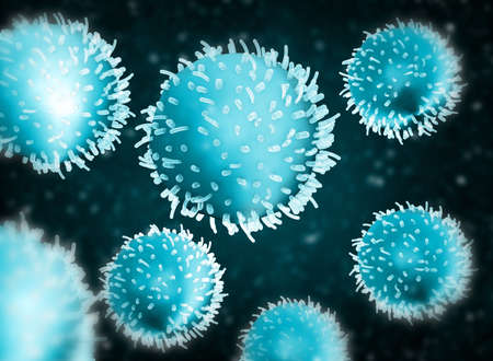
Viruses that cause infection in the human body. The study of cells under a microscope.
Коллекция по умолчанию
Коллекция по умолчанию
Создать новую
Animal cell- lymph node dog. Photo macro sections with high magnification with light microscope. Large format.
Коллекция по умолчанию
Коллекция по умолчанию
Создать новую

neutrophils. blood smear is often used as a follow-up test to abnormal results on a complete blood count (CBC) to evaluate the different types of blood cells.
Коллекция по умолчанию
Коллекция по умолчанию
Создать новую
Immature white blood cells in leukemia.Science concept.
Коллекция по умолчанию
Коллекция по умолчанию
Создать новую
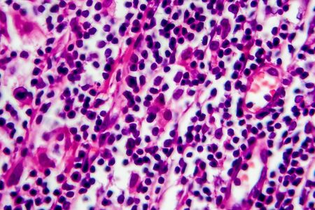
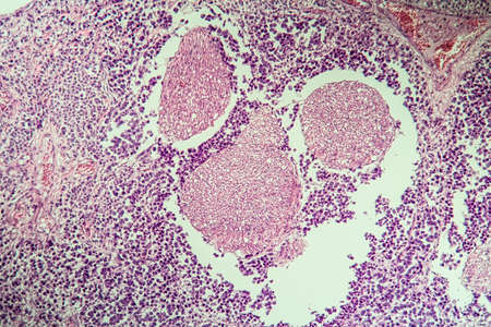
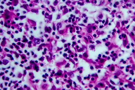
Hodgkins lymphoma, light micrograph, photo under microscope
Коллекция по умолчанию
Коллекция по умолчанию
Создать новую
Microscopic view of cells, bacteria and viruses. Pathogens and microscopic organisms. Vivid biomedical backdrop. Banner. Concept of microbiology, immunology, health research, infection
Коллекция по умолчанию
Коллекция по умолчанию
Создать новую
Chromosomes Human under the microscope for education.
Коллекция по умолчанию
Коллекция по умолчанию
Создать новую
pollen particles viewed with electronic microscope
Коллекция по умолчанию
Коллекция по умолчанию
Создать новую
Microscopic View Rendered Image of Abnormal, Diseased Cells in Biology and Medicine Illustration
Коллекция по умолчанию
Коллекция по умолчанию
Создать новую
destructive mushroom in wood fabric 100x
Коллекция по умолчанию
Коллекция по умолчанию
Создать новую
Spinal cord tissue section under the microscope 100x
Коллекция по умолчанию
Коллекция по умолчанию
Создать новую
taste buds under the microscope, hairs inside the intestines, gastric mucosa, body microflora. High quality photo
Коллекция по умолчанию
Коллекция по умолчанию
Создать новую
Leukemia cells
Коллекция по умолчанию
Коллекция по умолчанию
Создать новую

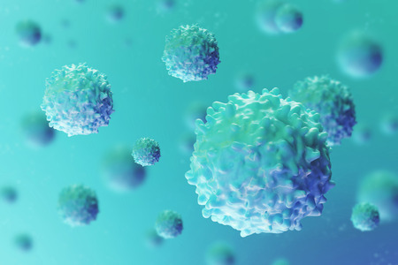
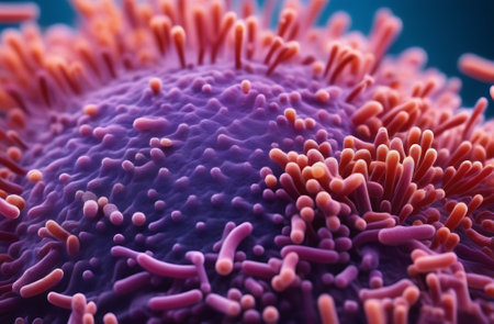
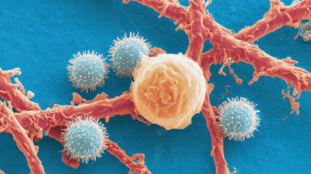
Single T-cell background
Коллекция по умолчанию
Коллекция по умолчанию
Создать новую
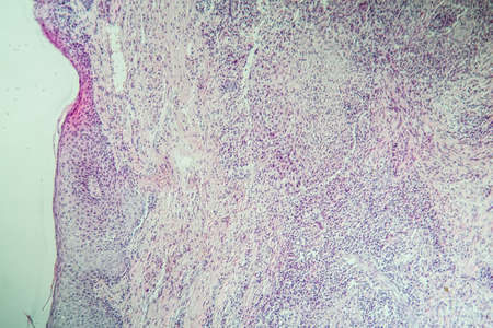
Hodgkins lymphoma, light micrograph, photo under microscope
Коллекция по умолчанию
Коллекция по умолчанию
Создать новую
Lungworm under the microscope 100x
Коллекция по умолчанию
Коллекция по умолчанию
Создать новую
Palatal tonsils transverse 100x under a microscope
Коллекция по умолчанию
Коллекция по умолчанию
Создать новую

Microscopic view of the yellow fever virus. 3D rendering
Коллекция по умолчанию
Коллекция по умолчанию
Создать новую
Virus cells. Viral disease outbreak. 3d illustration
Коллекция по умолчанию
Коллекция по умолчанию
Создать новую

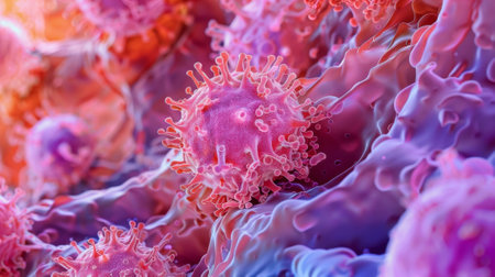
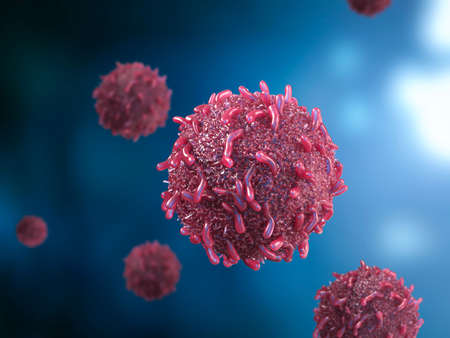
Cancer cells, malignant cells, scientific 3D illustration
Коллекция по умолчанию
Коллекция по умолчанию
Создать новую

Thyroid follicular carcinoma, light micrograph, photo under microscope
Коллекция по умолчанию
Коллекция по умолчанию
Создать новую
Leukocytes squeezing in between epithelial cells to reach the lining of an organ
Коллекция по умолчанию
Коллекция по умолчанию
Создать новую
3d rendering red cancer cell in blood
Коллекция по умолчанию
Коллекция по умолчанию
Создать новую
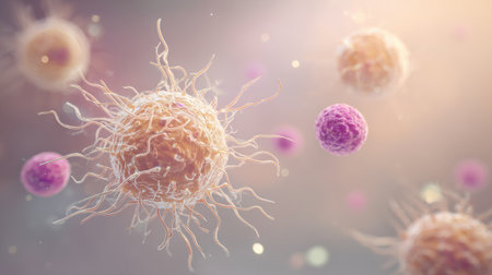
Immune cells are depicted in a dynamic interaction, showcasing their unique structures and behavior within a soft, illuminated background
Коллекция по умолчанию
Коллекция по умолчанию
Создать новую
Columnar epithelium of human gall bladder under the microscope in Lab.
Коллекция по умолчанию
Коллекция по умолчанию
Создать новую
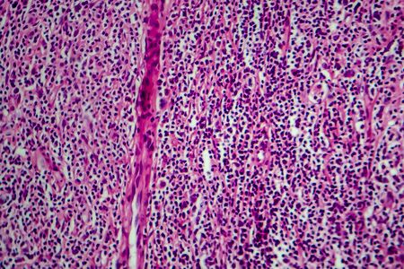
Hodgkin's lymphoma, light micrograph, photo under microscope. High magnification
Коллекция по умолчанию
Коллекция по умолчанию
Создать новую

SARS-CoV-2 coronavirus, the virus which causes COVID-19, scientifically accurate 3D illustration showing surface spikes of the virus
Коллекция по умолчанию
Коллекция по умолчанию
Создать новую
Cancer cells.3d illustration
Коллекция по умолчанию
Коллекция по умолчанию
Создать новую
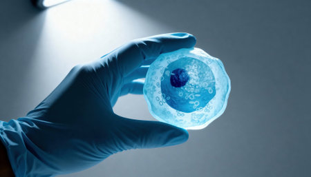
Gloved hand holds a translucent cell model under focused laboratory light with visible nucleus and organelle structures, suggesting scientific research and educational demonstration, with empty background space available for text
Коллекция по умолчанию
Коллекция по умолчанию
Создать новую
Hodgkins lymphoma, light micrograph, photo under microscope
Коллекция по умолчанию
Коллекция по умолчанию
Создать новую
AIDS with fungi 100x infected tissue
Коллекция по умолчанию
Коллекция по умолчанию
Создать новую
White blood cells of a human, photomicrograph panorama as seen under the microscope
Коллекция по умолчанию
Коллекция по умолчанию
Создать новую
Hodgkins lymphoma, light micrograph, photo under microscope. High magnification
Коллекция по умолчанию
Коллекция по умолчанию
Создать новую
Lymph node tissue under the microscope 100x
Коллекция по умолчанию
Коллекция по умолчанию
Создать новую

Leukemia cells
Коллекция по умолчанию
Коллекция по умолчанию
Создать новую
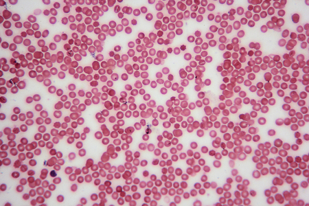
Blood smear with red blood cells in human body, medical background.
Коллекция по умолчанию
Коллекция по умолчанию
Создать новую

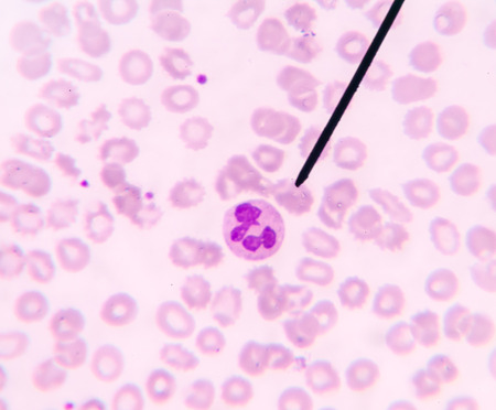
Blood smear showing, in the center, three neutrophil with hypersegmented nucleus. These cells appear in pathological situations such as megaloblastic anemias. Wright stain.
Коллекция по умолчанию
Коллекция по умолчанию
Создать новую
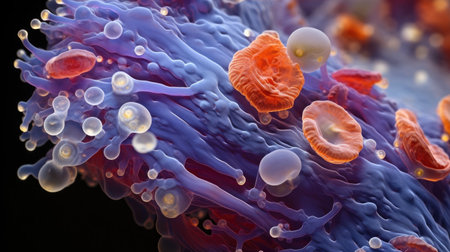
Close-up scanning electron micrograph showing immune cells, likely lymphocytes, interacting with the intricate network of a blood vessel.
Коллекция по умолчанию
Коллекция по умолчанию
Создать новую

Hodgkin's lymphoma, light micrograph, photo under microscope
Коллекция по умолчанию
Коллекция по умолчанию
Создать новую
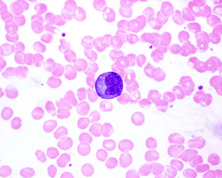
Human blood smear showing a monocyte with a basophilic cytoplasm in an infectious mononucleosis. It is the largest leukocyte (compare with red blood cell size).
Коллекция по умолчанию
Коллекция по умолчанию
Создать новую
white blood cells
Коллекция по умолчанию
Коллекция по умолчанию
Создать новую
a close up of a colorful structure
Коллекция по умолчанию
Коллекция по умолчанию
Создать новую

Itch mites under the skin cross-section 100x
Коллекция по умолчанию
Коллекция по умолчанию
Создать новую
Almond tonsils in cross section under the microscope 100x
Коллекция по умолчанию
Коллекция по умолчанию
Создать новую
Atrophy kidney tissue under the microscope 100x
Коллекция по умолчанию
Коллекция по умолчанию
Создать новую

Cancer cells, malignant cells, scientific 3D illustration
Коллекция по умолчанию
Коллекция по умолчанию
Создать новую
Hodgkin's lymphoma, light micrograph, photo under microscope. High magnification
Коллекция по умолчанию
Коллекция по умолчанию
Создать новую
Skin ulcer carcinoma enlarged 100x
Коллекция по умолчанию
Коллекция по умолчанию
Создать новую
Actinomyces in the jaw diseased tissue 200x
Коллекция по умолчанию
Коллекция по умолчанию
Создать новую

Coronavirus attacks blood cells. Close-up of a virus in the blood under a microscope. The concept of the SARS-CoV-2 COVID-19 pandemic. Realistic high quality medical 3D animation.
Коллекция по умолчанию
Коллекция по умолчанию
Создать новую

diseased liver with cirrhosis 100x under the microscope
Коллекция по умолчанию
Коллекция по умолчанию
Создать новую

Characteristics of Lichen, hyphae and Symbiotic algae under the microscope for education.
Коллекция по умолчанию
Коллекция по умолчанию
Создать новую
Neutrophil show in blood smear CBC test find with microscope.
Коллекция по умолчанию
Коллекция по умолчанию
Создать новую

Acute promyelocytic leukemia cells or APL, analyze by microscope, original magnification 1000x
Коллекция по умолчанию
Коллекция по умолчанию
Создать новую
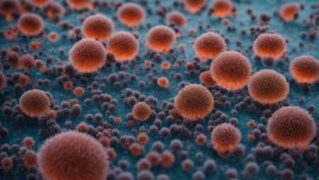
3d illustration of virus cells. Bacteria and microorganism disease causing chronic disease.
Коллекция по умолчанию
Коллекция по умолчанию
Создать новую
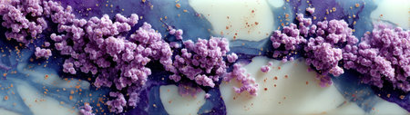
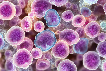
Abstract view of semiprecious resin surface with vivid grape-like purple clusters scattered across cloudy blue and white texture.
Коллекция по умолчанию
Коллекция по умолчанию
Создать новую
Squamous cell carcinoma of the uterus, light micrograph, photo under microscope
Коллекция по умолчанию
Коллекция по умолчанию
Создать новую
Lymphocytes and biological immune system, 3d rendering. 3D illustration.
Коллекция по умолчанию
Коллекция по умолчанию
Создать новую

Hodgkins lymphoma, light micrograph, photo under microscope. High magnification
Коллекция по умолчанию
Коллекция по умолчанию
Создать новую
complete blood count
Коллекция по умолчанию
Коллекция по умолчанию
Создать новую
Colorful abstract background
Коллекция по умолчанию
Коллекция по умолчанию
Создать новую
Red and white blood cells with platelets isometric view 3d illustration
Коллекция по умолчанию
Коллекция по умолчанию
Создать новую
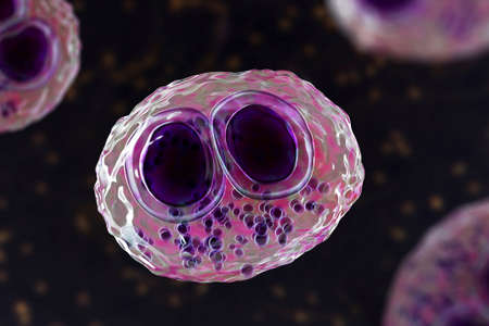
Cytomegalovirus CMV in a human cell, owl's eye inclusion in nucleus, multinucleated cell, 3D illustration. It is herpes virus, causes diseases in fetus, organ transplant patients, HIV infected people
Коллекция по умолчанию
Коллекция по умолчанию
Создать новую
multinucleated giant
Коллекция по умолчанию
Коллекция по умолчанию
Создать новую

Non-Hodgkin's lymphoma, light micrograph, photo under microscope
Коллекция по умолчанию
Коллекция по умолчанию
Создать новую
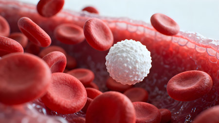
Ultra-detailed 3D render of a white blood cell floating among red blood cells in bloodstream, medical and scientific concept.
Коллекция по умолчанию
Коллекция по умолчанию
Создать новую
3d illustration of Neisseria gonorrhoeae bacteria cells
Коллекция по умолчанию
Коллекция по умолчанию
Создать новую
Tissue of Small intestine (Duodenum) and Vermiform appendix Human under the microscope in Lab.
Коллекция по умолчанию
Коллекция по умолчанию
Создать новую
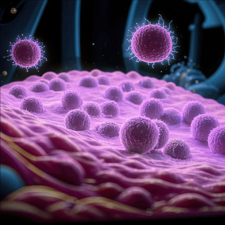
Illustration of a set of cells. Reproduction technologies. In vitro gametogenesis. This technique transforms skin cells into induced stem cells, which can then be turned into eggs and sperm.
Коллекция по умолчанию
Коллекция по умолчанию
Создать новую
Lung adenocarcinoma, light micrograph, photo under microscope
Коллекция по умолчанию
Коллекция по умолчанию
Создать новую

Medicine - Human Blood Cells photographed using a microscope.
Коллекция по умолчанию
Коллекция по умолчанию
Создать новую
Science plant cells by light microscope
Коллекция по умолчанию
Коллекция по умолчанию
Создать новую

Lymphocyte, 3D illustration. Closeup view of T-cell or B-cell
Коллекция по умолчанию
Коллекция по умолчанию
Создать новую

Characteristics Tissue of Olfactory epithelium Human under the microscope in Lab.
Коллекция по умолчанию
Коллекция по умолчанию
Создать новую
This mesmerizing abstract image features colorful organic cell structures in a dynamic environment, emphasizing intricate patterns and radiant hues for a stunning visual experience.
Коллекция по умолчанию
Коллекция по умолчанию
Создать новую

Bacterial infection in the blood. Viruses attack the human body. Microorganisms, germs and microbes. 3D colorful illustration on microbiology
Коллекция по умолчанию
Коллекция по умолчанию
Создать новую

Neutrophil cell (white blood cell) in peripheral blood smear
Коллекция по умолчанию
Коллекция по умолчанию
Создать новую

Villous colon adenocarcinoma, light micrograph, photo under microscope. High magnification
Коллекция по умолчанию
Коллекция по умолчанию
Создать новую
Abstract marbling floral pattern for fabric, tile design. background texture
Коллекция по умолчанию
Коллекция по умолчанию
Создать новую
nucleated red cell
Коллекция по умолчанию
Коллекция по умолчанию
Создать новую
Microscopic view of human cells under microscope.
Коллекция по умолчанию
Коллекция по умолчанию
Создать новую
3D Illustration of floating stem cells or cancer cells in the body
Коллекция по умолчанию
Коллекция по умолчанию
Создать новую
Liver cirrhosis tissue affected 100x after alcohol abuse
Коллекция по умолчанию
Коллекция по умолчанию
Создать новую
science medical anthropotomy physiology microscopic section of lymph gland tissue background
Коллекция по умолчанию
Коллекция по умолчанию
Создать новую
Histopathology of interstitial pneumonia, light micrograph, photo under microscope showing diffuse alveolar damage and fibrosis
Коллекция по умолчанию
Коллекция по умолчанию
Создать новую
Blood cells in human body under microscope view for education in laboratory.
Коллекция по умолчанию
Коллекция по умолчанию
Создать новую

Growing cancer cell concept image. 3d rendering
Коллекция по умолчанию
Коллекция по умолчанию
Создать новую
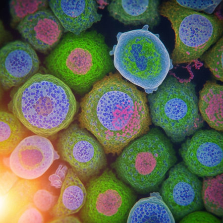
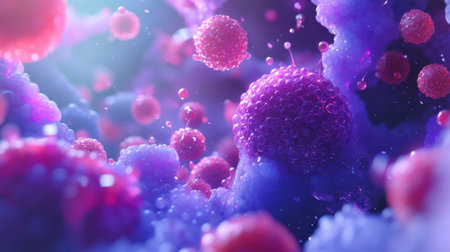
Close-up of a Cluster of Purple and Blue Cells
Коллекция по умолчанию
Коллекция по умолчанию
Создать новую
Legion-Media
Создайте свои проекты на основе качественных стоковых фотографий и видео.


Copyright © Legion-Media.