

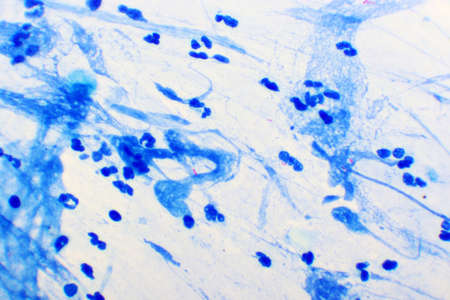
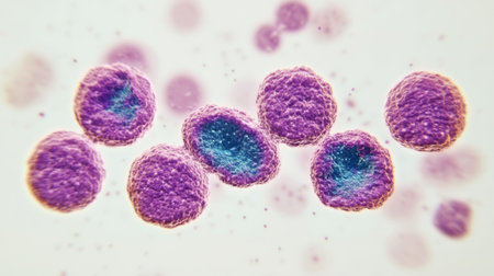
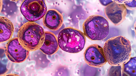
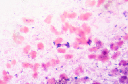
Picture of acute lymphocytic leukemia or ALL cells in blood smear, analyze by microscope, 400x
Коллекция по умолчанию
Коллекция по умолчанию
Создать новую
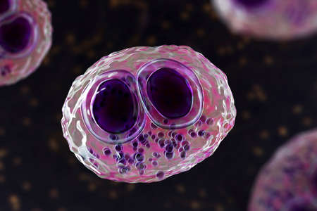
Cytomegalovirus CMV in a human cell, owl's eye inclusion in nucleus, multinucleated cell, 3D illustration. It is herpes virus, causes diseases in fetus, organ transplant patients, HIV infected people
Коллекция по умолчанию
Коллекция по умолчанию
Создать новую
Anatomy and Histological Ovary, Testis and Sperm human cells under microscope.
Коллекция по умолчанию
Коллекция по умолчанию
Создать новую

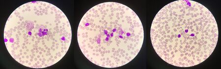
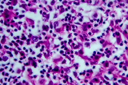
Chronic myeloid leukemia cells or CML, analyze by microscope, original magnification 1000x
Коллекция по умолчанию
Коллекция по умолчанию
Создать новую
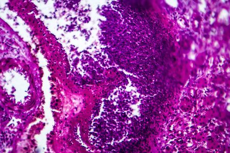
Lung adenocarcinoma, light micrograph, photo under microscope
Коллекция по умолчанию
Коллекция по умолчанию
Создать новую
Chaos ink texture background, ink in water pattern frost. Crystal winter design
Коллекция по умолчанию
Коллекция по умолчанию
Создать новую

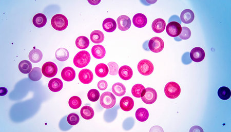
malaria parasite plasmodium falciparum on a thick blood smear reading under a microscope
Коллекция по умолчанию
Коллекция по умолчанию
Создать новую
Moderate Red cell on center Mycobacterium Tuberculosis bacteria.
Коллекция по умолчанию
Коллекция по умолчанию
Создать новую
Close up blue bacteria cells with microscope.
Коллекция по умолчанию
Коллекция по умолчанию
Создать новую
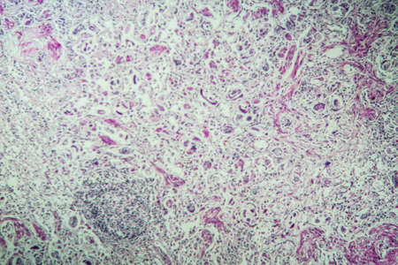
multinucleated giant
Коллекция по умолчанию
Коллекция по умолчанию
Создать новую
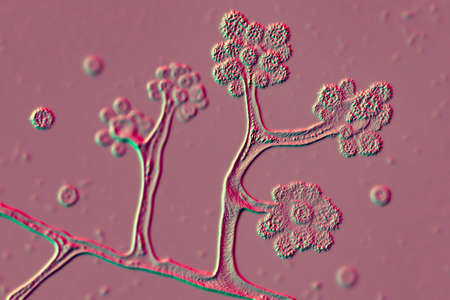
Characteristics of Lichen, hyphae and Symbiotic algae under the microscope for education.
Коллекция по умолчанию
Коллекция по умолчанию
Создать новую

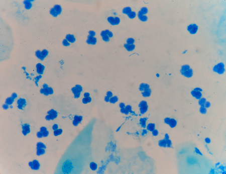
Bacteria gram staining
Коллекция по умолчанию
Коллекция по умолчанию
Создать новую
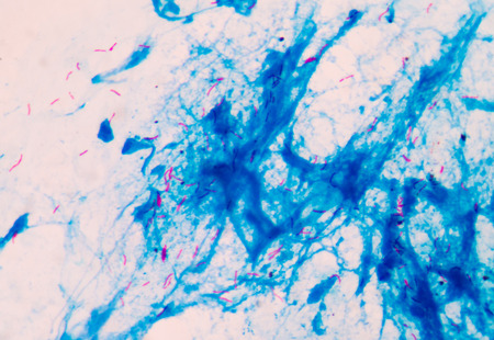
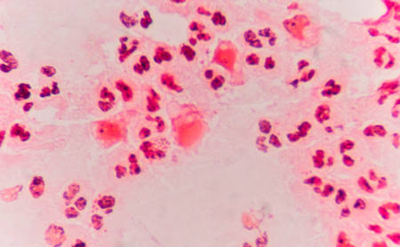
Mycobacterium tuberculosis positive (small red rod) in sputum smear, acid-fast stain, analyze by microscope 1000x
Коллекция по умолчанию
Коллекция по умолчанию
Создать новую
Anatomy and Histological Ovary, Testis and Sperm human cells under microscope.
Коллекция по умолчанию
Коллекция по умолчанию
Создать новую

Characteristics of Lichen, hyphae and Symbiotic algae under the microscope for education.
Коллекция по умолчанию
Коллекция по умолчанию
Создать новую
Malaria blood parasite infected red blood cells laboratory background.
Коллекция по умолчанию
Коллекция по умолчанию
Создать новую

Thyroid follicular carcinoma, light micrograph, photo under microscope
Коллекция по умолчанию
Коллекция по умолчанию
Создать новую
Smear of sputum gram's stained with gram positive cocci bacteria & WBC, under 100X light microscope.
Коллекция по умолчанию
Коллекция по умолчанию
Создать новую
Microscopic View Rendered Image of Abnormal, Diseased Cells in Biology and Medicine Illustration
Коллекция по умолчанию
Коллекция по умолчанию
Создать новую
Microscope with metal lens at laboratory. Medical equipment.
Коллекция по умолчанию
Коллекция по умолчанию
Создать новую

Microbiota, Probiotic Streptococcus bacteria on mucosa 3d illustration
Коллекция по умолчанию
Коллекция по умолчанию
Создать новую

Bladder cancer, light micrograph, photo under microscope
Коллекция по умолчанию
Коллекция по умолчанию
Создать новую

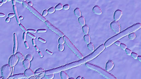
Fungi Paracoccidioides lutzii, a dimorphic fungus that causes paracoccidioidomycosis found in Brazil and Ecuador. Scientific 3D illustration showing characteristic morphology of the fungus
Коллекция по умолчанию
Коллекция по умолчанию
Создать новую
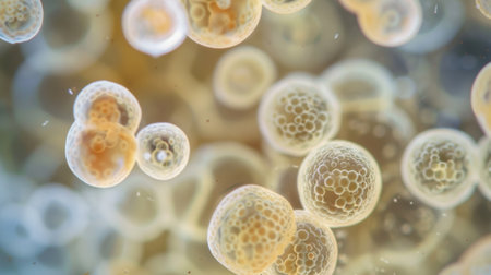
Macro shot of spherical, white, textured shapes clustered together, dark background
Коллекция по умолчанию
Коллекция по умолчанию
Создать новую
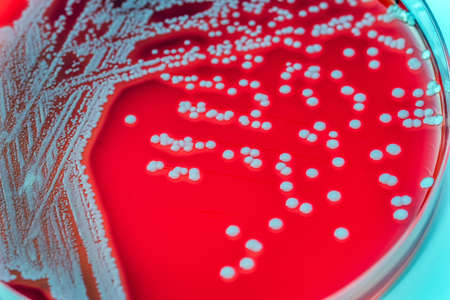
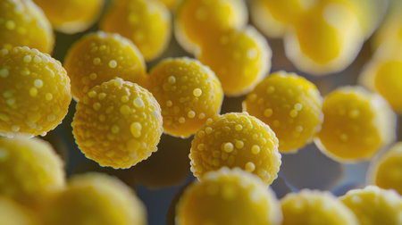
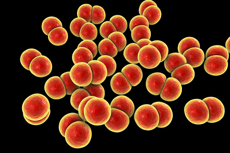
Staphylococcus aureus: Gram-positive, to Gram-variable, nonmotile, Coccus,beta haemolysis, saprotrophic bacterium that belongs to the family Staphylococcus growth on blood agar.
Коллекция по умолчанию
Коллекция по умолчанию
Создать новую
Histopathology of acute nephritis, light micrograph, photo under microscope
Коллекция по умолчанию
Коллекция по умолчанию
Создать новую
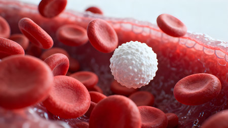
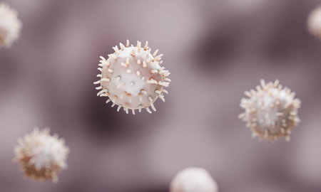
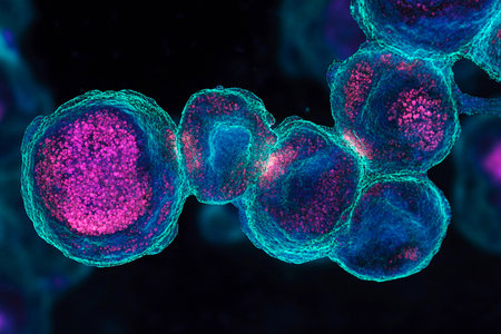
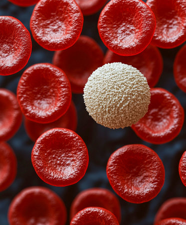
Ultra-detailed 3D render of a white blood cell floating among red blood cells in bloodstream, medical and scientific concept.
Коллекция по умолчанию
Коллекция по умолчанию
Создать новую
Microscopic fungi Trichosporon, 3D illustration shows septate hyphae, pseudohyphae, blastoconidia singly or in short chains, arthroconidia. Cause white piedra, superficial and invasive infections
Коллекция по умолчанию
Коллекция по умолчанию
Создать новую
Chromosomes Human under the microscope for education.
Коллекция по умолчанию
Коллекция по умолчанию
Создать новую
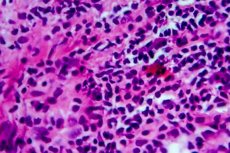
Immature white blood cells in leukemia.Science concept.
Коллекция по умолчанию
Коллекция по умолчанию
Создать новую

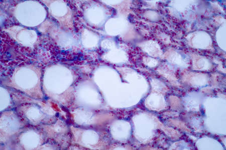
3D illustration of Inflamed Fat cells on Tissue in the human body.
Коллекция по умолчанию
Коллекция по умолчанию
Создать новую

Ovarian cancer, light micrograph, photo under microscope. Photograph shows a fragment of a cancerous tumor in the female ovary. Selective focus
Коллекция по умолчанию
Коллекция по умолчанию
Создать новую
Close up abnormal blood cells abstract background.
Коллекция по умолчанию
Коллекция по умолчанию
Создать новую
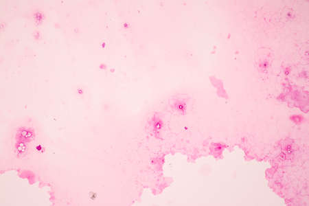
Smear of Acid-Fast bacilli AFB stained with WBC and mucous, under 100X light microscope.
Коллекция по умолчанию
Коллекция по умолчанию
Создать новую
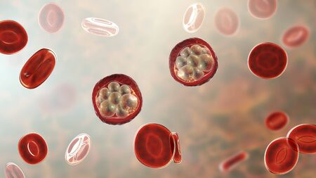
Red blood cells infected with malaria parasite, 3D illustration showing Plasmodium parasites inside red blood cells in the stage of schizont
Коллекция по умолчанию
Коллекция по умолчанию
Создать новую

Budding yeast cells with pseudohyphae in urine sample finding with microscope 100X
Коллекция по умолчанию
Коллекция по умолчанию
Создать новую
budding yeast with pseudohyphae in urine specimen
Коллекция по умолчанию
Коллекция по умолчанию
Создать новую
Blood smear with red blood cells in human body, medical background.
Коллекция по умолчанию
Коллекция по умолчанию
Создать новую
Bacteria methicillin-resistant Staphylococcus aureus MRSA, multidrug resistant bacteria, 3D illustration
Коллекция по умолчанию
Коллекция по умолчанию
Создать новую
Chromosomes Human under the microscope for education.
Коллекция по умолчанию
Коллекция по умолчанию
Создать новую
Bacteria Streptococcus pneumoniae, 3D illustration. Gram-positive diplococci, the causative agent of pneumonia
Коллекция по умолчанию
Коллекция по умолчанию
Создать новую
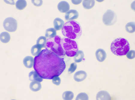
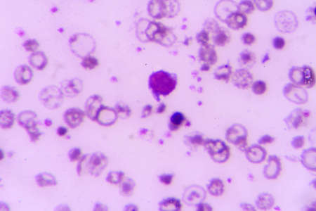
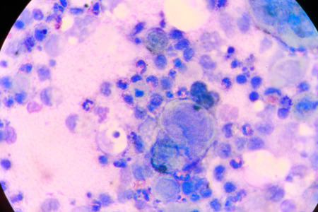
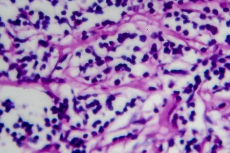
Immature cells in myeloid serie myelocyte metamyelocyte.
Коллекция по умолчанию
Коллекция по умолчанию
Создать новую
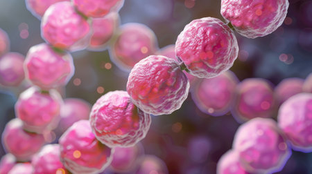
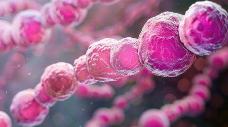
Streptococcus pyogenes bacteria. 3D computer illustration of Streptococcus pyogenes, or group-A Streptococcus, bacteria. S. pyogenes is a gram-positive spherical (coccus) bacteria
Коллекция по умолчанию
Коллекция по умолчанию
Создать новую
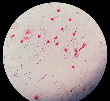
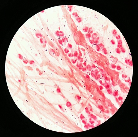
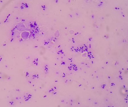
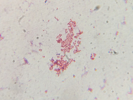
Smear of Gram's stained with gram positive cocci in chain bacteria, under 100X light microscope.
Коллекция по умолчанию
Коллекция по умолчанию
Создать новую
Sepsis or septicaemia is a life-threatening illness.
Коллекция по умолчанию
Коллекция по умолчанию
Создать новую
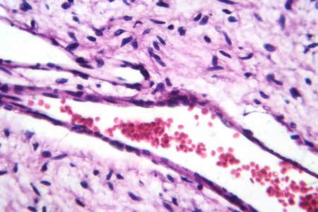
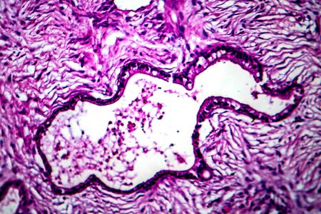
A small blood vessel with red blood cells in neurofibroma tissue sample, light photomicrograph.
Коллекция по умолчанию
Коллекция по умолчанию
Создать новую

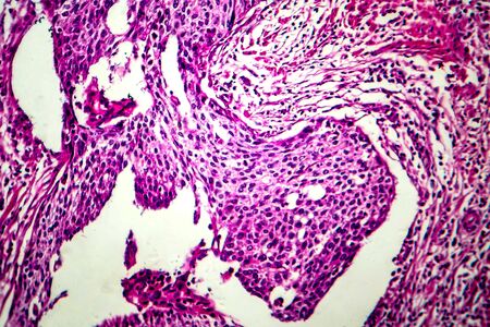
Villous colon adenocarcinoma, light micrograph, photo under microscope. High magnification
Коллекция по умолчанию
Коллекция по умолчанию
Создать новую
nucleated red cell
Коллекция по умолчанию
Коллекция по умолчанию
Создать новую
Streptococcus pyogenes bacteria. 3D computer illustration of Streptococcus pyogenes, or group-A Streptococcus, bacteria. S. pyogenes is a gram-positive spherical (coccus) bacteria
Коллекция по умолчанию
Коллекция по умолчанию
Создать новую
Microscopic view of cells, bacteria and viruses. Pathogens and microscopic organisms. Vivid biomedical backdrop. Banner. Concept of microbiology, immunology, health research, infection
Коллекция по умолчанию
Коллекция по умолчанию
Создать новую
Bacteria Enterococcus isolated on white background, 3D illustration. Gram-positive cocci which cause infant endocarditis and other infections
Коллекция по умолчанию
Коллекция по умолчанию
Создать новую
multinucleated giant
Коллекция по умолчанию
Коллекция по умолчанию
Создать новую
Microscopic view of human cells under microscope.
Коллекция по умолчанию
Коллекция по умолчанию
Создать новую
Numerous tiny white bubbles float gracefully on the surface of the water. The bubbles appear to be delicate and light, moving with the gentle current in a mesmerizing manner.
Коллекция по умолчанию
Коллекция по умолчанию
Создать новую
Blue cells background.Medical background concept.
Коллекция по умолчанию
Коллекция по умолчанию
Создать новую
Hodgkins lymphoma, light micrograph, photo under microscope. High magnification
Коллекция по умолчанию
Коллекция по умолчанию
Создать новую

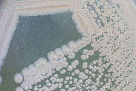
Backgrounds of Characteristics and Different shaped Colony of Bacteria and Mold growing on agar plates from Soil samples for education in Microbiology laboratory.
Коллекция по умолчанию
Коллекция по умолчанию
Создать новую
Nanoparticles Functionalization Therapeutics, Nanoparticles application in bioiechnology illustration
Коллекция по умолчанию
Коллекция по умолчанию
Создать новую
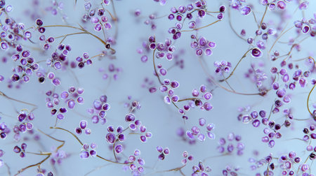
This image captures delicate purple flowers with fine stems set against a soft blue background, evoking a tranquil and ethereal feeling perfect for decor.
Коллекция по умолчанию
Коллекция по умолчанию
Создать новую

Apple pollen from a blossom in spring under the microscope
Коллекция по умолчанию
Коллекция по умолчанию
Создать новую
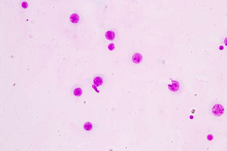
budding yeast cell with epithelial in gram stain method.
Коллекция по умолчанию
Коллекция по умолчанию
Создать новую
Microscopic fungi Cunninghamella, scientific 3D illustration. Pathogenic fungi from the order Mucorales, cause sinopulmonary and disseminated infections, one of the causative agents of mucormycosis
Коллекция по умолчанию
Коллекция по умолчанию
Создать новую

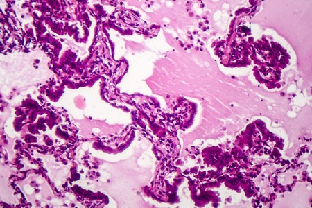
Histopathology of interstitial pneumonia, light micrograph, photo under microscope showing diffuse alveolar damage and fibrosis
Коллекция по умолчанию
Коллекция по умолчанию
Создать новую
microscope lens, viewing Trypanosoma cruzi parasitic protozoan, causer of Chagas disease
Коллекция по умолчанию
Коллекция по умолчанию
Создать новую

Human hyaline cartilage bone under microscope view for education pathology. Human tissue.
Коллекция по умолчанию
Коллекция по умолчанию
Создать новую
Lungworm under the microscope 100x
Коллекция по умолчанию
Коллекция по умолчанию
Создать новую

Fungus Sporothrix schenckii, the causative agent of sporotrichosis, especially common in florists and gardeners. 3D illustration showing fungal hyphae and spores
Коллекция по умолчанию
Коллекция по умолчанию
Создать новую
Pichia is a genus of yeasts in the family Saccharomycetaceae under the microscope for education.
Коллекция по умолчанию
Коллекция по умолчанию
Создать новую
Hodgkin's lymphoma, light micrograph, photo under microscope. High magnification
Коллекция по умолчанию
Коллекция по умолчанию
Создать новую
Bacterial infection tuberculosis.red cells in blue background.AFB 3+ fine with microscope.
Коллекция по умолчанию
Коллекция по умолчанию
Создать новую
Colorful cells are showcased under a microscope, revealing their unique shapes and structures. The vibrant blues and purples illustrate complex biological interactions at a microscopic level.
Коллекция по умолчанию
Коллекция по умолчанию
Создать новую
Uterine cancer, light micrograph, photo under microscope
Коллекция по умолчанию
Коллекция по умолчанию
Создать новую
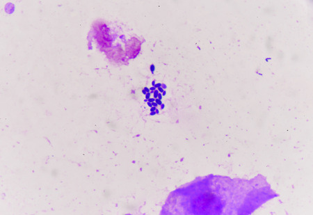
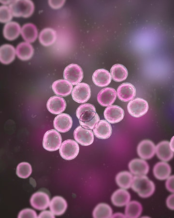
Microscopic View of Purple Cocci Bacteria Clustered in Grapelike Formations
Коллекция по умолчанию
Коллекция по умолчанию
Создать новую
Blood cells in human body under microscope view for education in laboratory.
Коллекция по умолчанию
Коллекция по умолчанию
Создать новую

Candida tropicalis yeasts, microscopic fungi that cause infections in immunocompromised patients. Scientific 3D illustration showing pseudohyphae and blastoconidia formed singly or in small groups
Коллекция по умолчанию
Коллекция по умолчанию
Создать новую
Bacteria Neisseria gonorrhoeae, gonoccoccus, diplococci which cause sexually transmitted infection gonorrhoea. 3D illustration
Коллекция по умолчанию
Коллекция по умолчанию
Создать новую

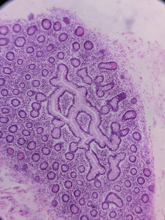
Showing Light micrograph of the Trachea, Thymus, Parathyroid gland and Tonsil human under the microscope for education in the laboratory.
Коллекция по умолчанию
Коллекция по умолчанию
Создать новую
Breast fibroadenosis, light micrograph, photo under microscope. Common benign hyperplastic process involving breast glands
Коллекция по умолчанию
Коллекция по умолчанию
Создать новую
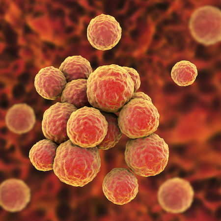
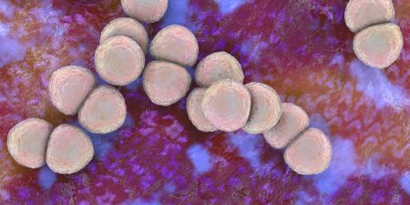
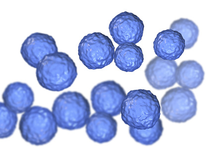
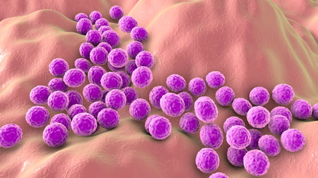
Staphylococcus bacteria, a genus of Gram-positive bacteria known for causing various infections in humans, 3D illustration.
Коллекция по умолчанию
Коллекция по умолчанию
Создать новую

budding yeast with pseudohyphae in urine specimen
Коллекция по умолчанию
Коллекция по умолчанию
Создать новую
Streptococcus pyogenes bacteria. 3D computer illustration of Streptococcus pyogenes, or group-A Streptococcus, bacteria. S. pyogenes is a gram-positive spherical (coccus) bacteria
Коллекция по умолчанию
Коллекция по умолчанию
Создать новую
White Blood Cells, Essential Components of the Immune System, Amidst Red Blood Cells.
Коллекция по умолчанию
Коллекция по умолчанию
Создать новую
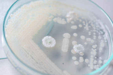
Yeast in petri dish, Microbiology for education in laboratories.
Коллекция по умолчанию
Коллекция по умолчанию
Создать новую
Squamous cell carcinoma of the uterus, light micrograph, photo under microscope
Коллекция по умолчанию
Коллекция по умолчанию
Создать новую
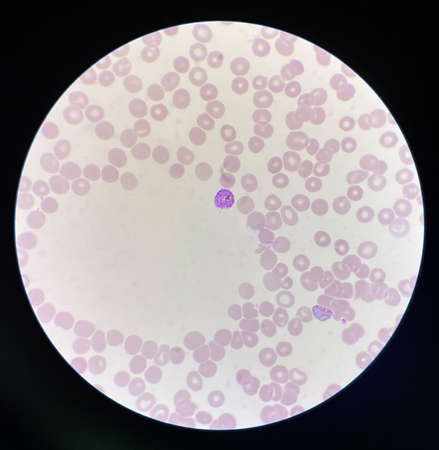
Neutrophil show in blood smear CBC test find with microscope.
Коллекция по умолчанию
Коллекция по умолчанию
Создать новую
blood smear is often used as a follow-up test to abnormal results on a complete blood count (CBC) to evaluate the different types of blood cells.
Коллекция по умолчанию
Коллекция по умолчанию
Создать новую
Burkitts lymphoma cells, a cancer of the lymphatic system, monoclonal B-cell tumor, 3D illustration
Коллекция по умолчанию
Коллекция по умолчанию
Создать новую
Hodgkin's lymphoma, light micrograph, photo under microscope. High magnification
Коллекция по умолчанию
Коллекция по умолчанию
Создать новую

Close up the media plate on hand medical technicians working on bacterial culture and drug resistance of pathogens in laboratory.
Коллекция по умолчанию
Коллекция по умолчанию
Создать новую
Moderate red white blood cells with gram negative diplococci intracellular Gram-negative coffee bean-shaped diplococci bacteria responsible for the sexually transmitted infection gonorrhea
Коллекция по умолчанию
Коллекция по умолчанию
Создать новую

Petri dish close up. Bacteria culture.
Коллекция по умолчанию
Коллекция по умолчанию
Создать новую
Human lung pathology under light microscope, The lungs is organs of the respiratory system in humans. Human pathology education. Haematoxylin and eosin staining technique slide.
Коллекция по умолчанию
Коллекция по умолчанию
Создать новую
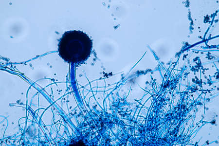
Aspergillus niger and Aspergillus oryzae (mold) under microscope for Microbiology in Lab.
Коллекция по умолчанию
Коллекция по умолчанию
Создать новую
Pancreas cancer cell under microscope view for medical education.
Коллекция по умолчанию
Коллекция по умолчанию
Создать новую
image through the microscope. zoom x 40
Коллекция по умолчанию
Коллекция по умолчанию
Создать новую
Bacteria Staphylococcus aureus, Staphylococcus epidermidis, MRSA, multidrug resistant bacteria, 3D illustration
Коллекция по умолчанию
Коллекция по умолчанию
Создать новую
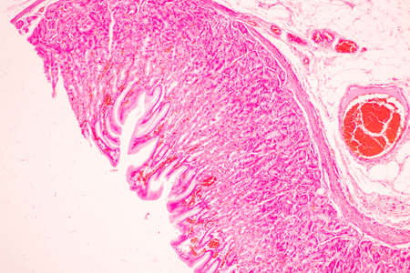
Tissue of Stomach Human under the microscope in Lab.
Коллекция по умолчанию
Коллекция по умолчанию
Создать новую
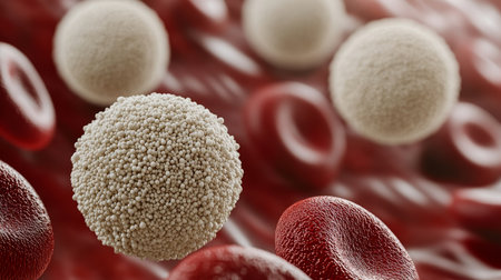
A white blood cell with a granular surface surrounded by many smooth red blood cells.
Коллекция по умолчанию
Коллекция по умолчанию
Создать новую
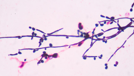
Branching budding yeast cells with pseudohyphae in urine gram stain fine with microscope.
Коллекция по умолчанию
Коллекция по умолчанию
Создать новую

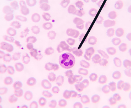
Blood smear showing, in the center, three neutrophil with hypersegmented nucleus. These cells appear in pathological situations such as megaloblastic anemias. Wright stain.
Коллекция по умолчанию
Коллекция по умолчанию
Создать новую
Legion-Media
Создайте свои проекты на основе качественных стоковых фотографий и видео.


Copyright © Legion-Media.