

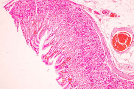
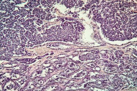
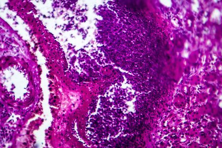
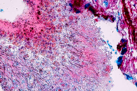
Ovarian cancer, light micrograph, photo under microscope. Photograph shows a fragment of a cancerous tumor in the female ovary. Selective focus
Коллекция по умолчанию
Коллекция по умолчанию
Создать новую
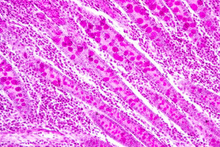
Bacillary dysentery, light micrograph, photo under microscope showing presence of bacteria and accumulation of inflammatory cells in intestinal epithelium
Коллекция по умолчанию
Коллекция по умолчанию
Создать новую

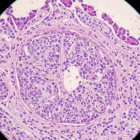
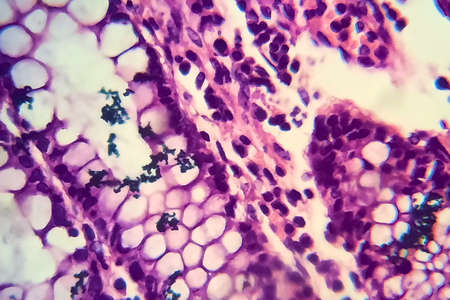
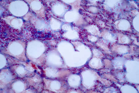
Thyroid follicular carcinoma, light micrograph, photo under microscope
Коллекция по умолчанию
Коллекция по умолчанию
Создать новую
Lungworm under the microscope 100x
Коллекция по умолчанию
Коллекция по умолчанию
Создать новую

Rows of microscope glass slide in the cells
Коллекция по умолчанию
Коллекция по умолчанию
Создать новую

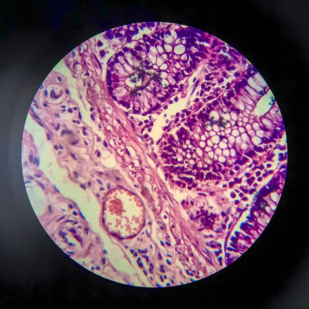
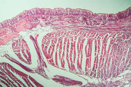
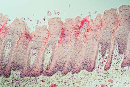
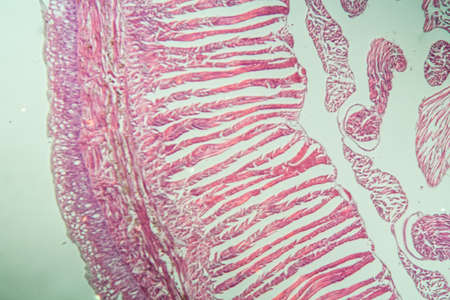
Stomach tissue under the microscope 100x
Коллекция по умолчанию
Коллекция по умолчанию
Создать новую
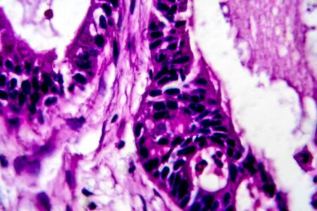
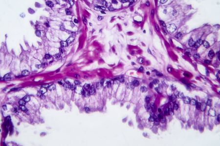
Columnar epithelium of human gall bladder under the microscope in Lab.
Коллекция по умолчанию
Коллекция по умолчанию
Создать новую

Characteristics of Lichen, hyphae and Symbiotic algae under the microscope for education.
Коллекция по умолчанию
Коллекция по умолчанию
Создать новую

Bladder cancer, light micrograph, photo under microscope
Коллекция по умолчанию
Коллекция по умолчанию
Создать новую
Tongue Tissue with taste buds across 200x
Коллекция по умолчанию
Коллекция по умолчанию
Создать новую
Earthworm histology cross section 10th segment 100x
Коллекция по умолчанию
Коллекция по умолчанию
Создать новую

Tongue with taste buds Papilla across 100x
Коллекция по умолчанию
Коллекция по умолчанию
Создать новую
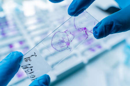
Hand in blue glove holding glass histology slides
Коллекция по умолчанию
Коллекция по умолчанию
Создать новую
Lung adenocarcinoma, light micrograph, photo under microscope
Коллекция по умолчанию
Коллекция по умолчанию
Создать новую
Ovarian cancer, light micrograph, photo under microscope. Photograph shows a fragment of a cancerous tumor in the female ovary. Selective focus
Коллекция по умолчанию
Коллекция по умолчанию
Создать новую
Characteristics of Lichen, hyphae and Symbiotic algae under the microscope for education.
Коллекция по умолчанию
Коллекция по умолчанию
Создать новую
Squamous cell carcinoma of the uterus, light micrograph, photo under microscope
Коллекция по умолчанию
Коллекция по умолчанию
Создать новую

Papillary thyroid carcinoma, light micrograph, photo under microscope. The most common type of thyroid cancer
Коллекция по умолчанию
Коллекция по умолчанию
Создать новую
Colon inflammation in Crohn's disease 100x
Коллекция по умолчанию
Коллекция по умолчанию
Создать новую

Rows of microscope glass slide in the cells
Коллекция по умолчанию
Коллекция по умолчанию
Создать новую
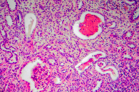
Fibrin deposits in the kidney, microscopy 100x
Коллекция по умолчанию
Коллекция по умолчанию
Создать новую
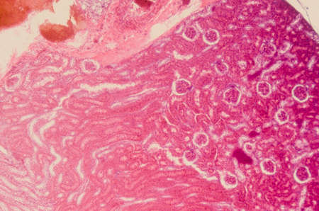
Chronic pyelonephritis, light micrograph, photo under microscope
Коллекция по умолчанию
Коллекция по умолчанию
Создать новую
Endometriosis, a disorder in which cells similar to those in the endometrium grow outside the uterus. Light micrograph, photo under microscope
Коллекция по умолчанию
Коллекция по умолчанию
Создать новую

Histopathology of human under microscope view for education in laboratory.
Коллекция по умолчанию
Коллекция по умолчанию
Создать новую
Education anatomy and Histological sample of Human under the microscope.
Коллекция по умолчанию
Коллекция по умолчанию
Создать новую
Atrophy kidney tissue under the microscope 100x
Коллекция по умолчанию
Коллекция по умолчанию
Создать новую
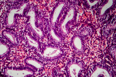
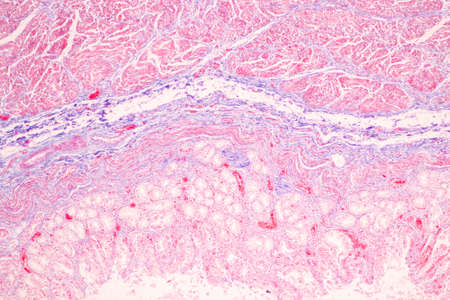
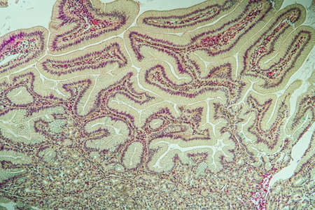
Tissue of Stomach Human under the microscope in Lab.
Коллекция по умолчанию
Коллекция по умолчанию
Создать новую
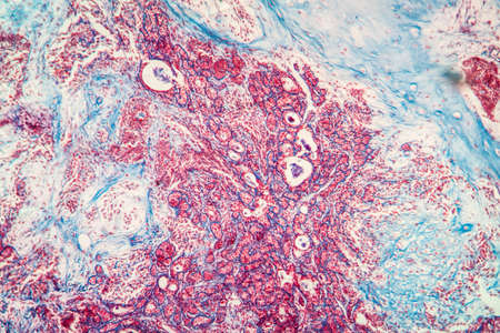
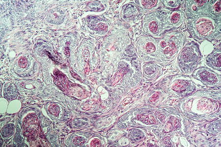
Histopathology of cirrhosis, light micrograph, photo under microscope
Коллекция по умолчанию
Коллекция по умолчанию
Создать новую
Colon tissue with diverticulum 100x
Коллекция по умолчанию
Коллекция по умолчанию
Создать новую
destructive mushroom in wood fabric 100x
Коллекция по умолчанию
Коллекция по умолчанию
Создать новую
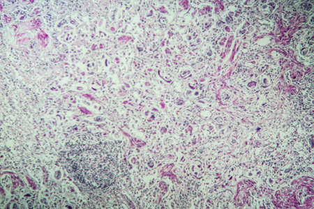
Metastases tumor diseased tissue 100x
Коллекция по умолчанию
Коллекция по умолчанию
Создать новую
Papillary thyroid carcinoma, light micrograph, photo under microscope. The most common type of thyroid cancer
Коллекция по умолчанию
Коллекция по умолчанию
Создать новую
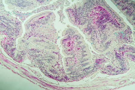
Colon carcinoma arising from adenoma, 100x
Коллекция по умолчанию
Коллекция по умолчанию
Создать новую
Breast fibroadenosis, light micrograph, photo under microscope. Common benign hyperplastic process involving breast glands
Коллекция по умолчанию
Коллекция по умолчанию
Создать новую
Bacillary dysentery, light micrograph, photo under microscope showing presence of bacteria and accumulation of inflammatory cells in intestinal epithelium
Коллекция по умолчанию
Коллекция по умолчанию
Создать новую
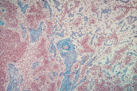
Lymph node tissue under the microscope 100x
Коллекция по умолчанию
Коллекция по умолчанию
Создать новую
Characteristics of Lichen, hyphae and Symbiotic algae under the microscope for education.
Коллекция по умолчанию
Коллекция по умолчанию
Создать новую
Bowel with goblet cells in the dark field 100x
Коллекция по умолчанию
Коллекция по умолчанию
Создать новую
Tissue of Stomach Human under the microscope in Lab.
Коллекция по умолчанию
Коллекция по умолчанию
Создать новую
Inguinal testicles gonadically diseased tissue 100x
Коллекция по умолчанию
Коллекция по умолчанию
Создать новую
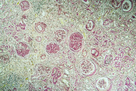
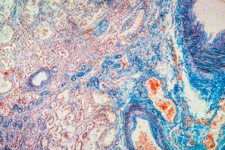
science medical anthropotomy physiology microscopic section of human kidney tissue background
Коллекция по умолчанию
Коллекция по умолчанию
Создать новую
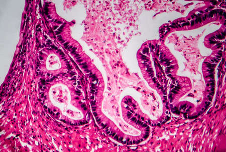
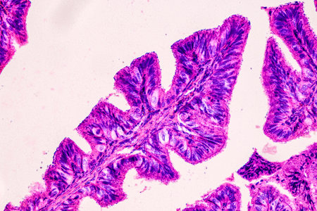
Cross-section through the intestine with glands 200x
Коллекция по умолчанию
Коллекция по умолчанию
Создать новую
Abstract ink design template mixed texture background. Blue abstract texture. Multicolored liquid marble pattern. Fluid Art
Коллекция по умолчанию
Коллекция по умолчанию
Создать новую
Uterine cancer, light micrograph, photo under microscope
Коллекция по умолчанию
Коллекция по умолчанию
Создать новую
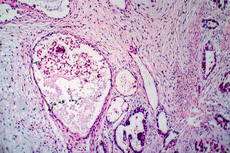
Wilms tumor, or nephroblastoma, light micrograph, photo under microscope. High magnification
Коллекция по умолчанию
Коллекция по умолчанию
Создать новую
Histology of human tissue, show tracheitis as seen under the microscope
Коллекция по умолчанию
Коллекция по умолчанию
Создать новую
Chaos ink texture background, ink in water pattern frost. Crystal winter design
Коллекция по умолчанию
Коллекция по умолчанию
Создать новую

Hand in blue glove holding glass histology slides
Коллекция по умолчанию
Коллекция по умолчанию
Создать новую

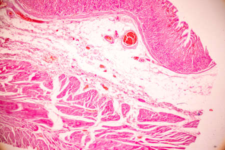
Fibromyoma of the uterus diseased tissue 100x
Коллекция по умолчанию
Коллекция по умолчанию
Создать новую
science medical anthropotomy physiology microscopic section of lymph gland tissue background
Коллекция по умолчанию
Коллекция по умолчанию
Создать новую

Condyloma acuminatum, also known as genital warts. Light micrograph, photo under microscope
Коллекция по умолчанию
Коллекция по умолчанию
Создать новую
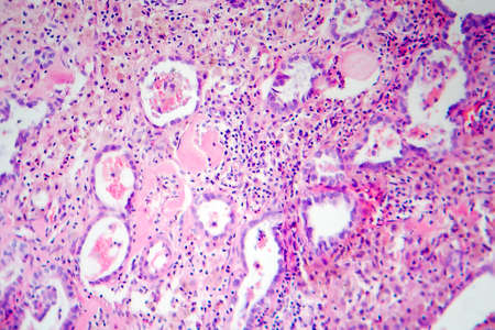
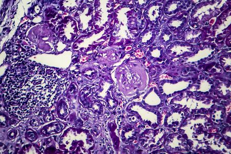
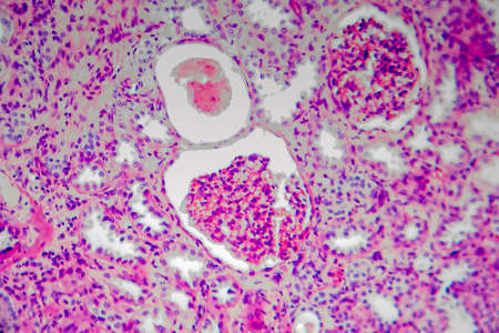
Diffuse proliferative glomerulonephritis, DPGN, light micrograph, photo under microscope. High magnification
Коллекция по умолчанию
Коллекция по умолчанию
Создать новую
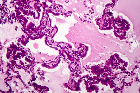
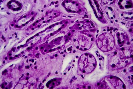
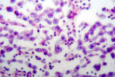
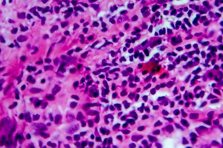
This detailed microscopic image showcases various cellular structures, highlighted in striking purple tones. The intricate patterns and textures reveal the complexity of biological tissues, making it a valuable resource for educational and scientific purposes
Коллекция по умолчанию
Коллекция по умолчанию
Создать новую
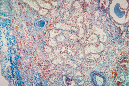
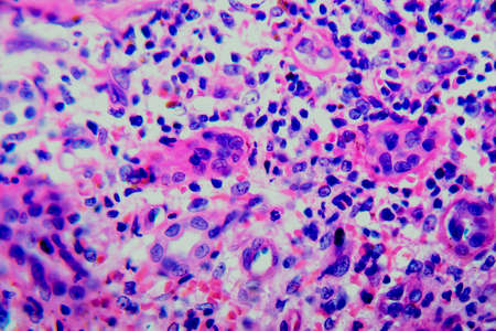
Histopathology of acute nephritis, light micrograph, photo under microscope
Коллекция по умолчанию
Коллекция по умолчанию
Создать новую
Papillary thyroid carcinoma, light micrograph, photo under microscope. The most common type of thyroid cancer
Коллекция по умолчанию
Коллекция по умолчанию
Создать новую

Lungworm under the microscope 100x
Коллекция по умолчанию
Коллекция по умолчанию
Создать новую
Fibroepithelium Diseased tissue 100x
Коллекция по умолчанию
Коллекция по умолчанию
Создать новую
Diffuse proliferative glomerulonephritis, light micrograph, photo under microscope. High magnification
Коллекция по умолчанию
Коллекция по умолчанию
Создать новую

Papillary thyroid carcinoma, light micrograph, photo under microscope. The most common type of thyroid cancer
Коллекция по умолчанию
Коллекция по умолчанию
Создать новую

microscopy micrograph plant tissue, corn embryo
Коллекция по умолчанию
Коллекция по умолчанию
Создать новую
Chronic nephritis, light micrograph, photo under microscope
Коллекция по умолчанию
Коллекция по умолчанию
Создать новую
Carcinoma in guinea pigs, tissue 100x
Коллекция по умолчанию
Коллекция по умолчанию
Создать новую
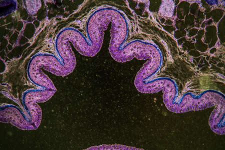
Palatal tonsils transverse 100x under a microscope
Коллекция по умолчанию
Коллекция по умолчанию
Создать новую
Blue and pink refill ink spilled onto the white washbasin and the ink mixed into abstract blobs and patterns.
Коллекция по умолчанию
Коллекция по умолчанию
Создать новую
Characteristics of Lichen, hyphae and Symbiotic algae under the microscope for education.
Коллекция по умолчанию
Коллекция по умолчанию
Создать новую
Uterine cancer, light micrograph, photo under microscope
Коллекция по умолчанию
Коллекция по умолчанию
Создать новую
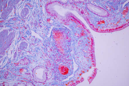
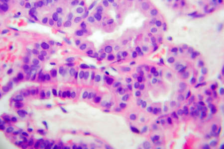
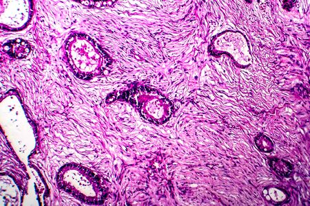
Benign prostatic hyperplasia. Micrograph shows dilated glands, papillary projections inside the lumen of the glands, cystic dilatation with accumulation of secretory material. Photo under microscope
Коллекция по умолчанию
Коллекция по умолчанию
Создать новую
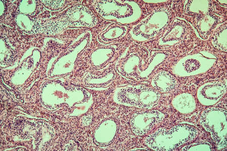
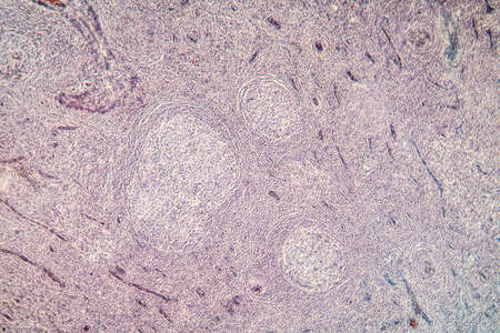
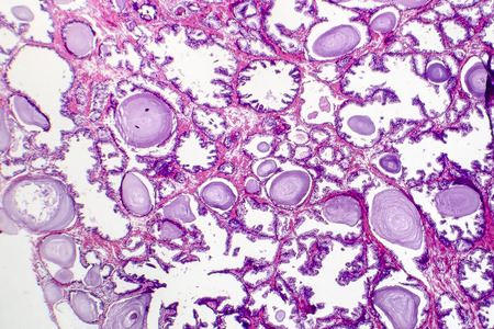
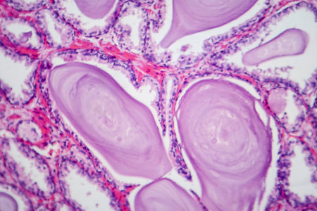
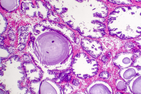
Endemic goiter, light micrograph, abnormal enlargement of the thyroid gland due to dietary iodine deficiency. Photomicrograph shows follicles of varying size, abundant colloid, lymphocytic infiltrate
Коллекция по умолчанию
Коллекция по умолчанию
Создать новую
serous gland tissue under the microscope 100x
Коллекция по умолчанию
Коллекция по умолчанию
Создать новую
Shrinked kidney diseased tissue under the microscope 100x
Коллекция по умолчанию
Коллекция по умолчанию
Создать новую
Testicular seminoma, light micrograph, photo under microscope. A most common germ cell tumor of the testis
Коллекция по умолчанию
Коллекция по умолчанию
Создать новую

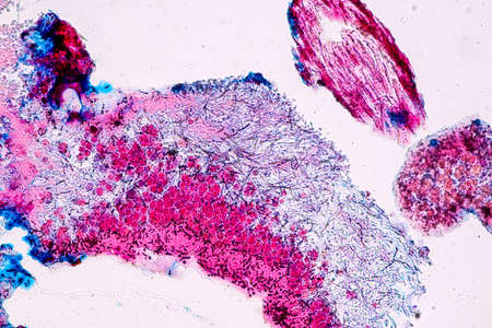
diseased liver with cirrhosis 100x under the microscope
Коллекция по умолчанию
Коллекция по умолчанию
Создать новую
Transitional epithelium tissue of the urinary bladder under microscope, light micrograph, hematoxylin eosin staining
Коллекция по умолчанию
Коллекция по умолчанию
Создать новую
Tongue Tissue with taste buds across 200x
Коллекция по умолчанию
Коллекция по умолчанию
Создать новую

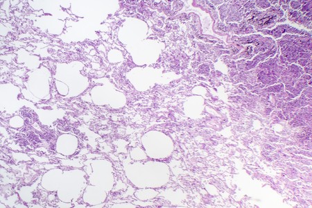
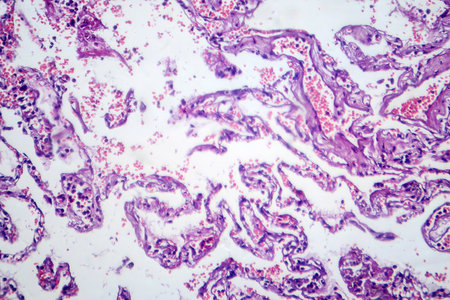
Histopathology of interstitial pneumonia, light micrograph, photo under microscope showing diffuse alveolar damage and fibrosis
Коллекция по умолчанию
Коллекция по умолчанию
Создать новую
Chronic pyelonephritis, light micrograph, photo under microscope
Коллекция по умолчанию
Коллекция по умолчанию
Создать новую
Light micrograph of teratoma, a tumor made up of several different types of tissue, such as hair, teeth, muscle, or bone. Teratoma is typically found in the ovary, testicle, or coccyx
Коллекция по умолчанию
Коллекция по умолчанию
Создать новую
Slow worm histology bowel transverse 100x
Коллекция по умолчанию
Коллекция по умолчанию
Создать новую
science medical anthropotomy physiology microscopic section of lymph gland tissue background
Коллекция по умолчанию
Коллекция по умолчанию
Создать новую
Earthworm histology cross section 10th segment 100x
Коллекция по умолчанию
Коллекция по умолчанию
Создать новую

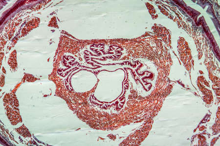
Characteristics Tissue of Olfactory epithelium Human under the microscope in Lab.
Коллекция по умолчанию
Коллекция по умолчанию
Создать новую
Uterine cancer, light micrograph, photo under microscope
Коллекция по умолчанию
Коллекция по умолчанию
Создать новую
Histopathology of interstitial pneumonia, light micrograph, photo under microscope showing diffuse alveolar damage and fibrosis
Коллекция по умолчанию
Коллекция по умолчанию
Создать новую
Hodgkin's lymphoma, light micrograph, photo under microscope. High magnification
Коллекция по умолчанию
Коллекция по умолчанию
Создать новую
Tissue of Stomach Human under the microscope in Lab.
Коллекция по умолчанию
Коллекция по умолчанию
Создать новую
Atrophy kidney tissue under the microscope 100x
Коллекция по умолчанию
Коллекция по умолчанию
Создать новую
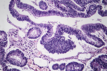
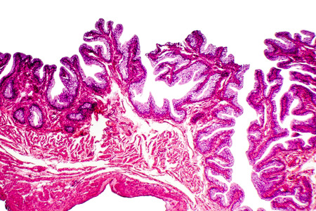
Histopathology of intestinal adenoma, light micrograph, photo under microscope
Коллекция по умолчанию
Коллекция по умолчанию
Создать новую
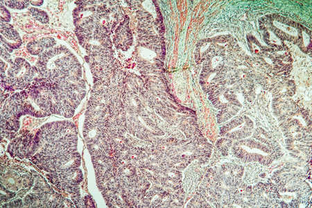
Prostate cancer, light micrograph, photo under microscope
Коллекция по умолчанию
Коллекция по умолчанию
Создать новую
Large intestine with diverticular tissue under the microscope 100x
Коллекция по умолчанию
Коллекция по умолчанию
Создать новую
Photomicrograph showing histological features of benign prostatic hyperplasia. Enlarged prostate gland with nodular proliferation of glandular and stromal components.
Коллекция по умолчанию
Коллекция по умолчанию
Создать новую

Scalp and hair follicles of human under the microscope in Lab.
Коллекция по умолчанию
Коллекция по умолчанию
Создать новую
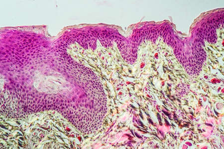
Basal cell cancer Diseased tissue 100x
Коллекция по умолчанию
Коллекция по умолчанию
Создать новую
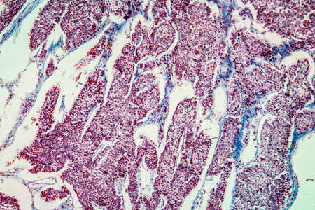
Photomicrograph of smoker's lung, revealing characteristic changes and damage associated with long-term smoking habits.
Коллекция по умолчанию
Коллекция по умолчанию
Создать новую
Benign prostatic hyperplasia. Micrograph shows dilated glands, papillary projections inside the lumen of the glands, cystic dilatation with accumulation of secretory material. Photo under microscope
Коллекция по умолчанию
Коллекция по умолчанию
Создать новую

Uterine cancer, light micrograph, photo under microscope
Коллекция по умолчанию
Коллекция по умолчанию
Создать новую
Cancer swollen Parotid diseased tissue 100x
Коллекция по умолчанию
Коллекция по умолчанию
Создать новую
Human lung pathology under light microscope, The lungs is organs of the respiratory system in humans. Human pathology education. Haematoxylin and eosin staining technique slide.
Коллекция по умолчанию
Коллекция по умолчанию
Создать новую
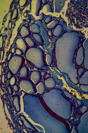
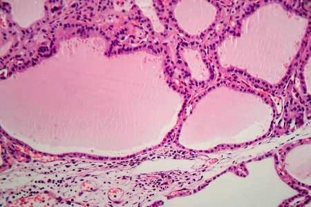
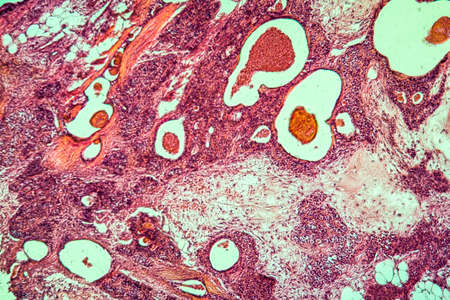
Goiter colloid goiter disease 100x
Коллекция по умолчанию
Коллекция по умолчанию
Создать новую
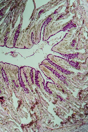
Tissue of Small intestine (Duodenum) and Vermiform appendix Human under the microscope in Lab.
Коллекция по умолчанию
Коллекция по умолчанию
Создать новую
Chronic pyelonephritis, light micrograph, photo under microscope
Коллекция по умолчанию
Коллекция по умолчанию
Создать новую
Legion-Media
Создайте свои проекты на основе качественных стоковых фотографий и видео.


Copyright © Legion-Media.