
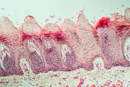
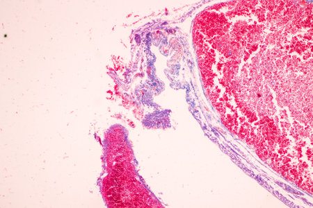
Tongue Tissue with taste buds across 200x
Коллекция по умолчанию
Коллекция по умолчанию
Создать новую
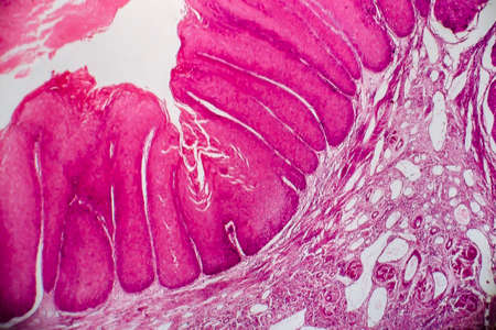
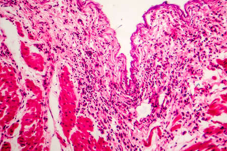
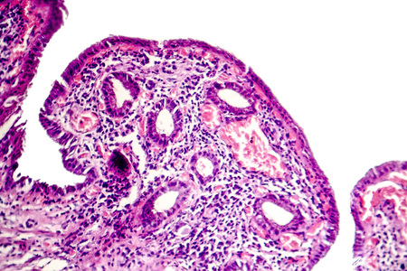
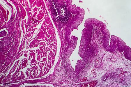
Condyloma acuminatum, also known as genital warts. Light micrograph, photo under microscope
Коллекция по умолчанию
Коллекция по умолчанию
Создать новую
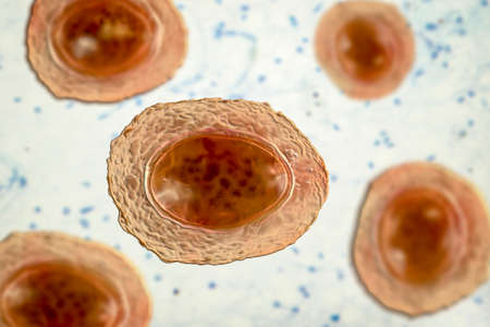
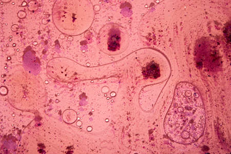
Ascaris lumbricoides, a large roundworm, unfertilized egg, 3D illustration
Коллекция по умолчанию
Коллекция по умолчанию
Создать новую
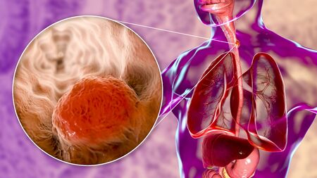
Esophageal cancer, 3D illustration showing malignant tumor in the human esophagus
Коллекция по умолчанию
Коллекция по умолчанию
Создать новую
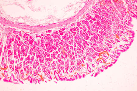
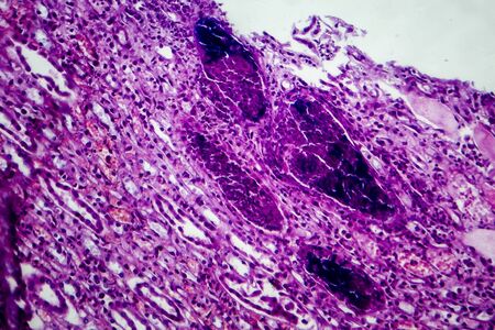
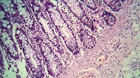
Signet ring cell carcinoma of the stomach, light micrograph, photo under microscope
Коллекция по умолчанию
Коллекция по умолчанию
Создать новую
Condyloma acuminatum, also known as genital warts. Light micrograph, photo under microscope
Коллекция по умолчанию
Коллекция по умолчанию
Создать новую

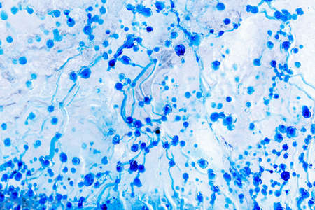
Bladder cat- cell nature background. Abstract- photo macro sections with high magnification with light microscope
Коллекция по умолчанию
Коллекция по умолчанию
Создать новую

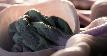
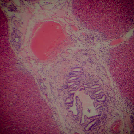
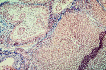
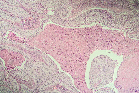
Ovarian cancer, light micrograph, photo under microscope. Photograph shows a fragment of a cancerous tumor in the female ovary. Selective focus
Коллекция по умолчанию
Коллекция по умолчанию
Создать новую
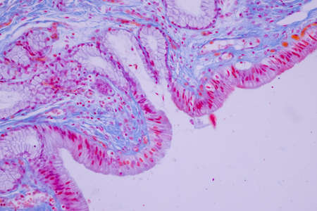
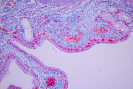
Columnar epithelium of human gall bladder under the microscope in Lab.
Коллекция по умолчанию
Коллекция по умолчанию
Создать новую
Microbes of different shapes, 3D illustration. Group of microorganisms. Digital art. Picture render by neural network.
Коллекция по умолчанию
Коллекция по умолчанию
Создать новую
fish caviar as a background. macro
Коллекция по умолчанию
Коллекция по умолчанию
Создать новую
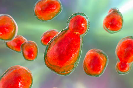
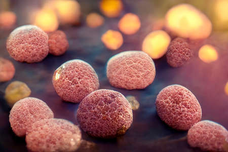
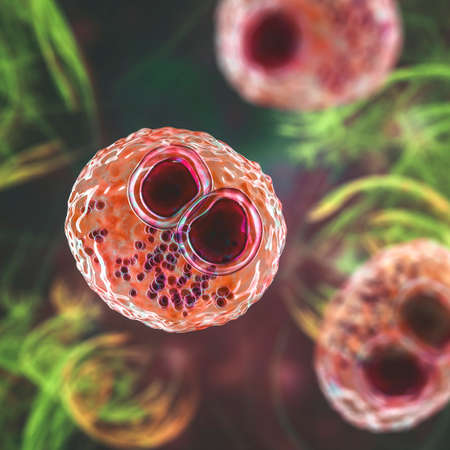
Pathogenic yeast fungus Cryptococcus neoformans that causes cryptococcal meningoencephalitis and lung disease in immunocompromised patients with AIDS
Коллекция по умолчанию
Коллекция по умолчанию
Создать новую
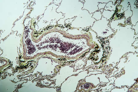
Lung tissue as dust lung under the microscope 100x
Коллекция по умолчанию
Коллекция по умолчанию
Создать новую

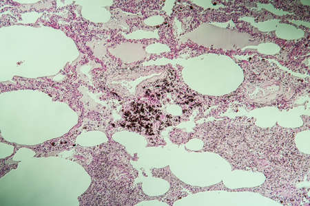
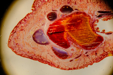
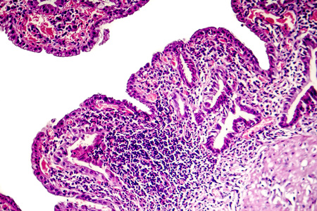
Histopathology of lung emphysema, light micrograph, photo under microscope showing enlargement of air spaces in lung tissue and destruction of alveolar septa
Коллекция по умолчанию
Коллекция по умолчанию
Создать новую

Histological Uterus human, Uterine tube human, Placenta human and Umbilical cord Human under the microscope for education.
Коллекция по умолчанию
Коллекция по умолчанию
Создать новую
Pathology and Histology Tissue of Mammals under microscope.
Коллекция по умолчанию
Коллекция по умолчанию
Создать новую
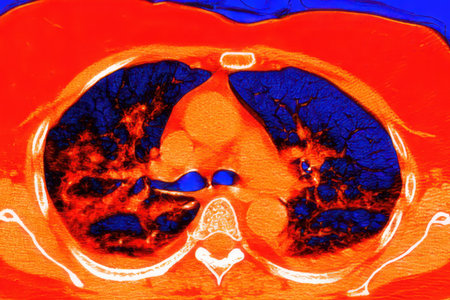
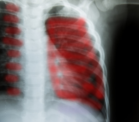
Human lungs with COVID-19 pneumonia, color-enhanced CT scan showing normal lung tissue in dark blue and affected bilateral areas with ground-glass opacities and crazy-paving patterns in red.
Коллекция по умолчанию
Коллекция по умолчанию
Создать новую

Cross section of human skin under microscope view for education in laboratory.
Коллекция по умолчанию
Коллекция по умолчанию
Создать новую
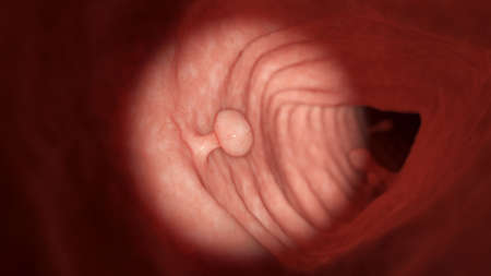
3d rendered illustration of a colon polyp
Коллекция по умолчанию
Коллекция по умолчанию
Создать новую
Cytomegalovirus CMV in a human cell, owl's eye inclusion in nucleus, multinucleated cell, 3D illustration. It is herpes virus, causes diseases in fetus, organ transplant patients, HIV infected people
Коллекция по умолчанию
Коллекция по умолчанию
Создать новую
Tissue of Stomach Human under the microscope in Lab.
Коллекция по умолчанию
Коллекция по умолчанию
Создать новую
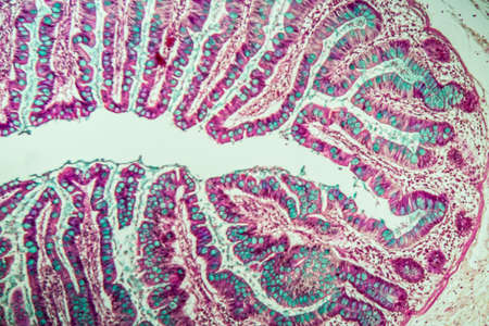
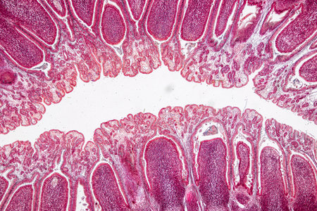
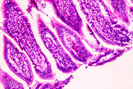
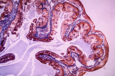
Small intestine with villi under the microscope 100x
Коллекция по умолчанию
Коллекция по умолчанию
Создать новую
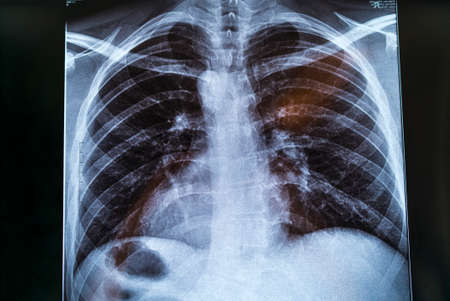
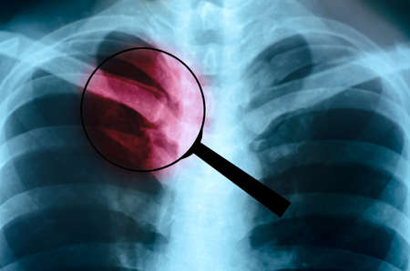
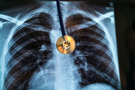
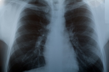
Chest X-ray image for physician's examination
Коллекция по умолчанию
Коллекция по умолчанию
Создать новую
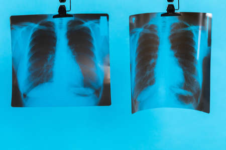
Lungs on a black and white photo taken on a photographic film of a special device.
Коллекция по умолчанию
Коллекция по умолчанию
Создать новую
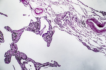
Histopathology of lung emphysema, light micrograph, photo under microscope showing enlargement of air spaces in lung tissue and destruction of alveolar septa
Коллекция по умолчанию
Коллекция по умолчанию
Создать новую
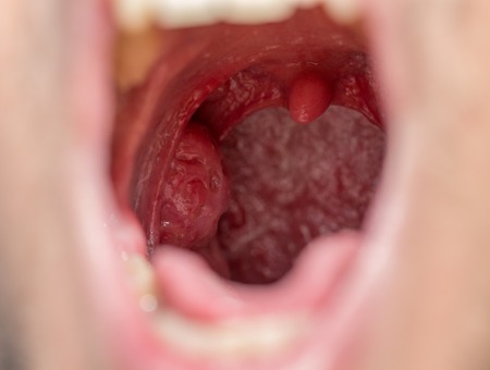
Open mouth view of tonsils
Коллекция по умолчанию
Коллекция по умолчанию
Создать новую
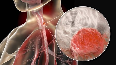
Esophageal cancer, 3D illustration showing malignant tumor in the human esophagus
Коллекция по умолчанию
Коллекция по умолчанию
Создать новую

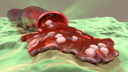
Hemorrhage, blood flowing from the damaged blood vessel, 3D illustration. Hemorrhagic stroke, traumatic injury or other diseases with bleeding
Коллекция по умолчанию
Коллекция по умолчанию
Создать новую

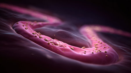
Blood cells in vein. Red blood cells circulating in blood vessels. medical health care. Vascular therapy. 3d illustration
Коллекция по умолчанию
Коллекция по умолчанию
Создать новую
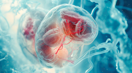
Embryonic Development: Microscopic View of a Translucent Embryo
Коллекция по умолчанию
Коллекция по умолчанию
Создать новую
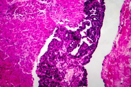
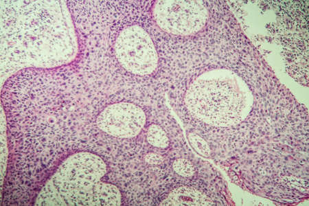
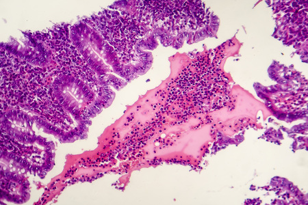
Papillary serous ovarian adenocarcinoma, cancer of ovary, light micrograph, photo under microscope
Коллекция по умолчанию
Коллекция по умолчанию
Создать новую

Parasitic worms in the lumen of intestine, 3D illustration. Ascaris lumbricoides, Enterobius vermicularis, and other round worms
Коллекция по умолчанию
Коллекция по умолчанию
Создать новую
A detailed description of chest pain shows its location and sensation of discomfort
Коллекция по умолчанию
Коллекция по умолчанию
Создать новую

Stomach cancer. Cancer attacking cell. Stomach disease concept. 3d illustration
Коллекция по умолчанию
Коллекция по умолчанию
Создать новую
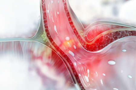
This 3D illustration shows a peptic ulcer affecting the stomach surface with visible tissue damage and surrounding elements captured in detail.
Коллекция по умолчанию
Коллекция по умолчанию
Создать новую
Pathology and Histology Tissue of Mammals under microscope.
Коллекция по умолчанию
Коллекция по умолчанию
Создать новую

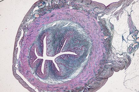
Cell- science background. Esophagus of the dog- cross section
Коллекция по умолчанию
Коллекция по умолчанию
Создать новую
Acute pyelonephritis, light micrograph, photo under microscope
Коллекция по умолчанию
Коллекция по умолчанию
Создать новую
Bacillary dysentery, light micrograph, photo under microscope showing presence of bacteria and accumulation of inflammatory cells in intestinal epithelium
Коллекция по умолчанию
Коллекция по умолчанию
Создать новую
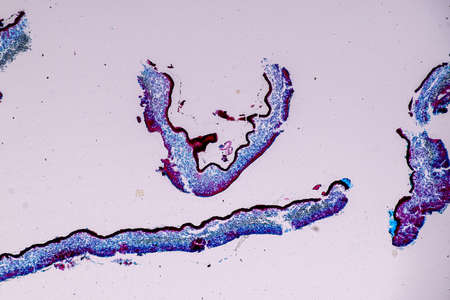
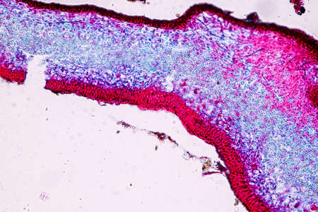
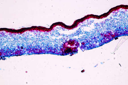
Characteristics of Lichen, hyphae and Symbiotic algae under the microscope for education.
Коллекция по умолчанию
Коллекция по умолчанию
Создать новую
Ice texture background, ink in water pattern frost. Crystal winter design
Коллекция по умолчанию
Коллекция по умолчанию
Создать новую

Endometriosis, a disorder in which cells similar to those in the endometrium grow outside the uterus. Light micrograph, photo under microscope
Коллекция по умолчанию
Коллекция по умолчанию
Создать новую
Extreme Close up of microscopic kidney Bowman's Capsule and Glomerulus
Коллекция по умолчанию
Коллекция по умолчанию
Создать новую
Asthma of the lungs diseased tissue under the microscope 100x
Коллекция по умолчанию
Коллекция по умолчанию
Создать новую
Leech cross section showing internal anatomical structures stained
Коллекция по умолчанию
Коллекция по умолчанию
Создать новую

3d illustration of red blood cells inside an artery, vein. The flow of blood inside a living organism. Scientific and medical microbiological concept. Enrichment with oxygen and important nutrients.
Коллекция по умолчанию
Коллекция по умолчанию
Создать новую
X-Ray Image Close up Of Human Chest
Коллекция по умолчанию
Коллекция по умолчанию
Создать новую

Columnar epithelium of human gall bladder under the microscope in Lab.
Коллекция по умолчанию
Коллекция по умолчанию
Создать новую
Stomach cancer. Cancer attacking cell. Stomach disease concept. 3d illustration
Коллекция по умолчанию
Коллекция по умолчанию
Создать новую

medical bacteria illustration of the legionella
Коллекция по умолчанию
Коллекция по умолчанию
Создать новую
Characteristics of Lichen, hyphae and Symbiotic algae under the microscope for education.
Коллекция по умолчанию
Коллекция по умолчанию
Создать новую
Chest X-ray image for physician's examination
Коллекция по умолчанию
Коллекция по умолчанию
Создать новую
X-Ray Image Of Human Chest for a medical diagnosis.
Коллекция по умолчанию
Коллекция по умолчанию
Создать новую
Characteristics of Lichen, hyphae and Symbiotic algae under the microscope for education.
Коллекция по умолчанию
Коллекция по умолчанию
Создать новую
Blood flowing from the damaged blood vessel, hemorrhage, 3D illustration
Коллекция по умолчанию
Коллекция по умолчанию
Создать новую

Cytomegalovirus CMV in a human cell, owl's eye inclusion in nucleus, multinucleated cell, 3D illustration. It is herpes virus, causes diseases in fetus, organ transplant patients, HIV infected people
Коллекция по умолчанию
Коллекция по умолчанию
Создать новую

Stomach cancer. Cancer attacking cell. Stomach disease concept. 3d illustration
Коллекция по умолчанию
Коллекция по умолчанию
Создать новую

3D Illustration Concept of Human Body Glands Lobes of Thyroid Gland Anatomy
Коллекция по умолчанию
Коллекция по умолчанию
Создать новую
Characteristics of Lichen, hyphae and Symbiotic algae under the microscope for education.
Коллекция по умолчанию
Коллекция по умолчанию
Создать новую
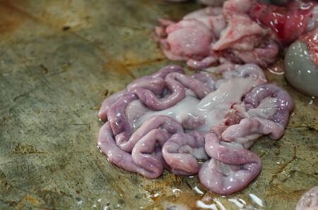
traditional home made pig slauhtering
Коллекция по умолчанию
Коллекция по умолчанию
Создать новую
Education anatomy and Histological sample of Human under the microscope.
Коллекция по умолчанию
Коллекция по умолчанию
Создать новую

Characteristics of Lichen, hyphae and Symbiotic algae under the microscope for education.
Коллекция по умолчанию
Коллекция по умолчанию
Создать новую
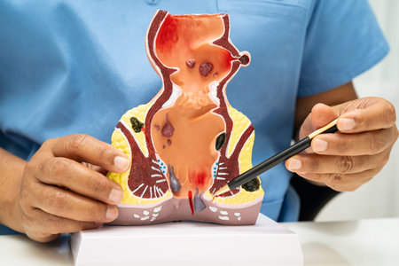
Asian doctor with rectum and hemorrhoid human anatomy model at hospital, inflamed vascular structure.
Коллекция по умолчанию
Коллекция по умолчанию
Создать новую
Soothing Pink Abstract background with little impurities
Коллекция по умолчанию
Коллекция по умолчанию
Создать новую
Stomach tissue under the microscope 100x
Коллекция по умолчанию
Коллекция по умолчанию
Создать новую

Histological Uterus human, Uterine tube human, Placenta human and Umbilical cord Human under the microscope for education
Коллекция по умолчанию
Коллекция по умолчанию
Создать новую
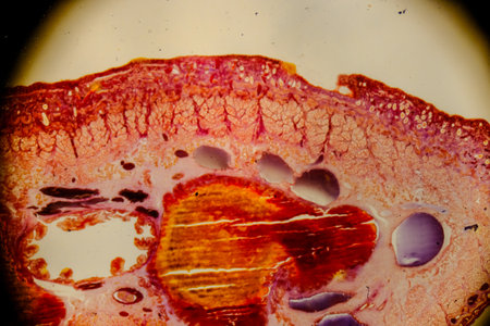
Leech cross section showing internal anatomical structures stained
Коллекция по умолчанию
Коллекция по умолчанию
Создать новую
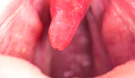
Uvulitis and sore throat in the oral cavity due to illness. Inflammation of the uvula and tonsils due to infections during illness, close-up
Коллекция по умолчанию
Коллекция по умолчанию
Создать новую
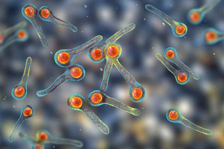
Clostridium tetani bacteria, the causative agent of tetanus, 3D illustration
Коллекция по умолчанию
Коллекция по умолчанию
Создать новую
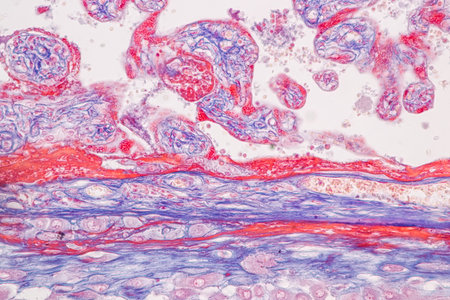
Histological Uterus human, Uterine tube human, Placenta human and Umbilical cord Human under the microscope for education.
Коллекция по умолчанию
Коллекция по умолчанию
Создать новую
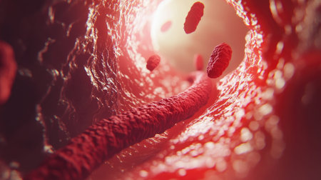
This image captures a detailed view of the interior of a blood vessel, showcasing the flow of red blood cells through a capillary tube in the human circulatory system.
Коллекция по умолчанию
Коллекция по умолчанию
Создать новую
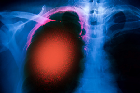
radiography of lungs
Коллекция по умолчанию
Коллекция по умолчанию
Создать новую
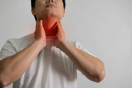
The man touches his sore throat, neck, Temperature, runny nose, illness, Pain,
Коллекция по умолчанию
Коллекция по умолчанию
Создать новую
Chronic cholecystitis, light micrograph, photo under microscope showing fibrosis and muscular hypertrophy of gallbladder wall, entrapped epithelial crypts, foamy macrophages
Коллекция по умолчанию
Коллекция по умолчанию
Создать новую
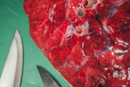
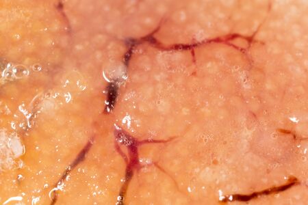
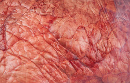
A piece of beef lung close-up. Meat for cooking. Selective focus
Коллекция по умолчанию
Коллекция по умолчанию
Создать новую
Columnar epithelium of human gall bladder under the microscope in Lab.
Коллекция по умолчанию
Коллекция по умолчанию
Создать новую
science medical anthropotomy physiology microscopic section of human liver tissue
Коллекция по умолчанию
Коллекция по умолчанию
Создать новую
Characteristics of Lichen, hyphae and Symbiotic algae under the microscope for education.
Коллекция по умолчанию
Коллекция по умолчанию
Создать новую
Chronic cholecystitis, light micrograph, photo under microscope showing fibrosis and muscular hypertrophy of gallbladder wall, entrapped epithelial crypts, foamy macrophages
Коллекция по умолчанию
Коллекция по умолчанию
Создать новую
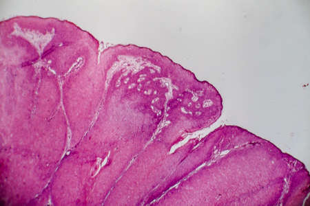
Bowen's Disease Tumor under the microscope 100x
Коллекция по умолчанию
Коллекция по умолчанию
Создать новую
X-Ray Image Of Human Chest for a medical diagnosis
Коллекция по умолчанию
Коллекция по умолчанию
Создать новую
Raw beef lungs on white backgroundу Top view
Коллекция по умолчанию
Коллекция по умолчанию
Создать новую

Female checking thyroid gland by herself. Close up of woman in white t- shirt touching neck with red spot. Thyroid disorder includes goiter, hyperthyroid, hypothyroid, tumor or cancer. Health care.
Коллекция по умолчанию
Коллекция по умолчанию
Создать новую
diseased ear tissue infected with Aspergillus 200x
Коллекция по умолчанию
Коллекция по умолчанию
Создать новую
Bladder cancer, light micrograph, photo under microscope
Коллекция по умолчанию
Коллекция по умолчанию
Создать новую
Coccidiosis of liver tissue under the microscope 100x
Коллекция по умолчанию
Коллекция по умолчанию
Создать новую
Characteristics of Lichen, hyphae and Symbiotic algae under the microscope for education.
Коллекция по умолчанию
Коллекция по умолчанию
Создать новую

Anatomy and Histological Bone, Elastic cartilage human and Joint of human foetus under the microscope for education.
Коллекция по умолчанию
Коллекция по умолчанию
Создать новую

Chronic nephritis, light micrograph, photo under microscope
Коллекция по умолчанию
Коллекция по умолчанию
Создать новую
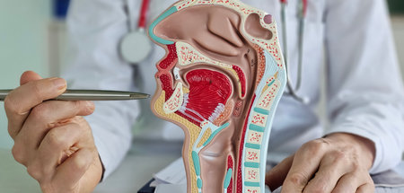
Education in the anatomy of ENT organs. The doctor explains the anatomy of the mucous membrane of the nose and throat using a model.
Коллекция по умолчанию
Коллекция по умолчанию
Создать новую
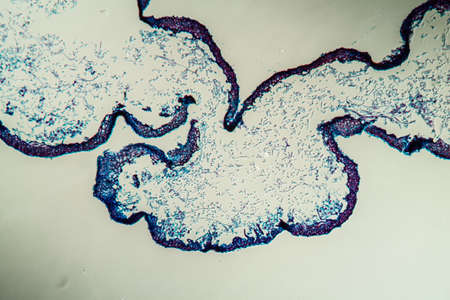
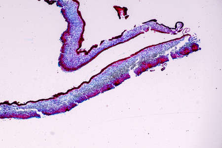
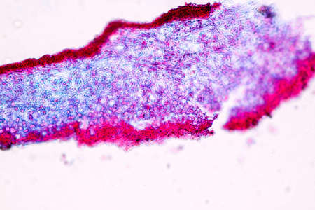
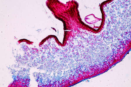
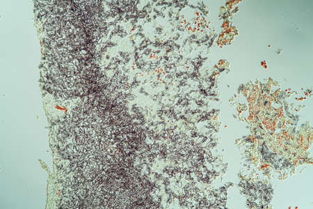
Cross-section through the lichen symbiote body 100x
Коллекция по умолчанию
Коллекция по умолчанию
Создать новую
Suppurative appendicitis, light micrograph, photo under microscope showing neutrophilic infiltrates of the appendix wall and lumen
Коллекция по умолчанию
Коллекция по умолчанию
Создать новую

Large roundworm in human intestine, The large roundworm (A. lumbricoides) lives in the intestines where it lays eggs. The larvae penetrate the intestine wall entering the blood stream and infestating several organs. About 1 billion people are infected worldwide with this parasite.
Коллекция по умолчанию
Коллекция по умолчанию
Создать новую

Education anatomy and Histological sample Spinal cord Tissue under the microscope.
Коллекция по умолчанию
Коллекция по умолчанию
Создать новую

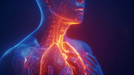
Human heart anatomy with internal organs. Anatomy of human body.
Коллекция по умолчанию
Коллекция по умолчанию
Создать новую

Columnar epithelium of human gall bladder under the microscope in Lab.
Коллекция по умолчанию
Коллекция по умолчанию
Создать новую

X-ray picture of a lung doctor holds a hand in a medical glove on a light background
Коллекция по умолчанию
Коллекция по умолчанию
Создать новую
Squamous cell carcinoma diseased tissue under the microscope 100x
Коллекция по умолчанию
Коллекция по умолчанию
Создать новую
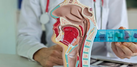
Doctor and anatomy of the nasopharynx with pills. Diseases of the throat, nose and thyroid gland and medications
Коллекция по умолчанию
Коллекция по умолчанию
Создать новую
Showing Light micrograph of the Trachea, Thymus, Parathyroid gland and Tonsil human under the microscope for education in the laboratory.
Коллекция по умолчанию
Коллекция по умолчанию
Создать новую
Legion-Media
Создайте свои проекты на основе качественных стоковых фотографий и видео.


Copyright © Legion-Media.