
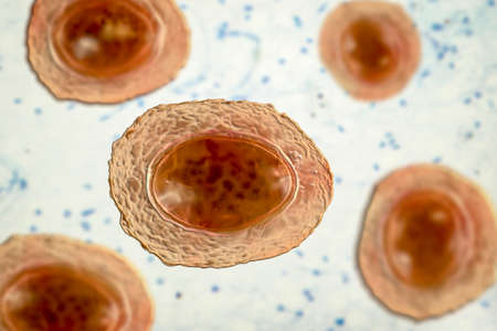
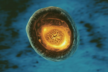
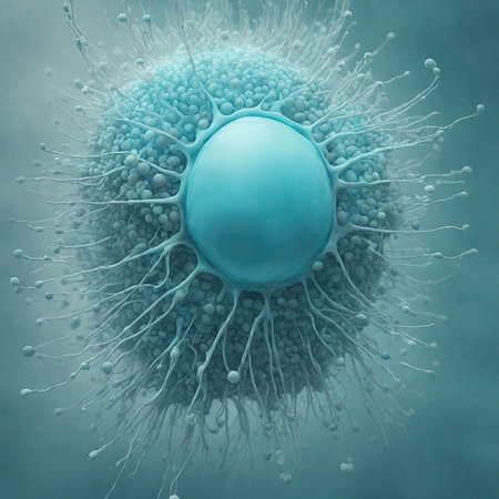
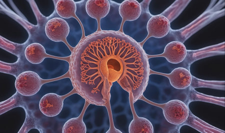
Ascaris lumbricoides, a large roundworm, unfertilized egg, 3D illustration
Коллекция по умолчанию
Коллекция по умолчанию
Создать новую
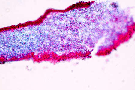
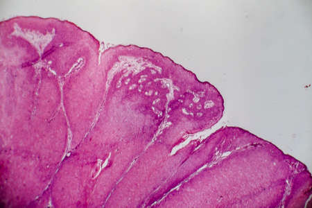
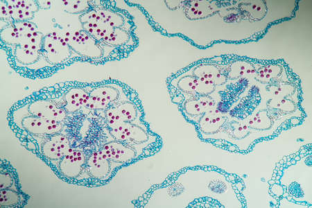
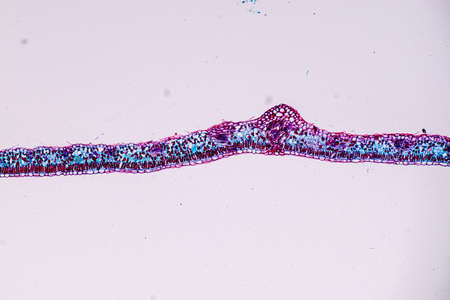
Heather leaf cross section under the microscope, 200x
Коллекция по умолчанию
Коллекция по умолчанию
Создать новую
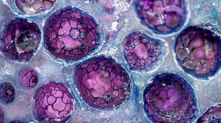
A close up of many small, round, clear objects with a lot of holes in them. The objects are all different sizes and are scattered throughout the image. Scene is one of curiosity and wonder
Коллекция по умолчанию
Коллекция по умолчанию
Создать новую
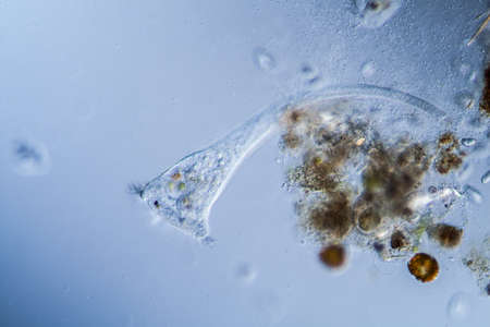
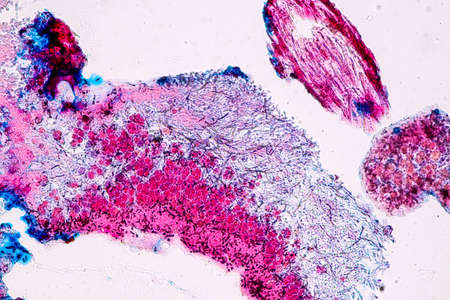
Trumpet animal as a microscopic plankton animal in drops of water
Коллекция по умолчанию
Коллекция по умолчанию
Создать новую
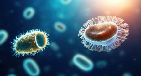
A highly detailed 3D illustration showing various bacteria and microbes in different colors, providing a close-up microscopic view of microorganisms
Коллекция по умолчанию
Коллекция по умолчанию
Создать новую
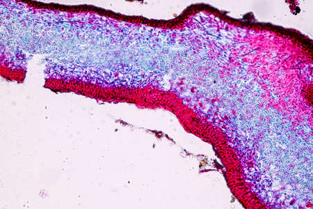
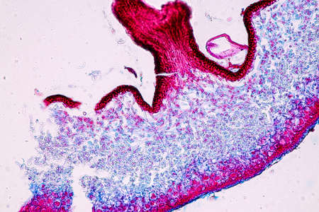
Cross-section through the lichen symbiote body 100x
Коллекция по умолчанию
Коллекция по умолчанию
Создать новую
Trumpet animal as a microscopic plankton animal in drops of water
Коллекция по умолчанию
Коллекция по умолчанию
Создать новую
Realistic Macro shot of different types of microbes. Virus cells and bacteria, by ai generative
Коллекция по умолчанию
Коллекция по умолчанию
Создать новую

Microscopic Fungi samples over white background
Коллекция по умолчанию
Коллекция по умолчанию
Создать новую

Host cells with spores (mold) are inside wood under the microscope for education.
Коллекция по умолчанию
Коллекция по умолчанию
Создать новую
Human egg cell, 3D illustration closeup
Коллекция по умолчанию
Коллекция по умолчанию
Создать новую
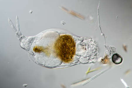
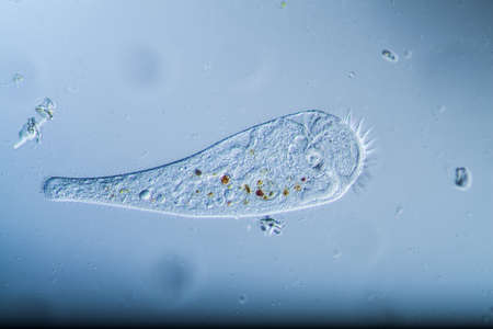
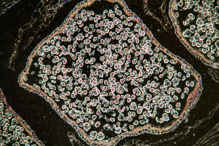
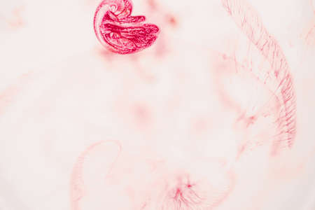
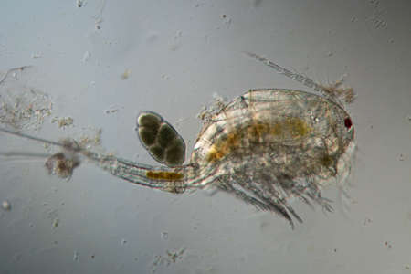
Rotifer foraging in the stream 200x
Коллекция по умолчанию
Коллекция по умолчанию
Создать новую
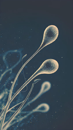
Spermatozoons floating in the air. 3d illustration
Коллекция по умолчанию
Коллекция по умолчанию
Создать новую
Cross-section through the lichen symbiote body 100x
Коллекция по умолчанию
Коллекция по умолчанию
Создать новую
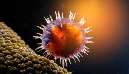
Red spherical cells with spiky outer layers float among textured surfaces, resembling microscopic biological structures or cellular formations, bathed in a soft, muted light
Коллекция по умолчанию
Коллекция по умолчанию
Создать новую

Aspergillus niger and Aspergillus oryzae (mold) under microscope for Microbiology in Lab.
Коллекция по умолчанию
Коллекция по умолчанию
Создать новую
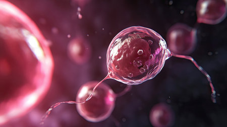
Microscopic sperm cells are seen swimming through a biological fluid, representing the cellular process of human reproduction, fertility, and the potential for a new life
Коллекция по умолчанию
Коллекция по умолчанию
Создать новую

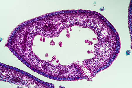
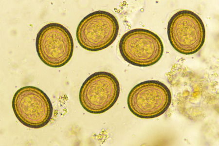
Egg of Ascaris lumbricoides (roundworm) in human stool, analyze by microscope, 400x
Коллекция по умолчанию
Коллекция по умолчанию
Создать новую
Clostridium tetani bacteria, the causative agent of tetanus, 3D illustration
Коллекция по умолчанию
Коллекция по умолчанию
Создать новую
Cytomegalovirus CMV in a human cell, owl's eye inclusion in nucleus, multinucleated cell, 3D illustration. It is herpes virus, causes diseases in fetus, organ transplant patients, HIV infected people
Коллекция по умолчанию
Коллекция по умолчанию
Создать новую

Jellyfish swimming in the water. 3D illustration. Jellyfish macro
Коллекция по умолчанию
Коллекция по умолчанию
Создать новую

Feeler snail's eye tissue under the microscope 200x
Коллекция по умолчанию
Коллекция по умолчанию
Создать новую
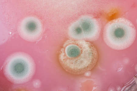
Mushroom cultivation. Macro. Fungi culture on petri dish plate, top view. Mycelium of mushrooms on agar in a petri dish
Коллекция по умолчанию
Коллекция по умолчанию
Создать новую
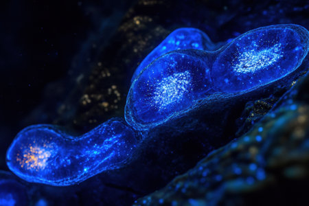
Under ultraviolet light, bacteria emit a vibrant glow, revealing their bioluminescent properties in a dark, captivating setting.
Коллекция по умолчанию
Коллекция по умолчанию
Создать новую
Vibrant cross-section of a developing seed under UV light, highlighting the embryo and endosperm in bright colors
Коллекция по умолчанию
Коллекция по умолчанию
Создать новую
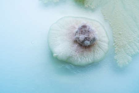
Rust fungus on milkweed plant, 200x
Коллекция по умолчанию
Коллекция по умолчанию
Создать новую
3d rendering of a virus in front of a blue background.
Коллекция по умолчанию
Коллекция по умолчанию
Создать новую
Ice texture background, ink in water pattern frost. Crystal winter design
Коллекция по умолчанию
Коллекция по умолчанию
Создать новую
Rare image of Ghost flatworm - Maricola (Planarian) triclad flatworms in reef aquarium glass
Коллекция по умолчанию
Коллекция по умолчанию
Создать новую
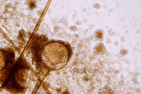
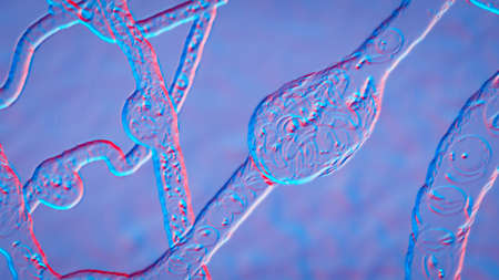
Characteristics of Lichen, hyphae and Symbiotic algae under the microscope for education.
Коллекция по умолчанию
Коллекция по умолчанию
Создать новую
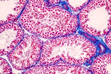
Anatomy and Histological Epididymis and Testis human cells under microscope.
Коллекция по умолчанию
Коллекция по умолчанию
Создать новую
Condyloma acuminatum, also known as genital warts. Light micrograph, photo under microscope
Коллекция по умолчанию
Коллекция по умолчанию
Создать новую
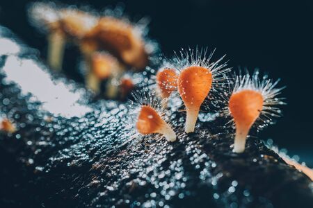
Orange mushroom ,Champagne mushroom or eyelash cup mushroom with sparkling droplets in the forest. Ecosystem or biological diversity concept.
Коллекция по умолчанию
Коллекция по умолчанию
Создать новую
Characteristics of Lichen, hyphae and Symbiotic algae under the microscope for education.
Коллекция по умолчанию
Коллекция по умолчанию
Создать новую

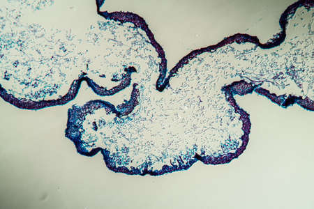
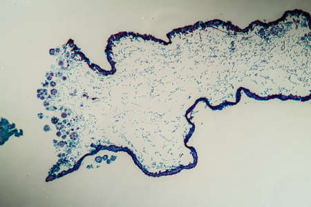
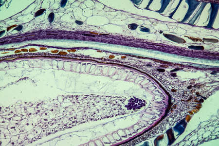
Cross-section leaf Plant of under the microscope for classroom education.
Коллекция по умолчанию
Коллекция по умолчанию
Создать новую
Cell division process, micro
Коллекция по умолчанию
Коллекция по умолчанию
Создать новую
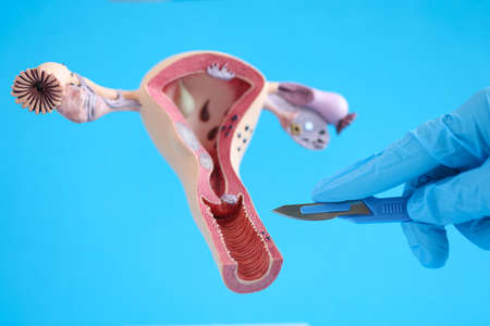
Female reproductive system anatomy uterus and scalpel closeup
Коллекция по умолчанию
Коллекция по умолчанию
Создать новую
Bacteria methicillin-resistant Staphylococcus aureus MRSA, multidrug resistant bacteria, 3D illustration
Коллекция по умолчанию
Коллекция по умолчанию
Создать новую
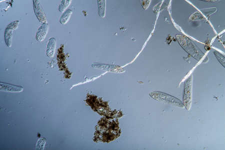
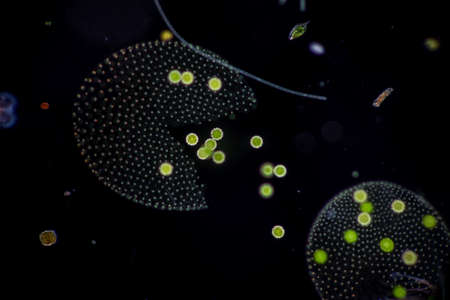
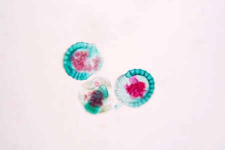
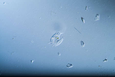
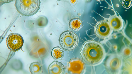
Plankton with microscopic ciliates
Коллекция по умолчанию
Коллекция по умолчанию
Создать новую
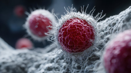
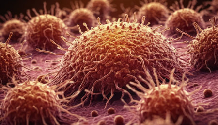
This close-up image depicts cancer cells under laboratory equipment, showcasing intricate details for medical analysis and health research. Ideal for educational use.
Коллекция по умолчанию
Коллекция по умолчанию
Создать новую
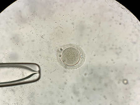
View through microscope at in vitro fertilization process
Коллекция по умолчанию
Коллекция по умолчанию
Создать новую
Johannes berry fruit cross 100x
Коллекция по умолчанию
Коллекция по умолчанию
Создать новую
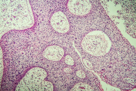
Coccidiosis of liver tissue under the microscope 100x
Коллекция по умолчанию
Коллекция по умолчанию
Создать новую

Mixed of bacteria colonies in Petri dish
Коллекция по умолчанию
Коллекция по умолчанию
Создать новую
a close-up portrait photograph showcasing the intricate details of unknown escherichia coli biological virus alien flowers. the image captures the brittle yet beautiful essence of these flowers, with dry and elegant petals. the rich and vivid contrast, along with the depth of field and black tones, creates a crisp and realistic depiction. shot on a 100mm lens at f/2.0, the natural lighting adds an impressive
Коллекция по умолчанию
Коллекция по умолчанию
Создать новую
Characteristics of Hair cell of human under microscope view for education in laboratory.
Коллекция по умолчанию
Коллекция по умолчанию
Создать новую
Volvox in drop of water under the microscope for classroom education.
Коллекция по умолчанию
Коллекция по умолчанию
Создать новую
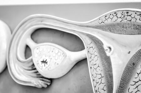
ovary of human with black and white color
Коллекция по умолчанию
Коллекция по умолчанию
Создать новую
Condyloma acuminatum, also known as genital warts. Light micrograph, photo under microscope
Коллекция по умолчанию
Коллекция по умолчанию
Создать новую
Bowen's Disease Tumor under the microscope 100x
Коллекция по умолчанию
Коллекция по умолчанию
Создать новую
Characteristics of Lichen, hyphae and Symbiotic algae under the microscope for education.
Коллекция по умолчанию
Коллекция по умолчанию
Создать новую
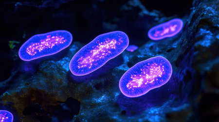
Close-up view of glowing bacteria and viruses with spiky exteriors, floating in a dark blue, luminous, and abstract background.
Коллекция по умолчанию
Коллекция по умолчанию
Создать новую

Histological Uterus human, Uterine tube human, Placenta human and Umbilical cord Human under the microscope for education.
Коллекция по умолчанию
Коллекция по умолчанию
Создать новую
Characteristics of Lichen, hyphae and Symbiotic algae under the microscope for education.
Коллекция по умолчанию
Коллекция по умолчанию
Создать новую
Club moss flower with seeds under the microscope 100x
Коллекция по умолчанию
Коллекция по умолчанию
Создать новую
Botanical study of fern spore under microscopic view. Plant physiology education.
Коллекция по умолчанию
Коллекция по умолчанию
Создать новую
Watercolor abstract. Red paint texture of watercolor pattern or splash ink stain for design isolated on water color background
Коллекция по умолчанию
Коллекция по умолчанию
Создать новую
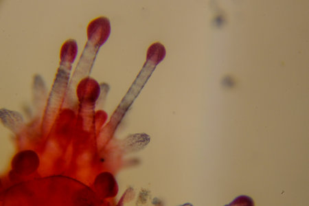
Characteristics of Rhizopus is a genus of common saprophytic fungi on Slide under the microscope for education.
Коллекция по умолчанию
Коллекция по умолчанию
Создать новую
sour and moldy food, top view close-up
Коллекция по умолчанию
Коллекция по умолчанию
Создать новую
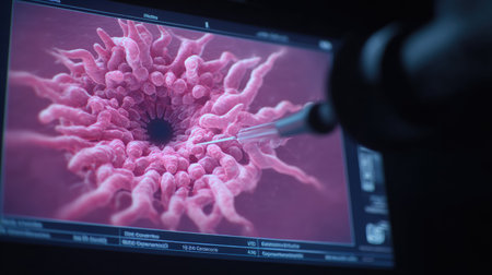
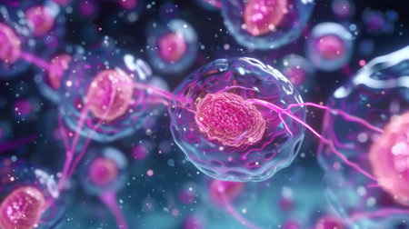
The intricate process of human fertilization is captured, showcasing the moment sperm encounters the egg. This depiction highlights cellular interaction in a scientific laboratory environment, emphasizing the complexity of reproductive biology and the beginning stages of life.
Коллекция по умолчанию
Коллекция по умолчанию
Создать новую

Stomach tissue under the microscope 100x
Коллекция по умолчанию
Коллекция по умолчанию
Создать новую
Microaneurysms, microscopic buldges in the artery walls filled with blood, 3D illustration. Found in the eye retina in diabetic retinopathy, and also in brain (Charcot-Bouchard aneurysms)
Коллекция по умолчанию
Коллекция по умолчанию
Создать новую

Histological Uterus human, Uterine tube human, Placenta human and Umbilical cord Human under the microscope for education
Коллекция по умолчанию
Коллекция по умолчанию
Создать новую
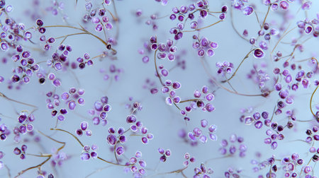
This image captures delicate purple flowers with fine stems set against a soft blue background, evoking a tranquil and ethereal feeling perfect for decor.
Коллекция по умолчанию
Коллекция по умолчанию
Создать новую
budding yeast cell structure fine with microscope in laboratory.
Коллекция по умолчанию
Коллекция по умолчанию
Создать новую
Microscopic view of plant cells with a spore and vascular tissue.
Коллекция по умолчанию
Коллекция по умолчанию
Создать новую
Entire stained starfish larva showing radial symmetry clearly
Коллекция по умолчанию
Коллекция по умолчанию
Создать новую
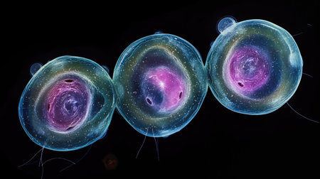
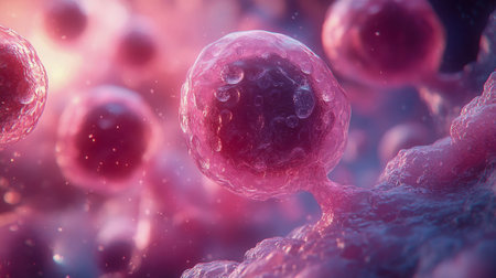
This vibrant depiction explores cellular structures under a microscope, showing intricate details in pink and blue hues during early morning research.
Коллекция по умолчанию
Коллекция по умолчанию
Создать новую
Viruses in blood, 3D illustration. Viruses in the blood
Коллекция по умолчанию
Коллекция по умолчанию
Создать новую

Paramecium caudatum is a genus of unicellular ciliated protozoan and Bacterium under the microscope.
Коллекция по умолчанию
Коллекция по умолчанию
Создать новую
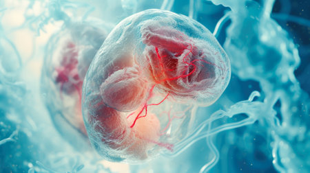
Embryonic Development: Microscopic View of a Translucent Embryo
Коллекция по умолчанию
Коллекция по умолчанию
Создать новую
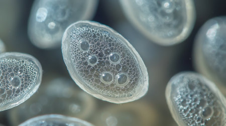
Eggs of Taenia in stool, analyze by microscope
Коллекция по умолчанию
Коллекция по умолчанию
Создать новую
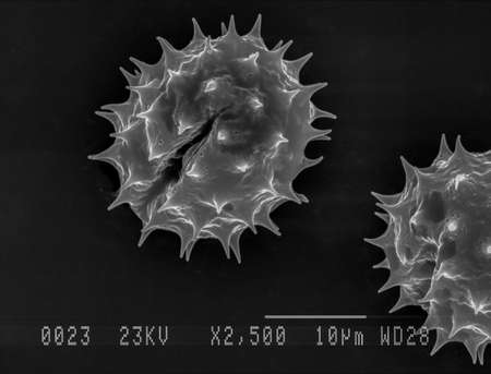
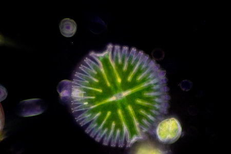
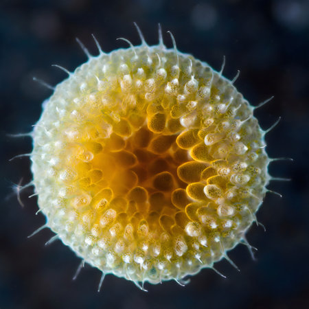
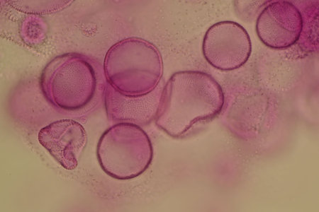
Scanning electron micrograph of one daisy pollen grain. Nottingham, UK
Коллекция по умолчанию
Коллекция по умолчанию
Создать новую
This high-resolution microscopy image provides a detailed view of a respiratory virus, focusing on its outer protein spikes, lipid envelope, and genetic material. The dark, scientific background enhances the contrast and detail, making it ideal for educational and medical contexts.
Коллекция по умолчанию
Коллекция по умолчанию
Создать новую

Cross-section through the lichen symbiote body 100x
Коллекция по умолчанию
Коллекция по умолчанию
Создать новую
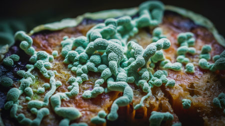
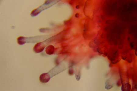
A close up of a fungus growing on an apple, AI
Коллекция по умолчанию
Коллекция по умолчанию
Создать новую
Entire stained starfish larva showing radial symmetry clearly
Коллекция по умолчанию
Коллекция по умолчанию
Создать новую
Protozoa and Green Algae in waste water under the microscope.
Коллекция по умолчанию
Коллекция по умолчанию
Создать новую
Human cell under microscope.
Коллекция по умолчанию
Коллекция по умолчанию
Создать новую

Cytomegalovirus CMV in a human cell, owl's eye inclusion in nucleus, multinucleated cell, 3D illustration. It is herpes virus, causes diseases in fetus, organ transplant patients, HIV infected people
Коллекция по умолчанию
Коллекция по умолчанию
Создать новую
Copepod crab swims through the water
Коллекция по умолчанию
Коллекция по умолчанию
Создать новую
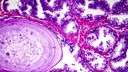
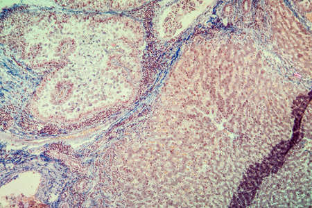
Histopathology of prostate gland hyperplasia, light micrograph, photo under microscope
Коллекция по умолчанию
Коллекция по умолчанию
Создать новую
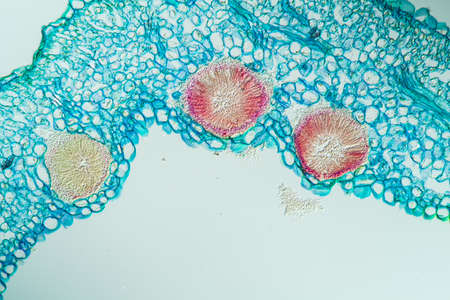
Tansy inflorescence in cross section 100x
Коллекция по умолчанию
Коллекция по умолчанию
Создать новую
AI generated macro shot of a virtual generic microorganism
Коллекция по умолчанию
Коллекция по умолчанию
Создать новую
Ciliates as single-cell plankton animals in drops of water
Коллекция по умолчанию
Коллекция по умолчанию
Создать новую
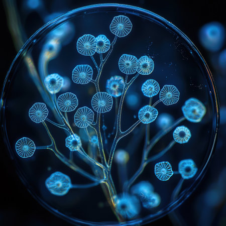
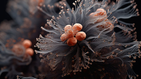
An intricate, illuminated plant structure thrives within a circular transparent vessel against a dark background. The detailed design and unique form are captivating.
Коллекция по умолчанию
Коллекция по умолчанию
Создать новую
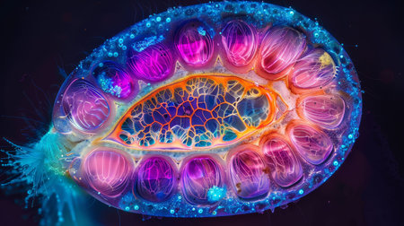
This is a microscopic image of a single-celled organism, specifically a radiolarian, which is a type of zooplankton
Коллекция по умолчанию
Коллекция по умолчанию
Создать новую
Microscopic view of the bone marrow stroma shows colonies of mesenchymal stem cells which have the ability to differentiate into various cell types
Коллекция по умолчанию
Коллекция по умолчанию
Создать новую
Host cells with spores (mold) are inside wood under the microscope for education.
Коллекция по умолчанию
Коллекция по умолчанию
Создать новую
Characteristics of Lichen, hyphae and Symbiotic algae under the microscope for education.
Коллекция по умолчанию
Коллекция по умолчанию
Создать новую
A microscope slide containing a sample of plankton viewed under high magnification to study its composition
Коллекция по умолчанию
Коллекция по умолчанию
Создать новую
Bacteria emit a brilliant glow under UV light in a dark setting, showing vibrant colors and intricate patterns.
Коллекция по умолчанию
Коллекция по умолчанию
Создать новую
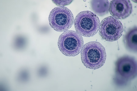
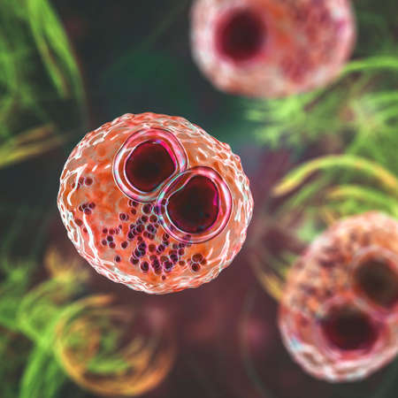
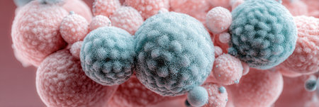
Close-up of pink circular cells under a microscope
Коллекция по умолчанию
Коллекция по умолчанию
Создать новую
A highly detailed microscopic view of streptococcus cells in vibrant pink hues, emphasizing microbiology and science visuals.
Коллекция по умолчанию
Коллекция по умолчанию
Создать новую
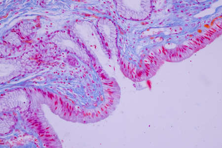
Columnar epithelium of human gall bladder under the microscope in Lab.
Коллекция по умолчанию
Коллекция по умолчанию
Создать новую
Entire stained starfish larva showing radial symmetry clearly
Коллекция по умолчанию
Коллекция по умолчанию
Создать новую
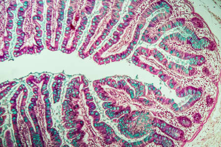
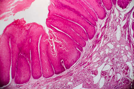
Small intestine with villi under the microscope 100x
Коллекция по умолчанию
Коллекция по умолчанию
Создать новую
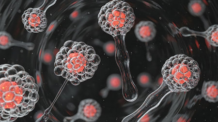
Mysterious Cellular Structures Unveiled
Коллекция по умолчанию
Коллекция по умолчанию
Создать новую
Leech cross section showing internal anatomical structures stained
Коллекция по умолчанию
Коллекция по умолчанию
Создать новую

Ovarian cancer, light micrograph, photo under microscope. Photograph shows a fragment of a cancerous tumor in the female ovary. Selective focus
Коллекция по умолчанию
Коллекция по умолчанию
Создать новую
Legion-Media
Создайте свои проекты на основе качественных стоковых фотографий и видео.


Copyright © Legion-Media.