
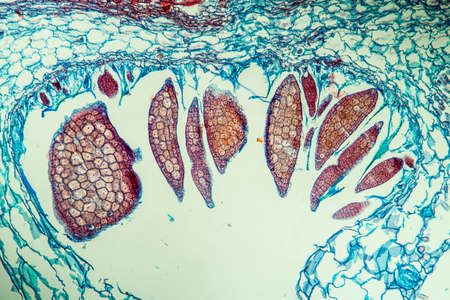
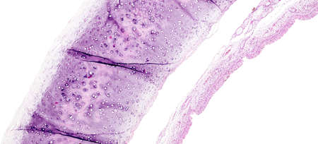
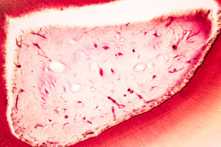
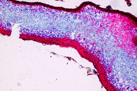
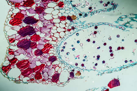
Nestwurz orchid root cross 100x
Коллекция по умолчанию
Коллекция по умолчанию
Создать новую
Abstract marbling floral pattern for fabric, tile design. background texture
Коллекция по умолчанию
Коллекция по умолчанию
Создать новую
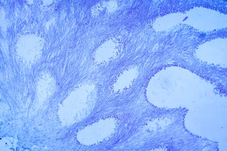
destructive mushroom in wood fabric 100x
Коллекция по умолчанию
Коллекция по умолчанию
Создать новую

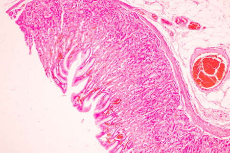
Scalp and hair follicles of human under the microscope in Lab.
Коллекция по умолчанию
Коллекция по умолчанию
Создать новую
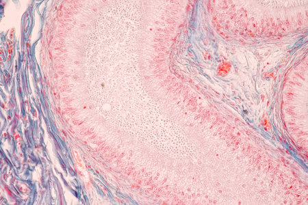
Tissue of Stomach Human under the microscope in Lab.
Коллекция по умолчанию
Коллекция по умолчанию
Создать новую
Histopathology of alveoli, light micrograph, photo under microscope
Коллекция по умолчанию
Коллекция по умолчанию
Создать новую
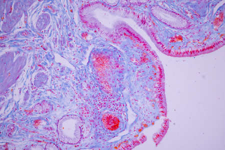
Columnar epithelium of human gall bladder under the microscope in Lab.
Коллекция по умолчанию
Коллекция по умолчанию
Создать новую
Tongue Tissue with taste buds across 200x
Коллекция по умолчанию
Коллекция по умолчанию
Создать новую
Bowel with goblet cells in the dark field 100x
Коллекция по умолчанию
Коллекция по умолчанию
Создать новую

Anatomy and Histological Epididymis and Testis human cells under microscope.
Коллекция по умолчанию
Коллекция по умолчанию
Создать новую
Inguinal testicles gonadically diseased tissue 100x
Коллекция по умолчанию
Коллекция по умолчанию
Создать новую
This detailed microscopic image showcases various cellular structures, highlighted in striking purple tones. The intricate patterns and textures reveal the complexity of biological tissues, making it a valuable resource for educational and scientific purposes
Коллекция по умолчанию
Коллекция по умолчанию
Создать новую
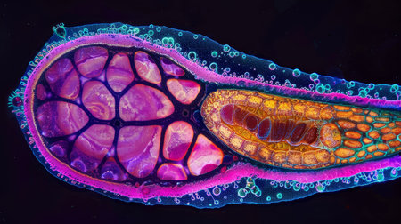
Embryo with endosperm across 100x
Коллекция по умолчанию
Коллекция по умолчанию
Создать новую
Structure of Tissue of Spleen Human, Liver Human and Kidney Human under the microscope in Lab.
Коллекция по умолчанию
Коллекция по умолчанию
Создать новую
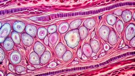
Palatal tonsils transverse 100x under a microscope
Коллекция по умолчанию
Коллекция по умолчанию
Создать новую
Microscopic view of human pancreas in laboratory. 3D rendering
Коллекция по умолчанию
Коллекция по умолчанию
Создать новую

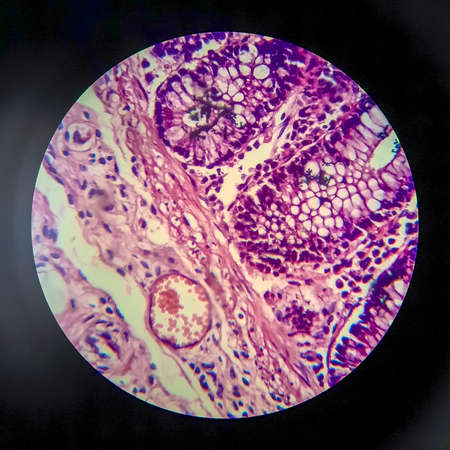
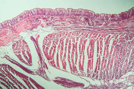
Stomach tissue under the microscope 100x
Коллекция по умолчанию
Коллекция по умолчанию
Создать новую
Characteristics of Lichen, hyphae and Symbiotic algae under the microscope for education.
Коллекция по умолчанию
Коллекция по умолчанию
Создать новую
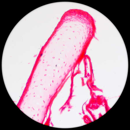
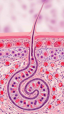
Scalp with hair roots tissue under the microscope 100x
Коллекция по умолчанию
Коллекция по умолчанию
Создать новую
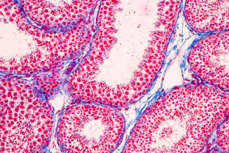
Anatomy and Histological Epididymis and Testis human cells under microscope.
Коллекция по умолчанию
Коллекция по умолчанию
Создать новую
Histopathology of cirrhosis, light micrograph, photo under microscope
Коллекция по умолчанию
Коллекция по умолчанию
Создать новую
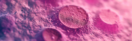
A close up of a pink and blue cell with a tree inside of it. The cell is surrounded by a pink and blue background
Коллекция по умолчанию
Коллекция по умолчанию
Создать новую
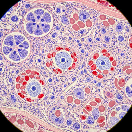
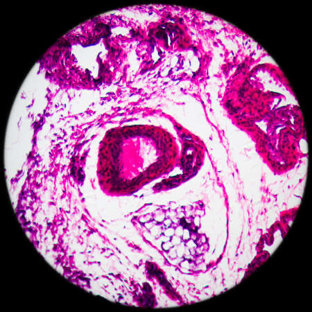
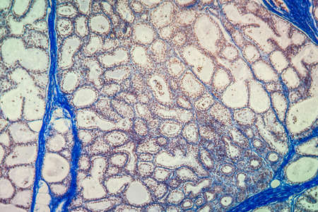
Goiter colloid goiter disease 100x
Коллекция по умолчанию
Коллекция по умолчанию
Создать новую
science medical anthropotomy physiology microscopic section of lymph gland tissue background
Коллекция по умолчанию
Коллекция по умолчанию
Создать новую
Cross section of human cell under microscope view for education in laboratory.
Коллекция по умолчанию
Коллекция по умолчанию
Создать новую
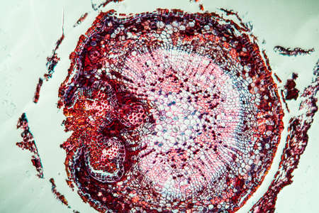
Gladiolus with root tip across 100x
Коллекция по умолчанию
Коллекция по умолчанию
Создать новую

Histopathology of human under microscope view for education in laboratory.
Коллекция по умолчанию
Коллекция по умолчанию
Создать новую
Actinomyces in the jaw diseased tissue 200x
Коллекция по умолчанию
Коллекция по умолчанию
Создать новую

Itch mites under the skin cross-section 100x
Коллекция по умолчанию
Коллекция по умолчанию
Создать новую
Breast tissue under the microscope 100x
Коллекция по умолчанию
Коллекция по умолчанию
Создать новую
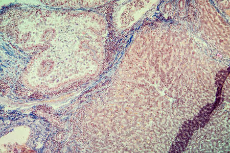
Lymph node tissue under the microscope 100x
Коллекция по умолчанию
Коллекция по умолчанию
Создать новую
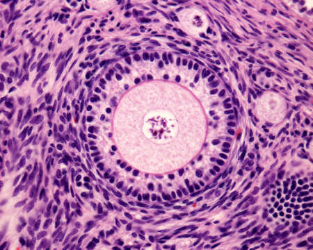
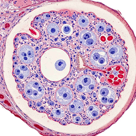
Light microscope micrograph of an ovary. Primary follicle with a big round ovocyte surrounded by the zona pellucida and two layers of granulosa cells.
Коллекция по умолчанию
Коллекция по умолчанию
Создать новую

Tongue with taste buds Papilla across 100x
Коллекция по умолчанию
Коллекция по умолчанию
Создать новую
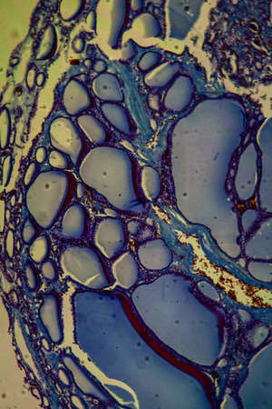
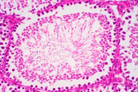
Light micrograph of a seminiferous tubule in the testis.A seminiferous tubule is a long, coiled tube in the testes where sperm is produced. The tubules are lined with Sertoli cells, which support and nourish the developing sperm cells. The tubules are also surrounded by Leydig cells, which produce testosterone.
Коллекция по умолчанию
Коллекция по умолчанию
Создать новую
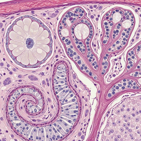
The study of tissue samples of Trachea of Cat, Epididymis, Prostate, Uterus with embryo of rat and Mammary gland cow under the microscope in Lab.
Коллекция по умолчанию
Коллекция по умолчанию
Создать новую
A microscopic view of tissue showing pink-stained cells and structures, indicative of biological samples, possibly related to histology or pathology
Коллекция по умолчанию
Коллекция по умолчанию
Создать новую
Fibroepithelium Diseased tissue 100x
Коллекция по умолчанию
Коллекция по умолчанию
Создать новую
serous gland tissue under the microscope 100x
Коллекция по умолчанию
Коллекция по умолчанию
Создать новую
Abstract ink design template mixed texture background. Blue abstract texture. Multicolored liquid marble pattern. Fluid Art
Коллекция по умолчанию
Коллекция по умолчанию
Создать новую
The study of tissue samples of Trachea of Cat, Epididymis, Prostate, Uterus with embryo of rat and Mammary gland cow under the microscope in Lab.
Коллекция по умолчанию
Коллекция по умолчанию
Создать новую
Histopathology of encephalitis in blood sample under microscope.
Коллекция по умолчанию
Коллекция по умолчанию
Создать новую
The study of tissue samples of Trachea of Cat, Epididymis, Prostate, Uterus with embryo of rat and Mammary gland cow under the microscope in Lab.
Коллекция по умолчанию
Коллекция по умолчанию
Создать новую
Education anatomy and Histological sample of Human under the microscope.
Коллекция по умолчанию
Коллекция по умолчанию
Создать новую
Characteristics of Lichen, hyphae and Symbiotic algae under the microscope for education.
Коллекция по умолчанию
Коллекция по умолчанию
Создать новую

Anatomy and Histological Epididymis and Testis human cells under microscope.
Коллекция по умолчанию
Коллекция по умолчанию
Создать новую
Anatomy and Histological Ovary, Testis and Sperm human cells under microscope.
Коллекция по умолчанию
Коллекция по умолчанию
Создать новую
Anatomy and Histological Epididymis and Testis human cells under microscope.
Коллекция по умолчанию
Коллекция по умолчанию
Создать новую
Histology of human tissue, show tracheitis as seen under the microscope
Коллекция по умолчанию
Коллекция по умолчанию
Создать новую
Light micrograph of human pancreas, light micrograph, photo under microscope
Коллекция по умолчанию
Коллекция по умолчанию
Создать новую

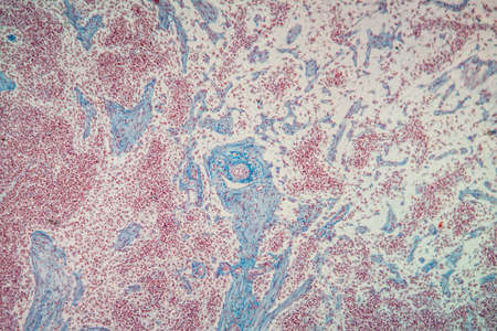
diseased liver with cirrhosis 100x under the microscope
Коллекция по умолчанию
Коллекция по умолчанию
Создать новую

Bladder cancer, light micrograph, photo under microscope
Коллекция по умолчанию
Коллекция по умолчанию
Создать новую
Abstract of fur, dreamy & artistic look background. Closeup, 3D rendering & illustration. Good for book, magazine, catalog cover & design.
Коллекция по умолчанию
Коллекция по умолчанию
Создать новую

Ovarian cancer, light micrograph, photo under microscope. Photograph shows a fragment of a cancerous tumor in the female ovary. Selective focus
Коллекция по умолчанию
Коллекция по умолчанию
Создать новую
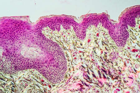
A microscopic view of skin tissue showing various layers and structures, with a focus on cellular details and staining patterns
Коллекция по умолчанию
Коллекция по умолчанию
Создать новую
Anatomy and Histological Epididymis and Testis human cells under microscope.
Коллекция по умолчанию
Коллекция по умолчанию
Создать новую
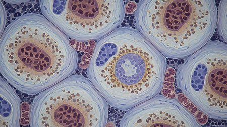
science micrograph of bone cell osteocyte
Коллекция по умолчанию
Коллекция по умолчанию
Создать новую
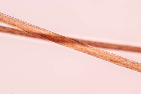
Characteristics of Hair cell of human under microscope view for education in laboratory.
Коллекция по умолчанию
Коллекция по умолчанию
Создать новую

Characteristics of Lichen, hyphae and Symbiotic algae under the microscope for education.
Коллекция по умолчанию
Коллекция по умолчанию
Создать новую
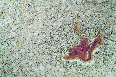
Coccidiosis of liver tissue under the microscope 100x
Коллекция по умолчанию
Коллекция по умолчанию
Создать новую

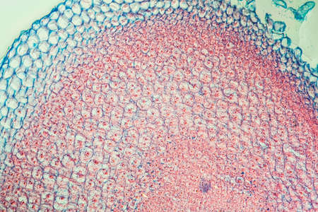
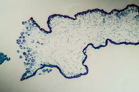
Vegetation cone of the water pest plant 100x
Коллекция по умолчанию
Коллекция по умолчанию
Создать новую
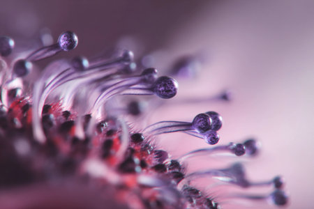
Delicate purple filaments emerge from a plant, showing intricate details in soft lighting.
Коллекция по умолчанию
Коллекция по умолчанию
Создать новую
Creative colorful sand flows on blue background. Place for text or product promotion. Copy space. High quality photo
Коллекция по умолчанию
Коллекция по умолчанию
Создать новую
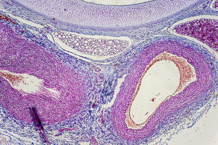
Detailed observation of a human arterys cross section showcasing colorful layers and cellular structures in a laboratory setting.
Коллекция по умолчанию
Коллекция по умолчанию
Создать новую
Histopathology of human liver under microscope view for medical education.
Коллекция по умолчанию
Коллекция по умолчанию
Создать новую
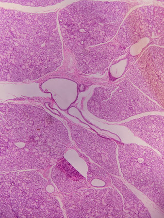
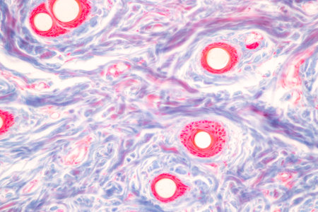
Microscopic view of the blood vessels of the human body under microscope.
Коллекция по умолчанию
Коллекция по умолчанию
Создать новую

Thyroid follicular carcinoma, light micrograph, photo under microscope
Коллекция по умолчанию
Коллекция по умолчанию
Создать новую
microscope slide with detailed view of plant stem, complete with cells and minutiae, created with generative ai
Коллекция по умолчанию
Коллекция по умолчанию
Создать новую
Tissue of Stomach Human under the microscope in Lab.
Коллекция по умолчанию
Коллекция по умолчанию
Создать новую

Condyloma acuminatum, also known as genital warts. Light micrograph, photo under microscope
Коллекция по умолчанию
Коллекция по умолчанию
Создать новую

The study of tissue samples of Trachea of Cat, Epididymis, Prostate, Uterus with embryo of rat and Mammary gland cow under the microscope in Lab.
Коллекция по умолчанию
Коллекция по умолчанию
Создать новую
Scalp and hair follicles of human under the microscope in Lab.
Коллекция по умолчанию
Коллекция по умолчанию
Создать новую

Spice plant with root tip across 100x
Коллекция по умолчанию
Коллекция по умолчанию
Создать новую
Colon tissue with diverticulum 100x
Коллекция по умолчанию
Коллекция по умолчанию
Создать новую
Fungus in the forest, growing in a field of cut grass
Коллекция по умолчанию
Коллекция по умолчанию
Создать новую
Carcinoma in guinea pigs, tissue 100x
Коллекция по умолчанию
Коллекция по умолчанию
Создать новую
Atrophy kidney tissue under the microscope 100x
Коллекция по умолчанию
Коллекция по умолчанию
Создать новую

Proglottid (body unit) of tapeworm Taenia saginata, 3D illustration. A flatworm parasitizing animal and human intestine. Proglottid contains uterus with 12-30 primary lateral branches filled with eggs
Коллекция по умолчанию
Коллекция по умолчанию
Создать новую
Bacillary dysentery, light micrograph, photo under microscope showing presence of bacteria and accumulation of inflammatory cells in intestinal epithelium
Коллекция по умолчанию
Коллекция по умолчанию
Создать новую
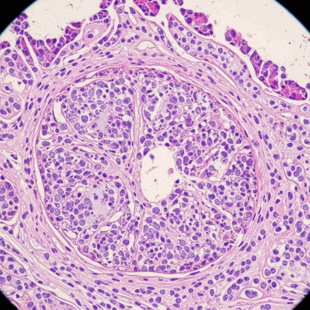
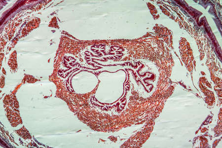
Cross section Human testis under microscope view. Shows spermatogonia, spermatocytes in meiosis, spermatids, and spermatozoa
Коллекция по умолчанию
Коллекция по умолчанию
Создать новую
Histopathology of fibroids, light micrograph, photo under microscope
Коллекция по умолчанию
Коллекция по умолчанию
Создать новую

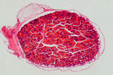
Yew tree with root across 100x
Коллекция по умолчанию
Коллекция по умолчанию
Создать новую
Cross-section through the lichen symbiote body 100x
Коллекция по умолчанию
Коллекция по умолчанию
Создать новую
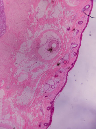
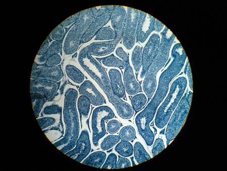
Earthworm histology cross section 10th segment 100x
Коллекция по умолчанию
Коллекция по умолчанию
Создать новую

Johannes berry fruit cross 100x
Коллекция по умолчанию
Коллекция по умолчанию
Создать новую
Human sperm in the testis morphology under microscope. Micrograph showing spermatozoon for pathology education.
Коллекция по умолчанию
Коллекция по умолчанию
Создать новую
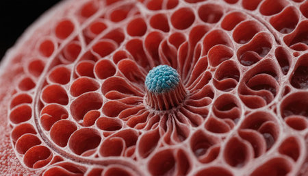
A close-up of a red, honeycomb-like structure with a blue center
Коллекция по умолчанию
Коллекция по умолчанию
Создать новую
A close up of a plant organ with blue and red colors. The organ is surrounded by a white background
Коллекция по умолчанию
Коллекция по умолчанию
Создать новую
Tooth development from human under microscope view for education.
Коллекция по умолчанию
Коллекция по умолчанию
Создать новую
Vibrant cross-section of a developing seed under UV light, highlighting the embryo and endosperm in bright colors
Коллекция по умолчанию
Коллекция по умолчанию
Создать новую
Oak tree with root across 100x
Коллекция по умолчанию
Коллекция по умолчанию
Создать новую
Abstract background of natural stone. Texture of natural stone. The surface of the stone.
Коллекция по умолчанию
Коллекция по умолчанию
Создать новую
Characteristics of Lichen, hyphae and Symbiotic algae under the microscope for education.
Коллекция по умолчанию
Коллекция по умолчанию
Создать новую
Vibrant cross-section of a developing seed under UV light, highlighting the embryo and endosperm in bright colors
Коллекция по умолчанию
Коллекция по умолчанию
Создать новую
Pathology and Histology Tissue of Mammals under microscope.
Коллекция по умолчанию
Коллекция по умолчанию
Создать новую
Microscopic view of human cells under microscope.
Коллекция по умолчанию
Коллекция по умолчанию
Создать новую
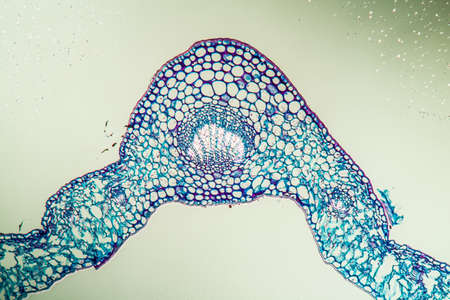
Horsetail sprout in cross section 100x
Коллекция по умолчанию
Коллекция по умолчанию
Создать новую
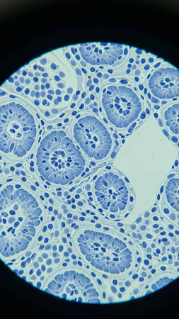
science medical anthropotomy physiology microscopic section of human thyroid gland background
Коллекция по умолчанию
Коллекция по умолчанию
Создать новую
Histopathology of human under microscope view for education in laboratory.
Коллекция по умолчанию
Коллекция по умолчанию
Создать новую
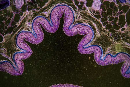
Linguster leaf cross section under the microscope 100x
Коллекция по умолчанию
Коллекция по умолчанию
Создать новую
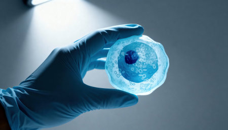
Gloved hand holds a translucent cell model under focused laboratory light with visible nucleus and organelle structures, suggesting scientific research and educational demonstration, with empty background space available for text
Коллекция по умолчанию
Коллекция по умолчанию
Создать новую
Legion-Media
Создайте свои проекты на основе качественных стоковых фотографий и видео.


Copyright © Legion-Media.