

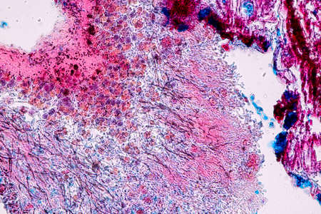
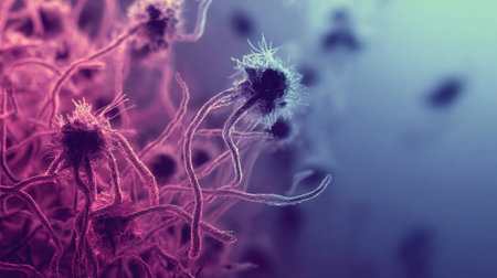
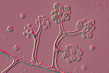
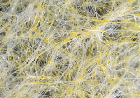
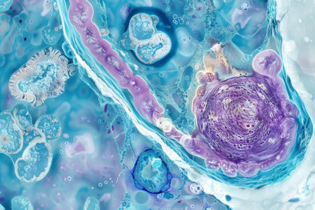
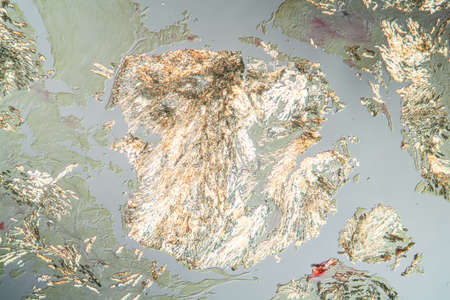
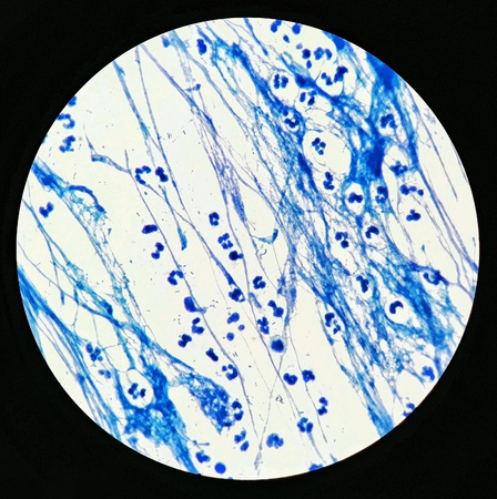
Characteristics of Lichen, hyphae and Symbiotic algae under the microscope for education.
Коллекция по умолчанию
Коллекция по умолчанию
Создать новую
Colon tissue with diverticulum 100x
Коллекция по умолчанию
Коллекция по умолчанию
Создать новую
Tongue Tissue with taste buds across 200x
Коллекция по умолчанию
Коллекция по умолчанию
Создать новую
hearth with amyloid deposits of sick tissue under the microscope 200x
Коллекция по умолчанию
Коллекция по умолчанию
Создать новую

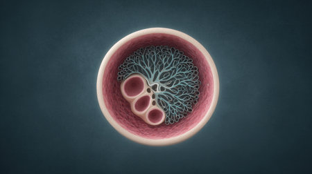
Blood vessel with flowing blood cells, 3D illustration. Small blood vessels, capillaries
Коллекция по умолчанию
Коллекция по умолчанию
Создать новую
Characteristics of Lichen, hyphae and Symbiotic algae under the microscope for education.
Коллекция по умолчанию
Коллекция по умолчанию
Создать новую

Fungus Sporothrix schenckii, the causative agent of sporotrichosis, especially common in florists and gardeners. 3D illustration showing fungal hyphae and spores
Коллекция по умолчанию
Коллекция по умолчанию
Создать новую
Characteristics of Lichen, hyphae and Symbiotic algae under the microscope for education.
Коллекция по умолчанию
Коллекция по умолчанию
Создать новую
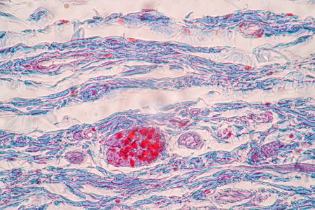
Breast fibroadenosis, light micrograph, photo under microscope. Common benign hyperplastic process involving breast glands
Коллекция по умолчанию
Коллекция по умолчанию
Создать новую
Actinomyces in the jaw diseased tissue 200x
Коллекция по умолчанию
Коллекция по умолчанию
Создать новую

Thyroid follicular carcinoma, light micrograph, photo under microscope
Коллекция по умолчанию
Коллекция по умолчанию
Создать новую
The activation of TLRs leading to the production of proinflammatory cytokines as seen in a micrograph of immune cells
Коллекция по умолчанию
Коллекция по умолчанию
Создать новую
Chaos ink texture background, ink in water pattern frost. Crystal winter design
Коллекция по умолчанию
Коллекция по умолчанию
Создать новую
A closeup examination of a cultured cell on a nanoengineered substrate with bright staining revealing cellular morphology and interaction with the surface at the nanoscale
Коллекция по умолчанию
Коллекция по умолчанию
Создать новую
Lung adenocarcinoma, light micrograph, photo under microscope
Коллекция по умолчанию
Коллекция по умолчанию
Создать новую
destructive mushroom in wood fabric 100x
Коллекция по умолчанию
Коллекция по умолчанию
Создать новую
Lungworm under the microscope 100x
Коллекция по умолчанию
Коллекция по умолчанию
Создать новую

Tooth development from human under microscope view for education.
Коллекция по умолчанию
Коллекция по умолчанию
Создать новую
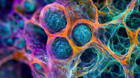
A detailed illustration showcases a colorful cell with filaments and textures, highlighting its complex structures and biological features in a laboratory environment.
Коллекция по умолчанию
Коллекция по умолчанию
Создать новую

Bladder cancer, light micrograph, photo under microscope
Коллекция по умолчанию
Коллекция по умолчанию
Создать новую
Stunning microscopic image of colorful cellular structures demonstrates the intricate details and vibrant colors found in the world of microbiology and life forms.
Коллекция по умолчанию
Коллекция по умолчанию
Создать новую
Abstract of fur, dreamy & artistic look background. Closeup, 3D rendering & illustration. Good for book, magazine, catalog cover & design.
Коллекция по умолчанию
Коллекция по умолчанию
Создать новую
Abstract ink design template mixed texture background. Blue abstract texture. Multicolored liquid marble pattern. Fluid Art
Коллекция по умолчанию
Коллекция по умолчанию
Создать новую
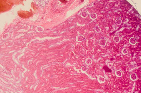
Atrophy kidney tissue under the microscope 100x
Коллекция по умолчанию
Коллекция по умолчанию
Создать новую
Fibroepithelium Diseased tissue 100x
Коллекция по умолчанию
Коллекция по умолчанию
Создать новую
close up shot of wires showcasing a textured image in the style of caffenol developing. the composition features elements like bryce 3d, explosive pigmentation, layered fibers, and cellular formations. the color palette consists of light brown and pink, revealing intricate details of the anatomy. ai generated
Коллекция по умолчанию
Коллекция по умолчанию
Создать новую
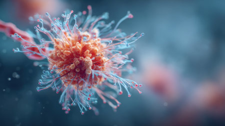
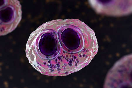
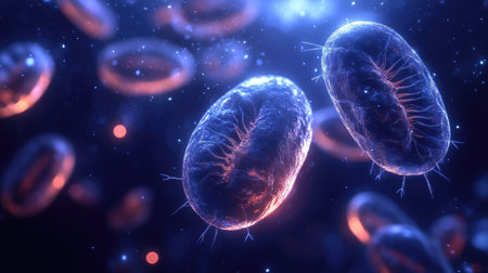
Microscopic view of botulinum bacteria illustrating their structure in vibrant colors, highlighting their relevance to therapy and medical applications.
Коллекция по умолчанию
Коллекция по умолчанию
Создать новую
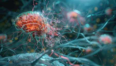
Capture the essence of Parkinson disease with this precise 3D model, showcasing the impact on neural pathways and regions in the brain.
Коллекция по умолчанию
Коллекция по умолчанию
Создать новую
A close-up of a colorful cellular structure featuring fine filaments and textures. This detailed representation highlights the complexity of microscopic life forms in a scientific setting.
Коллекция по умолчанию
Коллекция по умолчанию
Создать новую
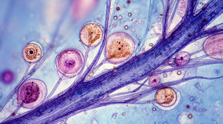
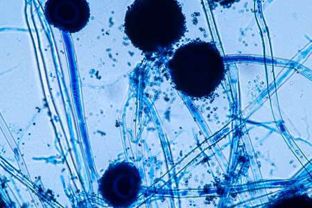
Aspergillus niger and Aspergillus oryzae (mold) under microscope for Microbiology in Lab.
Коллекция по умолчанию
Коллекция по умолчанию
Создать новую
Microscopic fungi Cunninghamella, scientific 3D illustration. Pathogenic fungi from the order Mucorales, cause sinopulmonary and disseminated infections, one of the causative agents of mucormycosis
Коллекция по умолчанию
Коллекция по умолчанию
Создать новую
Explore a stunning microscopic view featuring colorful cells and thin filaments against a vibrant blue background, showcasing scientific beauty in detail.
Коллекция по умолчанию
Коллекция по умолчанию
Создать новую
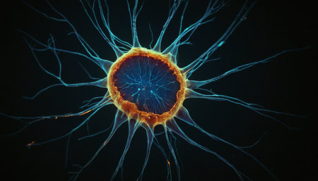
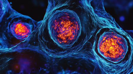
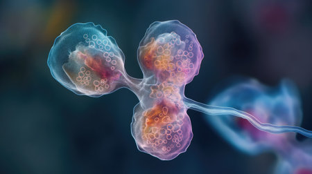
Radiant Neuron Structure
Коллекция по умолчанию
Коллекция по умолчанию
Создать новую
Columnar epithelium of human gall bladder under the microscope in Lab.
Коллекция по умолчанию
Коллекция по умолчанию
Создать новую
A highresolution image of tight junctions in epithelial cells displaying the various types and arrangements of proteins that make up these junctions
Коллекция по умолчанию
Коллекция по умолчанию
Создать новую
Unique X-ray texture reveals mottled and organic patterns in varied formations
Коллекция по умолчанию
Коллекция по умолчанию
Создать новую

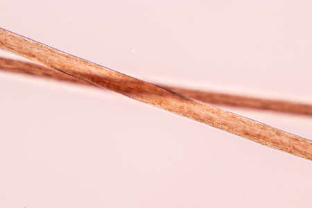
Scalp and hair follicles of human under the microscope in Lab.
Коллекция по умолчанию
Коллекция по умолчанию
Создать новую

Condyloma acuminatum, also known as genital warts. Light micrograph, photo under microscope
Коллекция по умолчанию
Коллекция по умолчанию
Создать новую

Tongue with taste buds Papilla across 100x
Коллекция по умолчанию
Коллекция по умолчанию
Создать новую
A close up of a pink and blue cell with a tree inside of it. The cell is surrounded by a pink and blue background
Коллекция по умолчанию
Коллекция по умолчанию
Создать новую
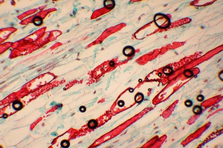
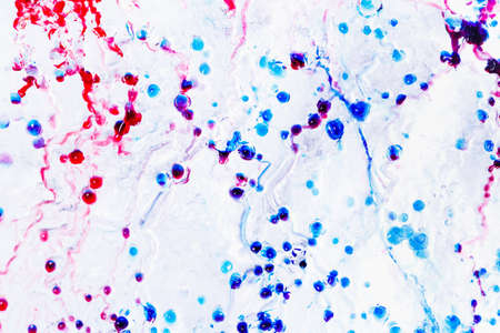
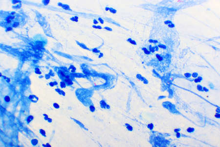
Photo of the microorganism increased in 1000 in the microscope
Коллекция по умолчанию
Коллекция по умолчанию
Создать новую
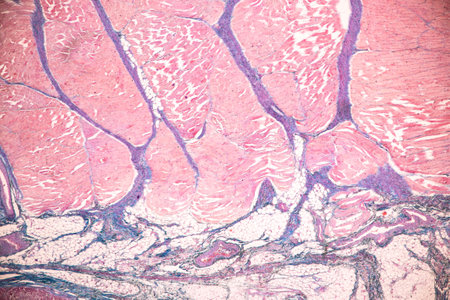
Fibrin deposits in the kidney, microscopy 100x
Коллекция по умолчанию
Коллекция по умолчанию
Создать новую
Lymph node tissue under the microscope 100x
Коллекция по умолчанию
Коллекция по умолчанию
Создать новую
beautiful electronic microscopy of bacteria fungi fantasy microbiology in blue tones microscopic life generative ai
Коллекция по умолчанию
Коллекция по умолчанию
Создать новую

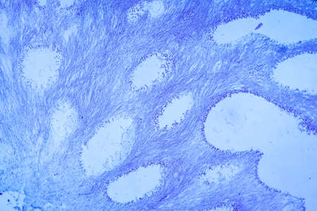
Stomach tissue under the microscope 100x
Коллекция по умолчанию
Коллекция по умолчанию
Создать новую
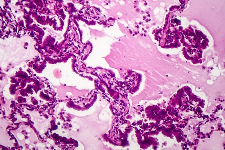
Human lung pathology under light microscope, The lungs is organs of the respiratory system in humans. Human pathology education. Haematoxylin and eosin staining technique slide.
Коллекция по умолчанию
Коллекция по умолчанию
Создать новую
The Microscopic World. Fabric under the microscope.
Коллекция по умолчанию
Коллекция по умолчанию
Создать новую
Mycobacterium tuberculosis positive (small red rod) in sputum smear, acid-fast stain, analyze by microscope 1000x
Коллекция по умолчанию
Коллекция по умолчанию
Создать новую
Discover a vibrant abstract microstructure showcasing cells and biological elements, illuminated with stunning colors and intricate patterns that evoke creativity.
Коллекция по умолчанию
Коллекция по умолчанию
Создать новую
Abstract Metallic Wireframe Mesh in Chrome and Black A futuristic, tech-inspired composition with interwoven metallic wireframe patterns in reflective chrome
Коллекция по умолчанию
Коллекция по умолчанию
Создать новую
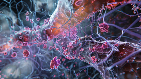
Immune cells actively fight against bacteria while glowing particles surround them, depicting a dynamic scientific process in action.
Коллекция по умолчанию
Коллекция по умолчанию
Создать новую
Close-up of microscopic cells
Коллекция по умолчанию
Коллекция по умолчанию
Создать новую
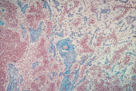
Carcinoma in guinea pigs, tissue 100x
Коллекция по умолчанию
Коллекция по умолчанию
Создать новую

Ovarian cancer, light micrograph, photo under microscope. Photograph shows a fragment of a cancerous tumor in the female ovary. Selective focus
Коллекция по умолчанию
Коллекция по умолчанию
Создать новую
Palatal tonsils transverse 100x under a microscope
Коллекция по умолчанию
Коллекция по умолчанию
Создать новую
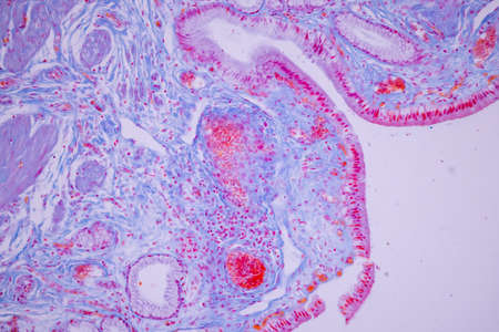
Histological Uterus human, Uterine tube human, Placenta human and Umbilical cord Human under the microscope for education.
Коллекция по умолчанию
Коллекция по умолчанию
Создать новую
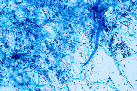
Aspergillus niger and Aspergillus oryzae (mold) under microscope for Microbiology in Lab.
Коллекция по умолчанию
Коллекция по умолчанию
Создать новую
An intricate web of ion channels and pumps working together to create an action potential
Коллекция по умолчанию
Коллекция по умолчанию
Создать новую

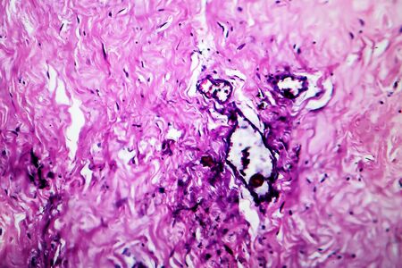
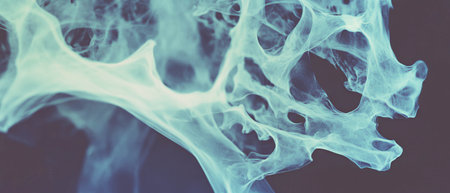
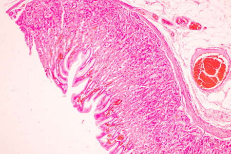
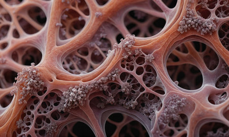
Human hyaline cartilage bone under microscope view for education pathology. Human tissue.
Коллекция по умолчанию
Коллекция по умолчанию
Создать новую
This view reveals colorful cells and structures, highlighting their intricate patterns and vibrant hues under a microscope.
Коллекция по умолчанию
Коллекция по умолчанию
Создать новую

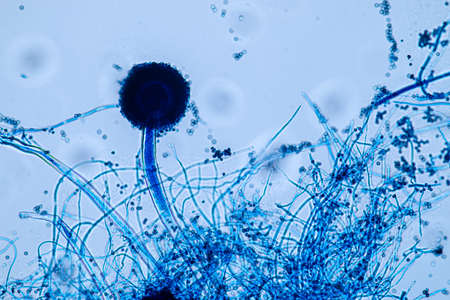
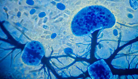
Microscope view of mold sporangia.
Коллекция по умолчанию
Коллекция по умолчанию
Создать новую
Shrinked kidney diseased tissue under the microscope 100x
Коллекция по умолчанию
Коллекция по умолчанию
Создать новую
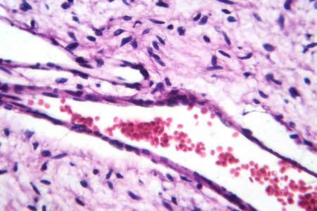
A small blood vessel with red blood cells in neurofibroma tissue sample, light photomicrograph.
Коллекция по умолчанию
Коллекция по умолчанию
Создать новую
A microcosm of motion as cilia on different cells move in different directions yet work together to maintain healthy bodily functions
Коллекция по умолчанию
Коллекция по умолчанию
Создать новую
Tissue of Stomach Human under the microscope in Lab.
Коллекция по умолчанию
Коллекция по умолчанию
Создать новую
A colorful image of a cell with a purple and blue blob in the center. The image is abstract and has a mood of curiosity and wonder
Коллекция по умолчанию
Коллекция по умолчанию
Создать новую
Characteristics of Hair cell of human under microscope view for education in laboratory.
Коллекция по умолчанию
Коллекция по умолчанию
Создать новую

Dry Man Hand With Skin Peeling Off
Коллекция по умолчанию
Коллекция по умолчанию
Создать новую
Cell division process, micro
Коллекция по умолчанию
Коллекция по умолчанию
Создать новую
Aspergillus niger and Aspergillus oryzae (mold) under microscope for Microbiology in Lab.
Коллекция по умолчанию
Коллекция по умолчанию
Создать новую
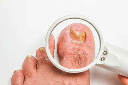
A toenail fungus
Коллекция по умолчанию
Коллекция по умолчанию
Создать новую

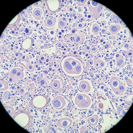
Trichinella spiralis larvae in muscle tissue under the microscope. Trichinella spiralis is a nematode parasite responsible for trichosis and affecting mammals.
Коллекция по умолчанию
Коллекция по умолчанию
Создать новую
Cytomegalovirus CMV in a human cell, owl's eye inclusion in nucleus, multinucleated cell, 3D illustration. It is herpes virus, causes diseases in fetus, organ transplant patients, HIV infected people
Коллекция по умолчанию
Коллекция по умолчанию
Создать новую
This vivid depiction shows coral polyps extending from their skeletons during high tide in a vibrant marine ecosystem.
Коллекция по умолчанию
Коллекция по умолчанию
Создать новую
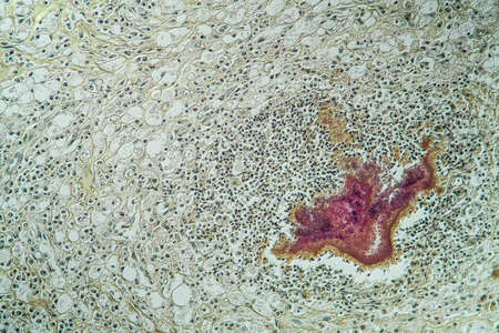
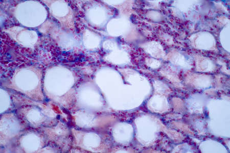
Goiter colloid goiter disease 100x
Коллекция по умолчанию
Коллекция по умолчанию
Создать новую
Microscopic Cellular Fibers enmeshed in network of bubbles
Коллекция по умолчанию
Коллекция по умолчанию
Создать новую
Nestwurz orchid root cross 100x
Коллекция по умолчанию
Коллекция по умолчанию
Создать новую
a close-up portrait photograph showcasing the intricate details of unknown escherichia coli biological virus alien flowers. the image captures the brittle yet beautiful essence of these flowers, with dry and elegant petals. the rich and vivid contrast, along with the depth of field and black tones, creates a crisp and realistic depiction. shot on a 100mm lens with an aperture of f/2.0, the natural lighting
Коллекция по умолчанию
Коллекция по умолчанию
Создать новую
Sunset in the mountains with grass and flowers in the foreground.
Коллекция по умолчанию
Коллекция по умолчанию
Создать новую
Histopathology of lymph nodal tuberculosis, light micrograph, hematoxylin and eosin staining
Коллекция по умолчанию
Коллекция по умолчанию
Создать новую
Microscopic view of human cells under microscope.
Коллекция по умолчанию
Коллекция по умолчанию
Создать новую

Education anatomy and Histological sample of Human under the microscope.
Коллекция по умолчанию
Коллекция по умолчанию
Создать новую
Cervical cancer cells, 3D illustration. Malignant tumor of cervix uteri
Коллекция по умолчанию
Коллекция по умолчанию
Создать новую
Tooth development from human under microscope view for education.
Коллекция по умолчанию
Коллекция по умолчанию
Создать новую
Arthritis gout crystals in the cells tissue section 100x
Коллекция по умолчанию
Коллекция по умолчанию
Создать новую

Cells in reproductive female cytology and histology education concept.
Коллекция по умолчанию
Коллекция по умолчанию
Создать новую
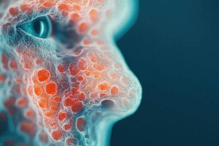
Abstract representation of a human face with colorful textures and patterns, highlighting emotions and features in a surreal style
Коллекция по умолчанию
Коллекция по умолчанию
Создать новую
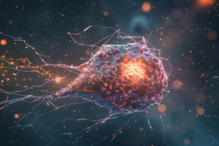
An intricate digital representation of bacteria displaying vibrant colors and glowing elements, highlighting the fascinating world of microscopic life in a stunning abstract setting.
Коллекция по умолчанию
Коллекция по умолчанию
Создать новую
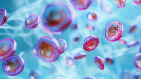
A microscopic image shows a cluster of pink and orange cells, with light refracting off their surfaces. The cells are suspended in a blue liquid, with a fine network of pale blue lines extending throughout the background.
Коллекция по умолчанию
Коллекция по умолчанию
Создать новую
Smear of Acid-Fast bacilli AFB stained with WBC and mucous, under 100X light microscope.
Коллекция по умолчанию
Коллекция по умолчанию
Создать новую
Characteristics of Lichen, hyphae and Symbiotic algae under the microscope for education.
Коллекция по умолчанию
Коллекция по умолчанию
Создать новую
An abstract macro shot showcasing water droplets forming intricate patterns on a textured surface, creating a visually captivating display.
Коллекция по умолчанию
Коллекция по умолчанию
Создать новую
A close up of a pink and blue wire like structure. The pink and blue colors are very bright and the structure is very detailed
Коллекция по умолчанию
Коллекция по умолчанию
Создать новую
Anatomy and Histological Bone, Elastic cartilage human and Joint of human foetus under the microscope for education.
Коллекция по умолчанию
Коллекция по умолчанию
Создать новую
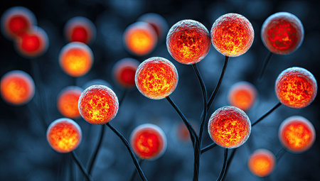
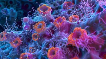
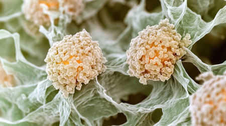
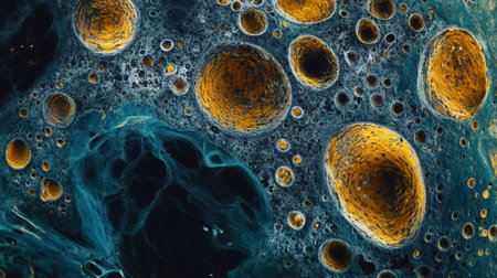
This image presents an abstract illustration featuring numerous spherical structures. They exhibit vibrant orange and red hues. The composition suggests an organic environment. The artwork incorporates a dark background, creating a visual contrast. Suitable for educational, scientific, or conceptual projects, it may be utilized in various commercial or editorial contexts.
Коллекция по умолчанию
Коллекция по умолчанию
Создать новую
A glowing, glowing, glowing blob of light in the sky. It's a bit like a jellyfish, but it's not
Коллекция по умолчанию
Коллекция по умолчанию
Создать новую
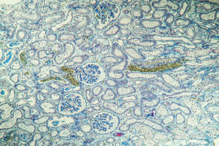
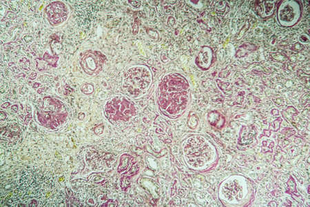
science medical anthropotomy physiology microscopic section of human kidney tissue background
Коллекция по умолчанию
Коллекция по умолчанию
Создать новую
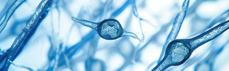
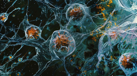
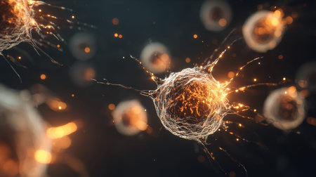
Neuron cell, 3D illustration. Neuron cell under microscope view
Коллекция по умолчанию
Коллекция по умолчанию
Создать новую
Histopathology of human liver under microscope view for medical education.
Коллекция по умолчанию
Коллекция по умолчанию
Создать новую
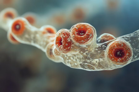
A close-up view of a corals intricate structure, revealing the tiny polyps that make up its colony
Коллекция по умолчанию
Коллекция по умолчанию
Создать новую
Legion-Media
Создайте свои проекты на основе качественных стоковых фотографий и видео.


Copyright © Legion-Media.