
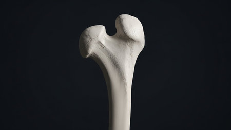
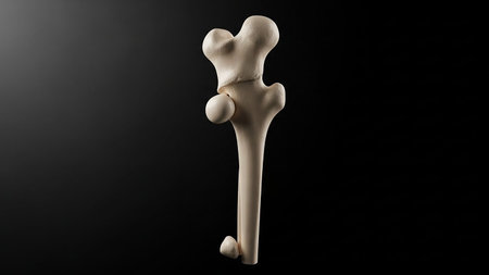
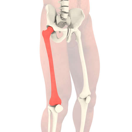
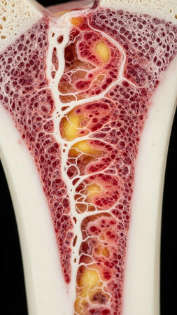
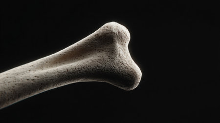
Detailed view of a human femur bone against a dark background, highlighting its anatomy and structure.
Коллекция по умолчанию
Коллекция по умолчанию
Создать новую
Human skeleton anatomy Femur Bone 3D Rendering For Medical Concept
Коллекция по умолчанию
Коллекция по умолчанию
Создать новую
Human skeleton anatomy Femur Bone 3D Rendering For Medical Concept
Коллекция по умолчанию
Коллекция по умолчанию
Создать новую
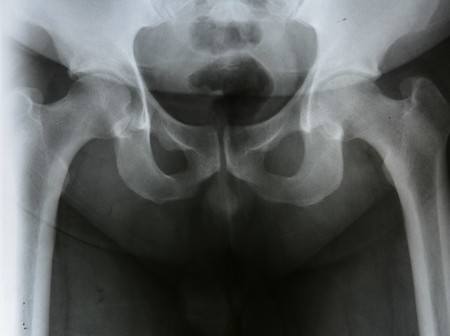
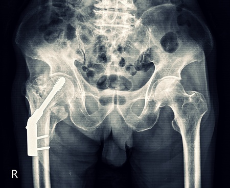
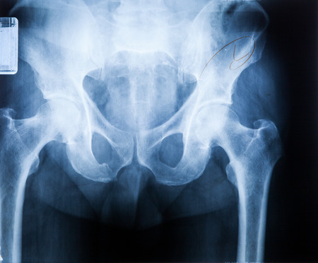
Detailed X-ray image showing a pelvis with an endoprosthesis, specifically designed for hip joint support in rheumatic diseases like Rheumatoid Arthritis.
Коллекция по умолчанию
Коллекция по умолчанию
Создать новую
Detailed view of a human femur bone against a dark background, highlighting its structure
Коллекция по умолчанию
Коллекция по умолчанию
Создать новую
3D Illustration Human Skeleton Anatomy Bones of Femur
Коллекция по умолчанию
Коллекция по умолчанию
Создать новую

X-ray of the pelvic bones of a man. A doctor radiologist is studying an x-ray examination. A hip joint is placed on the patient’s body
Коллекция по умолчанию
Коллекция по умолчанию
Создать новую
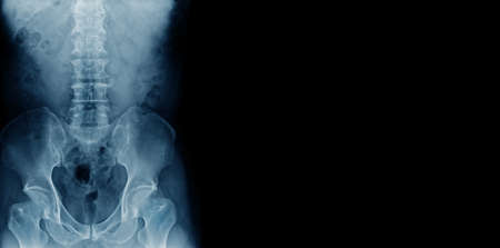
x ray image of pelvic bone with part of lumbar spine
Коллекция по умолчанию
Коллекция по умолчанию
Создать новую
Human skeleton anatomy Femur Bone 3D Rendering For Medical Concept
Коллекция по умолчанию
Коллекция по умолчанию
Создать новую
X-ray of the pelvis and sacrum. X-ray image.
Коллекция по умолчанию
Коллекция по умолчанию
Создать новую
Osteoarthritis of the knee and hand. Healthcare concept image
Коллекция по умолчанию
Коллекция по умолчанию
Создать новую
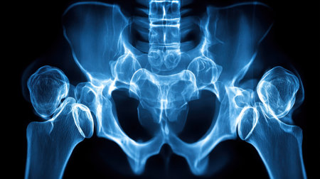
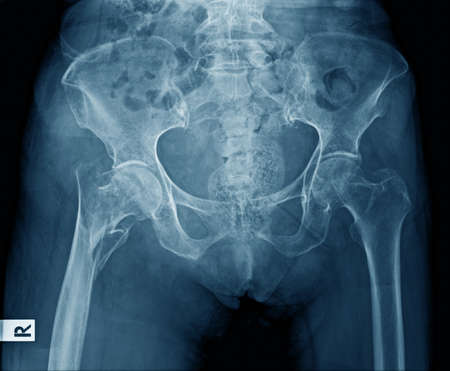
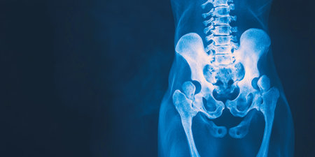
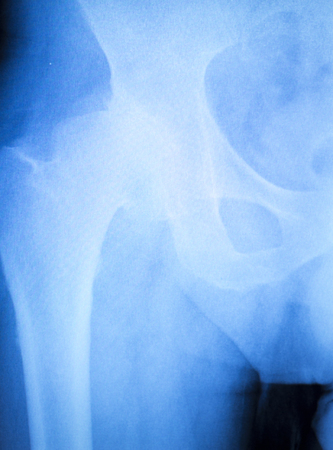
This high-resolution X-ray image of the human pelvis and hip joint displays detailed anatomical structures in stunning blue tones. Ideal for educational and medical purposes.
Коллекция по умолчанию
Коллекция по умолчанию
Создать новую

Banner with hand holding shoulder, clavicle X-ray image. Acromion, acromial end fracture. Health care, medical examination, arm injury detection concept. Copy space. High quality photo
Коллекция по умолчанию
Коллекция по умолчанию
Создать новую

X-ray of the pelvis and spinal column.
Коллекция по умолчанию
Коллекция по умолчанию
Создать новую
x-ray film broken bone at hip install plate with screw
Коллекция по умолчанию
Коллекция по умолчанию
Создать новую
Lumbar Spine and Pelvis XRay Highlighting Skeletal Structure and Medical Imaging Techniques.
Коллекция по умолчанию
Коллекция по умолчанию
Создать новую
pork bone isolated on white background - close-up
Коллекция по умолчанию
Коллекция по умолчанию
Создать новую
This X-ray reveals a detailed view of the human pelvis, highlighting the hip joints and surrounding bone structure in a clinical environment, showcasing alignment and health.
Коллекция по умолчанию
Коллекция по умолчанию
Создать новую
This image displays crossed femur bones arranged elegantly on a smooth white background. Ideal for medical illustrations, educational content, and anatomical studies.
Коллекция по умолчанию
Коллекция по умолчанию
Создать новую
A large, empty arena stage is bathed in blue and white lights, creating a dramatic and inviting atmosphere. The center of the stage is illuminated with a circular ring of lights, ready for a performance or event.
Коллекция по умолчанию
Коллекция по умолчанию
Создать новую
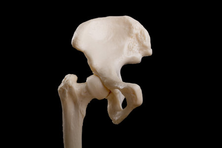
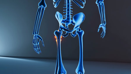
Human hip anatomy isolated on black background
Коллекция по умолчанию
Коллекция по умолчанию
Создать новую
intertrochanteric ( Neck of femur ) fracture left femur ( Thigh bone ) and blank area at right side
Коллекция по умолчанию
Коллекция по умолчанию
Создать новую
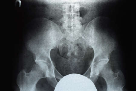
X-ray normal pelvis and hip joint . Blank area at right side .
Коллекция по умолчанию
Коллекция по умолчанию
Создать новую
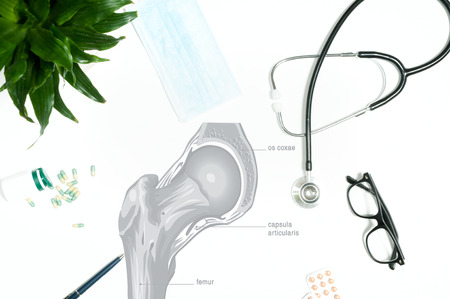
Workplace of doctor - stethoscope, medical items
Коллекция по умолчанию
Коллекция по умолчанию
Создать новую
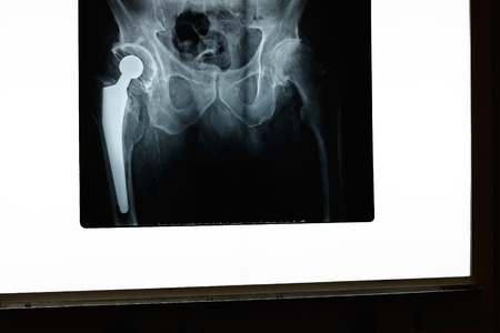
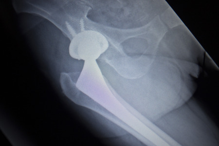
A x-ray of a hip and prosthesis on a x-ray screen
Коллекция по умолчанию
Коллекция по умолчанию
Создать новую
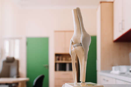
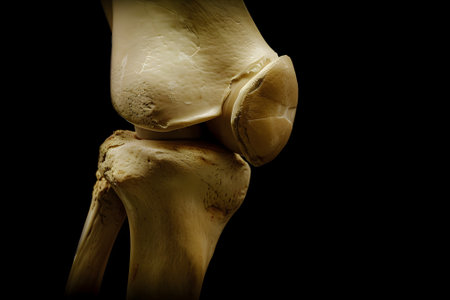
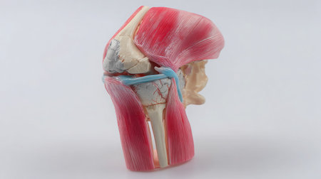
Model of Human knee and leg for medical education
Коллекция по умолчанию
Коллекция по умолчанию
Создать новую

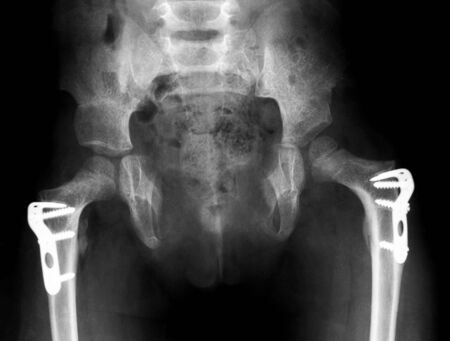
colorful x-rays image show broken hip joint with implant ( plate and screw )
Коллекция по умолчанию
Коллекция по умолчанию
Создать новую
Realistic illustration of knee and leg bones with glass shield. Media to hospitals and doctors, bone nourishing vitamins to protect your bones. 3D realistic modern.
Коллекция по умолчанию
Коллекция по умолчанию
Создать новую
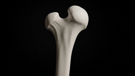
Detailed view of a human femur bone against a dark background
Коллекция по умолчанию
Коллекция по умолчанию
Создать новую
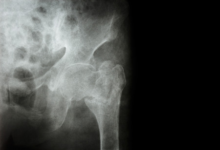
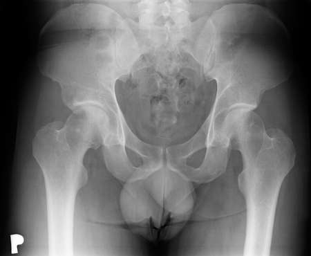
Rontgen picture of male pelvis
Коллекция по умолчанию
Коллекция по умолчанию
Создать новую
Detailed close-up x-ray image of a human knee joint showing the skeletal structure and ligaments on a black background, perfect for medical concepts and educational purposes.
Коллекция по умолчанию
Коллекция по умолчанию
Создать новую

Knee joint meniscus x-ray test scan results photo showing injury and pain for orthopedic surgery and Traumatology surgical treatment.
Коллекция по умолчанию
Коллекция по умолчанию
Создать новую

Human skeleton: pelvis and sacrum. Xray view. Medically accurate 3D illustration
Коллекция по умолчанию
Коллекция по умолчанию
Создать новую

MRI Knee joint (AP,LATERAL)Views a female 15 year old showing Osteosarcoma.Medical healthcare concept.
Коллекция по умолчанию
Коллекция по умолчанию
Создать новую

xray of hip
Коллекция по умолчанию
Коллекция по умолчанию
Создать новую
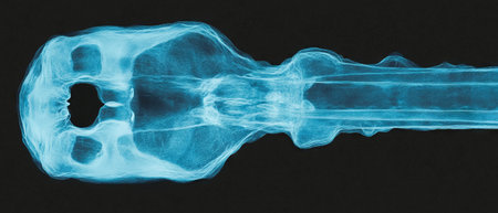
X-ray view of intricate antique key showcasing its unique teeth and locking mechanism details
Коллекция по умолчанию
Коллекция по умолчанию
Создать новую
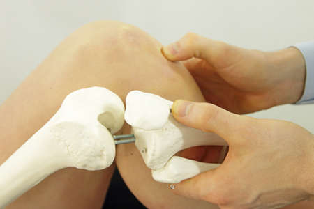
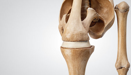
Full view of a D plastic human knee joint used for teaching on a bright white table
Коллекция по умолчанию
Коллекция по умолчанию
Создать новую
x-ray banner of lumbar spine with degenerative change and spur
Коллекция по умолчанию
Коллекция по умолчанию
Создать новую

3d rendered illustration of hip
Коллекция по умолчанию
Коллекция по умолчанию
Создать новую
two surgeons are sitting in a private office at a hospital discussing an upcoming major knee ligament surgery, there was a discussion between the two doctors about the knee joint before the surgery
Коллекция по умолчанию
Коллекция по умолчанию
Создать новую
Transparent skeleton Isolated render on a white background
Коллекция по умолчанию
Коллекция по умолчанию
Создать новую
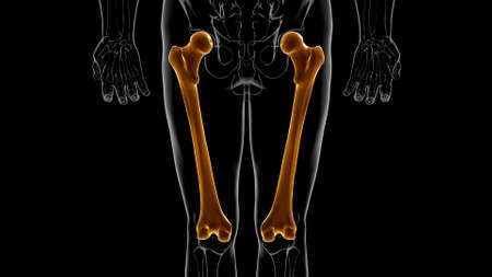
Human skeleton anatomy Femur Bone 3D Rendering For Medical Concept
Коллекция по умолчанию
Коллекция по умолчанию
Создать новую
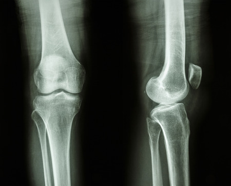
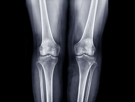
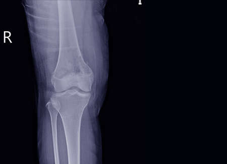
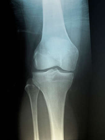
Film x-ray knee AP/lateral : show normal human's knee
Коллекция по умолчанию
Коллекция по умолчанию
Создать новую
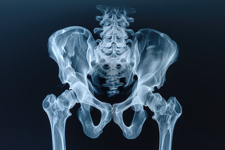
X-ray of the pelvis and spinal column
Коллекция по умолчанию
Коллекция по умолчанию
Создать новую
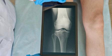
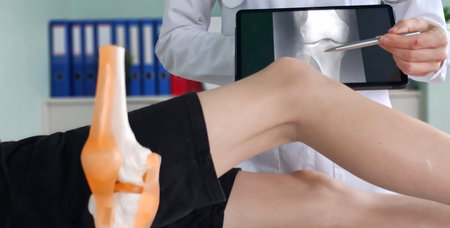
Doctor examines a knee x-ray on tablet in a medical facility. Injury sprained knee child teenager
Коллекция по умолчанию
Коллекция по умолчанию
Создать новую
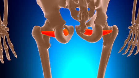
Obturator externus Muscle Anatomy For Medical Concept 3D Illustration
Коллекция по умолчанию
Коллекция по умолчанию
Создать новую

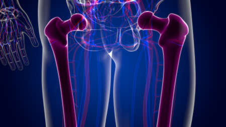
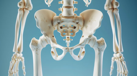
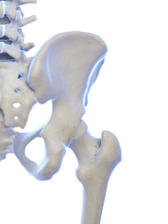
medically accurate 3d illustration of the human hip
Коллекция по умолчанию
Коллекция по умолчанию
Создать новую
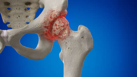
3d rendered illustration of an arthritic hip joint
Коллекция по умолчанию
Коллекция по умолчанию
Создать новую

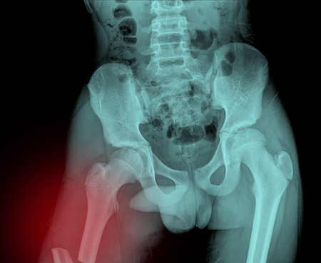
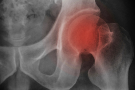
frontal x-ray right hip fracture
Коллекция по умолчанию
Коллекция по умолчанию
Создать новую
some parts of human body
Коллекция по умолчанию
Коллекция по умолчанию
Создать новую
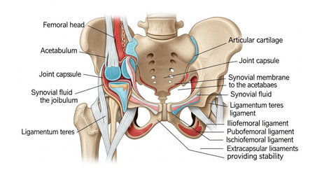
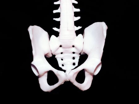
Human pelvic bones annotated
Коллекция по умолчанию
Коллекция по умолчанию
Создать новую
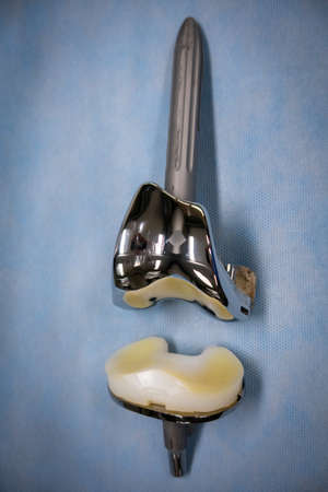
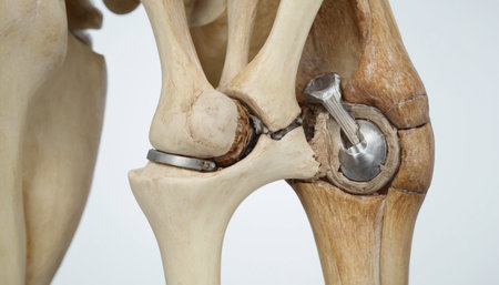
a complete explanted knee prosthesis consisting of distal and proximal parts
Коллекция по умолчанию
Коллекция по умолчанию
Создать новую

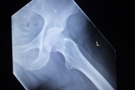
Hip xray
Коллекция по умолчанию
Коллекция по умолчанию
Создать новую

hip dysplasia of an 14 month old dog
Коллекция по умолчанию
Коллекция по умолчанию
Создать новую
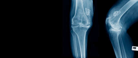
x-ray image of both knee AP view for detect Osteoarthritis Knee or OA Knee .
Коллекция по умолчанию
Коллекция по умолчанию
Создать новую
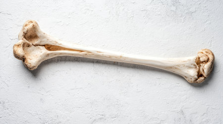
Close up Thigh bone x-ray medical science background
Коллекция по умолчанию
Коллекция по умолчанию
Создать новую
Medical professional examines knee joint using digital X-ray during patient consultation in clinical setting
Коллекция по умолчанию
Коллекция по умолчанию
Создать новую
X-ray scan image of hip joints with orthopedic hip joint replacement implant head and screws in human skeleton in blue gray tones. Scanned in orthopedics traumatology surgery hospital clinic.
Коллекция по умолчанию
Коллекция по умолчанию
Создать новую
Knee joint and ligament, Knee joint, Knee Joint
Коллекция по умолчанию
Коллекция по умолчанию
Создать новую
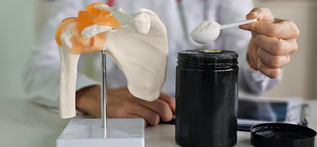
Doctor demonstrates the use of supplement powder next to a shoulder anatomy model in a clinical setting
Коллекция по умолчанию
Коллекция по умолчанию
Создать новую
X-ray image of both hip, AP view, show osteoarthritis Degenerative arthritis of the hip
Коллекция по умолчанию
Коллекция по умолчанию
Создать новую
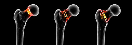
Femur neck fracture types: subcapital, transcervical, and basicervical, shown in sequence from left to right, 3D illustration.
Коллекция по умолчанию
Коллекция по умолчанию
Создать новую
X-Ray Skeleton. Human anatomy exploring skeletal system, joints, and bones of hip joint. Human Knee Joint X-ray: detailed X-ray image of human knee joint, intricate structure of bones and cartilage
Коллекция по умолчанию
Коллекция по умолчанию
Создать новую

Green tree isolated on white background with post cut out original background and white background replaced for easy to selection with clippings path inside, alpha channel for make brush and selection
Коллекция по умолчанию
Коллекция по умолчанию
Создать новую
Structure of the human spine. Abdominal pain. Isolated on black background
Коллекция по умолчанию
Коллекция по умолчанию
Создать новую
X-ray Knee join a female 15 year old Showing large osteolytic lesuion of medial aspect of right distal femur.with soft tissure mass.and malignant bone tumor,osteosarcoma is suspected.
Коллекция по умолчанию
Коллекция по умолчанию
Создать новую
removal of benign tumor on the dogs paw by surgery
Коллекция по умолчанию
Коллекция по умолчанию
Создать новую
X-ray image of human foot isolated on a black background.
Коллекция по умолчанию
Коллекция по умолчанию
Создать новую
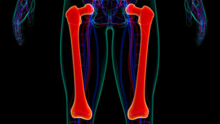
X-ray of human leg on a black and white background.
Коллекция по умолчанию
Коллекция по умолчанию
Создать новую
X-ray Hip Pelvis
Коллекция по умолчанию
Коллекция по умолчанию
Создать новую

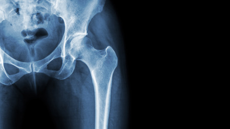
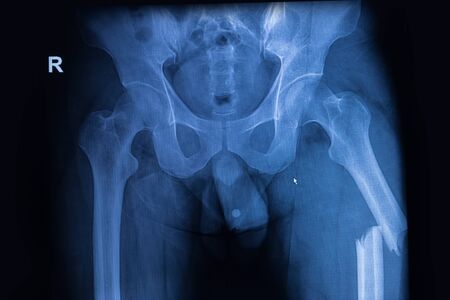
X-ray image pelvis and hip of a man
Коллекция по умолчанию
Коллекция по умолчанию
Создать новую
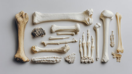
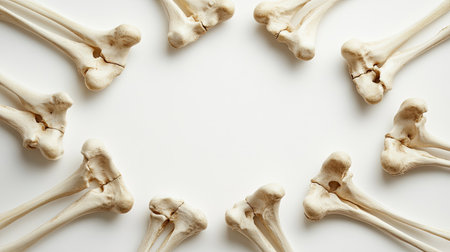
A visually informative arrangement of assorted animal bones showcasing various shapes and sizes, ideal for educational materials, scientific exploration, or forensic analysis.
Коллекция по умолчанию
Коллекция по умолчанию
Создать новую
X-ray image of both hip showing femur fracture at left side
Коллекция по умолчанию
Коллекция по умолчанию
Создать новую

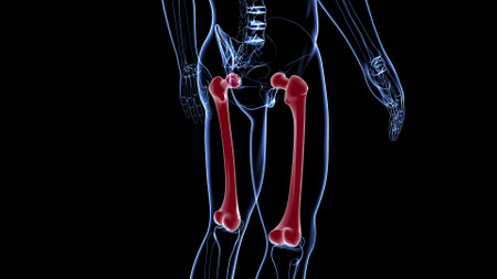
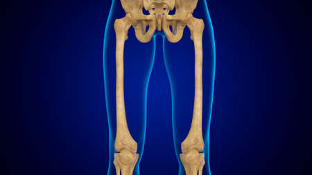
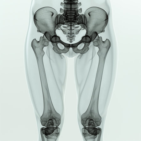
Human Skeleton System Lower Limbs Skeletal Anatomy 3D Illustration
Коллекция по умолчанию
Коллекция по умолчанию
Создать новую
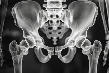
This x-ray image provides a detailed view of the human skeleton, revealing the intricate structure and positioning of bones, Three-dimensional X-ray film of human pelvic bones, AI Generated
Коллекция по умолчанию
Коллекция по умолчанию
Создать новую
x-ray OA knee with structure like calcium
Коллекция по умолчанию
Коллекция по умолчанию
Создать новую
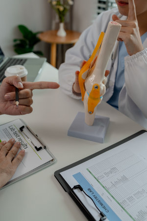
human knee and model comparison
Коллекция по умолчанию
Коллекция по умолчанию
Создать новую
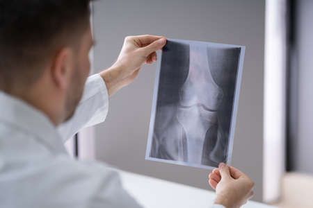
Close-up Of A Male Doctor's Hand Holding Knee X-ray
Коллекция по умолчанию
Коллекция по умолчанию
Создать новую
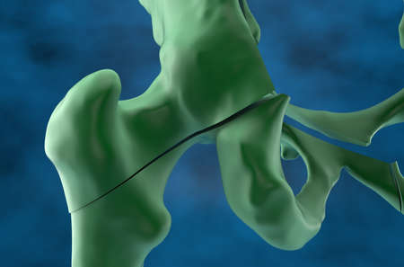
Fractured pelvis and femur in osteoporosis - closeup view 3d illustration
Коллекция по умолчанию
Коллекция по умолчанию
Создать новую

Medical knee joint meniscus plastic demonstration teaching model against plain black studio background. The knee joint meniscus femur thigh bone and tibia shin traumatology medical plastic model is used to show articulation and possible injuries.
Коллекция по умолчанию
Коллекция по умолчанию
Создать новую
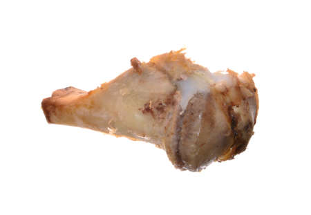
A single bone fragment placed diagonally on a textured white surface, showcasing its natural shape and detail in a clean, minimalistic style.
Коллекция по умолчанию
Коллекция по умолчанию
Создать новую
3d rendered illustration of a painful hip joint
Коллекция по умолчанию
Коллекция по умолчанию
Создать новую

hip xray
Коллекция по умолчанию
Коллекция по умолчанию
Создать новую
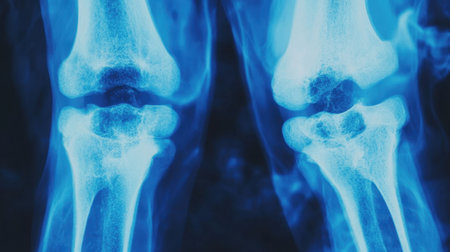
Blue-toned X-ray image of both knee joints, displaying signs of mild osteoarthritis, offering a detailed medical perspective
Коллекция по умолчанию
Коллекция по умолчанию
Создать новую
Undernourished Teenager with Visible Ribs and Sunken Stomach
Коллекция по умолчанию
Коллекция по умолчанию
Создать новую
Realistic 3D Render of Human Skeleton anatomy - Back view
Коллекция по умолчанию
Коллекция по умолчанию
Создать новую

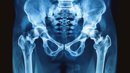
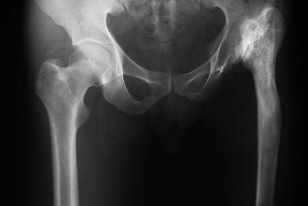
Film x-ray of normal human pelvis and hip joints .
Коллекция по умолчанию
Коллекция по умолчанию
Создать новую
X ray MRI - Image of Spine pain and Hip bone
Коллекция по умолчанию
Коллекция по умолчанию
Создать новую

X-ray Knee
Коллекция по умолчанию
Коллекция по умолчанию
Создать новую

Hip Pain close-up illustration. Blue Human Anatomy Body 3D Scan render on blue background
Коллекция по умолчанию
Коллекция по умолчанию
Создать новую
3d rendered medically accurate illustration of the hip joint
Коллекция по умолчанию
Коллекция по умолчанию
Создать новую

x-ray knee banner for website, hight quality of knee in flexion position
Коллекция по умолчанию
Коллекция по умолчанию
Создать новую
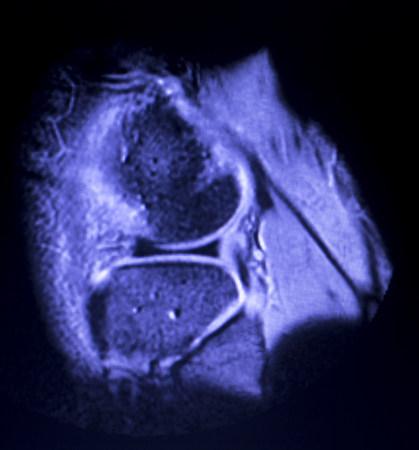
Magnetic resonance imaging MRI knee posterior horn medial meniscus tear scantest results.
Коллекция по умолчанию
Коллекция по умолчанию
Создать новую
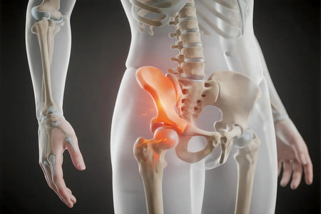
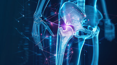
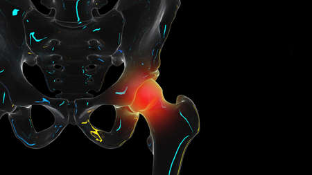
Human skeleton highlighting hip joint pain in a medical visualization showing inflammation and discomfort, emphasizing the importance of healthcare and treatment.
Коллекция по умолчанию
Коллекция по умолчанию
Создать новую
Preventive and control medical examination. X-ray of the Ilium and hip bones.
Коллекция по умолчанию
Коллекция по умолчанию
Создать новую
X-ray scan image of hip joints human skeleton in blue gray tones. Scanned in orthopedics traumatology surgery hospital clinic.
Коллекция по умолчанию
Коллекция по умолчанию
Создать новую
Arthritic hip xray test scan orthopedic and Traumatology results.
Коллекция по умолчанию
Коллекция по умолчанию
Создать новую
normal knee x-ray of a 58 year old man
Коллекция по умолчанию
Коллекция по умолчанию
Создать новую
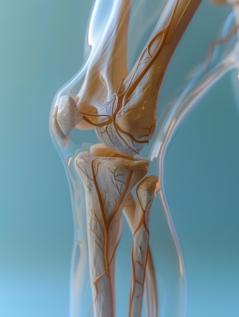
Close up of human body skeleton with visible veins and arteries
Коллекция по умолчанию
Коллекция по умолчанию
Создать новую
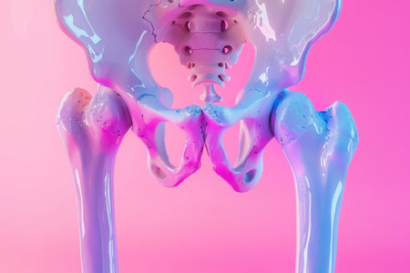
A pink background with a blue and purple skeleton. The skeleton is in the middle of the image and is the main focus
Коллекция по умолчанию
Коллекция по умолчанию
Создать новую
Legion-Media
Создайте свои проекты на основе качественных стоковых фотографий и видео.


Copyright © Legion-Media.