

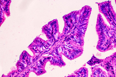
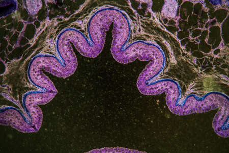
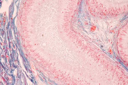
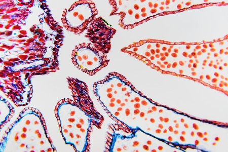
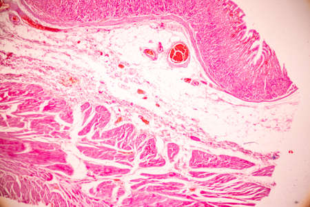
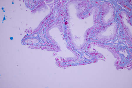
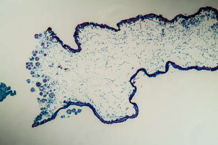
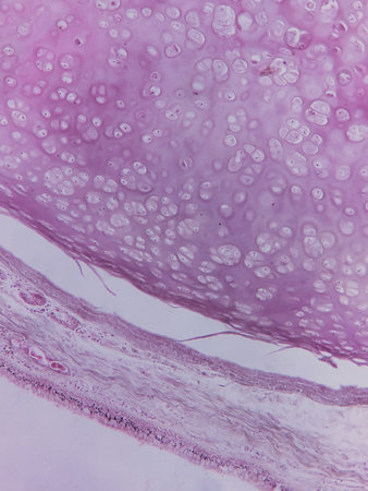
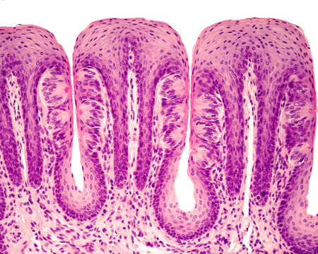
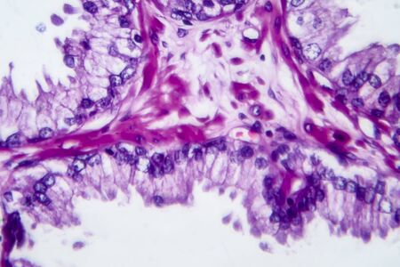
Stomach tissue under the microscope 100x
Коллекция по умолчанию
Коллекция по умолчанию
Создать новую
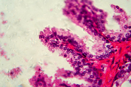
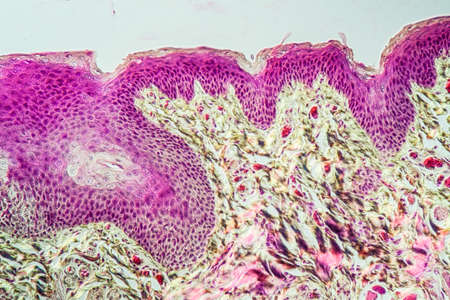
Education anatomy and Histological sample of Human under the microscope.
Коллекция по умолчанию
Коллекция по умолчанию
Создать новую
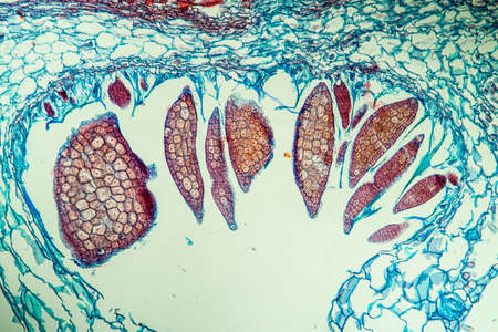
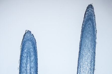
Nestwurz orchid root cross 100x
Коллекция по умолчанию
Коллекция по умолчанию
Создать новую
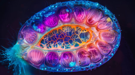
Vibrant cross-section of a developing seed under UV light, highlighting the embryo and endosperm in bright colors
Коллекция по умолчанию
Коллекция по умолчанию
Создать новую
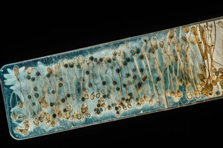
destructive mushroom in wood fabric 100x
Коллекция по умолчанию
Коллекция по умолчанию
Создать новую
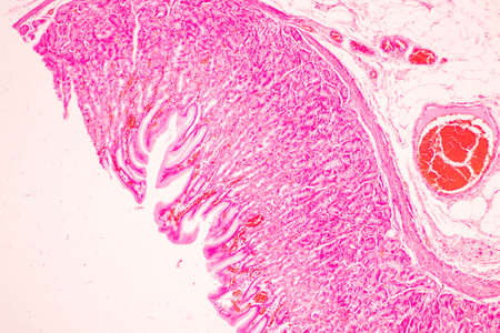
Tissue of Stomach Human under the microscope in Lab.
Коллекция по умолчанию
Коллекция по умолчанию
Создать новую

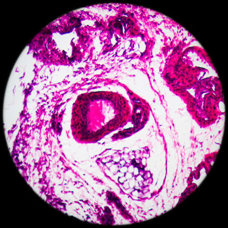
Ovarian cancer, light micrograph, photo under microscope. Photograph shows a fragment of a cancerous tumor in the female ovary. Selective focus
Коллекция по умолчанию
Коллекция по умолчанию
Создать новую
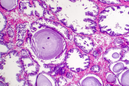
Photomicrograph showing histological features of benign prostatic hyperplasia. Enlarged prostate gland with nodular proliferation of glandular and stromal components. High-resolution histology image.
Коллекция по умолчанию
Коллекция по умолчанию
Создать новую

Bee compound eye enlarged 100x along
Коллекция по умолчанию
Коллекция по умолчанию
Создать новую
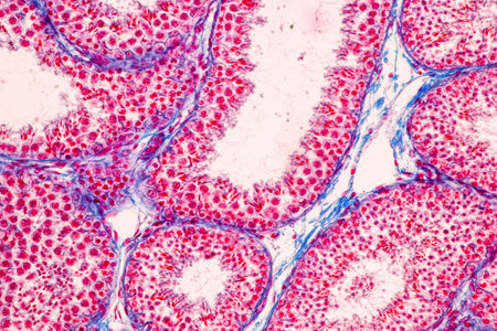
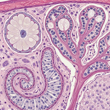
Anatomy and Histological Epididymis and Testis human cells under microscope.
Коллекция по умолчанию
Коллекция по умолчанию
Создать новую
Characteristics of Lichen, hyphae and Symbiotic algae under the microscope for education.
Коллекция по умолчанию
Коллекция по умолчанию
Создать новую
science medical anthropotomy physiology microscopic section of lymph gland tissue background
Коллекция по умолчанию
Коллекция по умолчанию
Создать новую
Inguinal testicles gonadically diseased tissue 100x
Коллекция по умолчанию
Коллекция по умолчанию
Создать новую
Columnar epithelium of human gall bladder under the microscope in Lab.
Коллекция по умолчанию
Коллекция по умолчанию
Создать новую
A close up of a plant organ with blue and red colors. The organ is surrounded by a white background
Коллекция по умолчанию
Коллекция по умолчанию
Создать новую
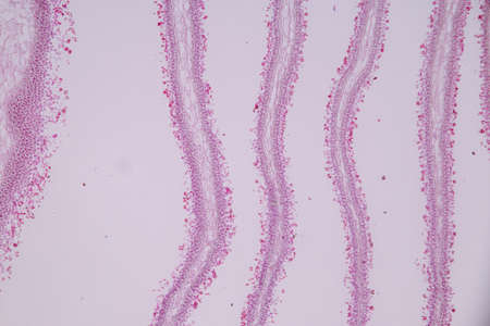
Bowel with goblet cells in the dark field 100x
Коллекция по умолчанию
Коллекция по умолчанию
Создать новую
Vibrant cross-section of a developing seed under UV light, highlighting the embryo and endosperm in bright colors
Коллекция по умолчанию
Коллекция по умолчанию
Создать новую

Scalp and hair follicles of human under the microscope in Lab.
Коллекция по умолчанию
Коллекция по умолчанию
Создать новую

Vegetation cone of the water pest plant 100x
Коллекция по умолчанию
Коллекция по умолчанию
Создать новую
Close-up of a Pink and Blue Wavy Surface with Bumps and Bubbles
Коллекция по умолчанию
Коллекция по умолчанию
Создать новую
The study of tissue samples of Trachea of Cat, Epididymis, Prostate, Uterus with embryo of rat and Mammary gland cow under the microscope in Lab.
Коллекция по умолчанию
Коллекция по умолчанию
Создать новую
serous gland tissue under the microscope 100x
Коллекция по умолчанию
Коллекция по умолчанию
Создать новую
Characteristics of Lichen, hyphae and Symbiotic algae under the microscope for education.
Коллекция по умолчанию
Коллекция по умолчанию
Создать новую
Cross section of the Cerebellum and Nerve human under the microscope for education in Lab.
Коллекция по умолчанию
Коллекция по умолчанию
Создать новую
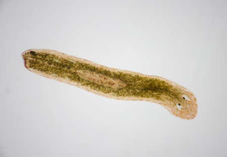
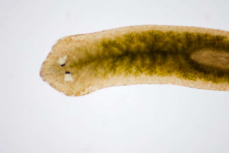
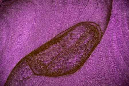
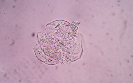
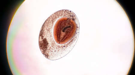
Planarian parasite (flatworm) under microscope view.
Коллекция по умолчанию
Коллекция по умолчанию
Создать новую
microscope slide with detailed view of plant stem, complete with cells and minutiae, created with generative ai
Коллекция по умолчанию
Коллекция по умолчанию
Создать новую
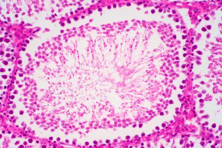
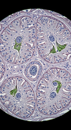
Cross section Human testis under microscope view. Shows spermatogonia, spermatocytes in meiosis, spermatids, and spermatozoa
Коллекция по умолчанию
Коллекция по умолчанию
Создать новую

Proglottid (body unit) of tapeworm Taenia saginata, 3D illustration. A flatworm parasitizing animal and human intestine. Proglottid contains uterus with 12-30 primary lateral branches filled with eggs
Коллекция по умолчанию
Коллекция по умолчанию
Создать новую
Anatomy and Histological Epididymis and Testis human cells under microscope.
Коллекция по умолчанию
Коллекция по умолчанию
Создать новую

Microscopic close up view of virus cell, 3D rendering
Коллекция по умолчанию
Коллекция по умолчанию
Создать новую
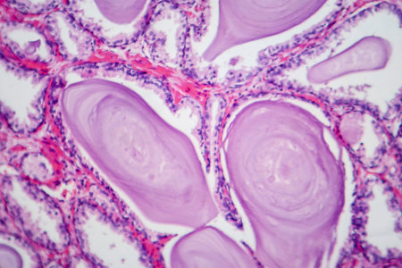
Photomicrograph showing histological features of benign prostatic hyperplasia. Enlarged prostate gland with nodular proliferation of glandular and stromal components.
Коллекция по умолчанию
Коллекция по умолчанию
Создать новую
Planarian parasite (flatworm) under microscope view.
Коллекция по умолчанию
Коллекция по умолчанию
Создать новую
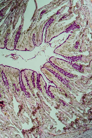
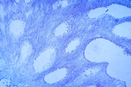
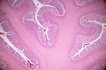
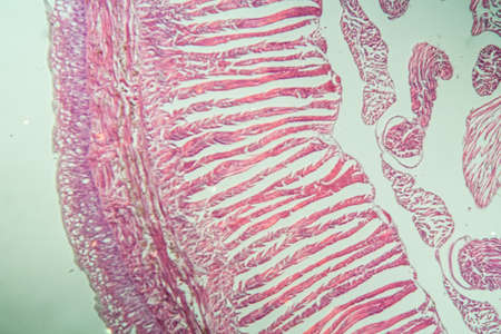
Cross-section through the intestine with glands 200x
Коллекция по умолчанию
Коллекция по умолчанию
Создать новую
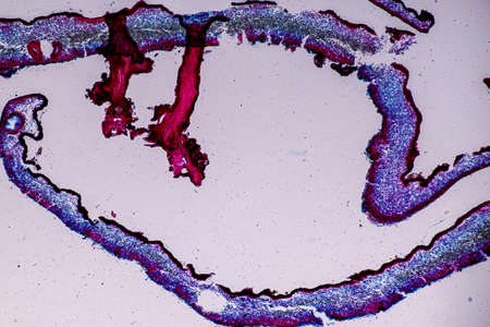
Transitional epithelium tissue of the urinary bladder under microscope, light micrograph, hematoxylin eosin staining
Коллекция по умолчанию
Коллекция по умолчанию
Создать новую
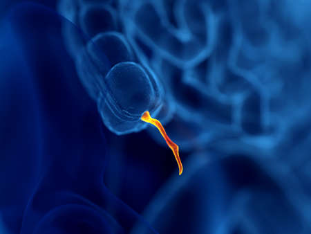
Human sperm in the testis morphology under microscope. Micrograph showing spermatozoon for pathology education.
Коллекция по умолчанию
Коллекция по умолчанию
Создать новую
Swirls of alcohol ink in lilac, lavender, violet are reminiscent of marble and ripples of agate. Fluid modern Art banners, ethereal graphic design.
Коллекция по умолчанию
Коллекция по умолчанию
Создать новую
Tooth development from human under microscope view for education.
Коллекция по умолчанию
Коллекция по умолчанию
Создать новую
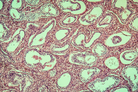
Cross section of human pancreas under microscope view for education in laboratory.
Коллекция по умолчанию
Коллекция по умолчанию
Создать новую

Anatomy and Histological Epididymis and Testis human cells under microscope.
Коллекция по умолчанию
Коллекция по умолчанию
Создать новую

science microscopy micrograph earthworm crosscutting
Коллекция по умолчанию
Коллекция по умолчанию
Создать новую
Reproductive parts: stigma, style, stamens, filament, petal. Structure floral- male reproductive organ of the flower of angiosperms
Коллекция по умолчанию
Коллекция по умолчанию
Создать новую

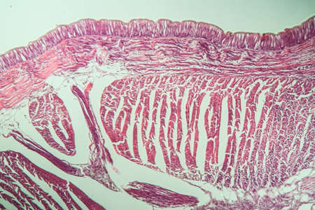
Education anatomy and Histological sample of Human under the microscope.
Коллекция по умолчанию
Коллекция по умолчанию
Создать новую

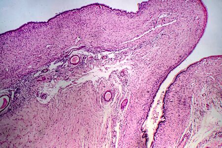
Histopathology of human under microscope view for education in laboratory.
Коллекция по умолчанию
Коллекция по умолчанию
Создать новую
Histopathology of cirrhosis, light micrograph, photo under microscope
Коллекция по умолчанию
Коллекция по умолчанию
Создать новую
Capture a close-up view of intricate microscopic structures within a high-efficiency solar cell, revealing the science behind capturing light --chaos 30 --ar 16:9 --v 6.1 Job ID: f7ac216d-5ddb-4e61-aef8-d497ef59608a
Коллекция по умолчанию
Коллекция по умолчанию
Создать новую
Uterine cancer, light micrograph, photo under microscope
Коллекция по умолчанию
Коллекция по умолчанию
Создать новую

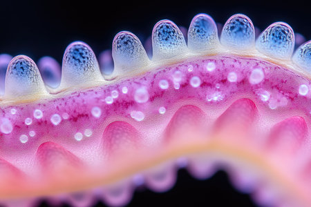
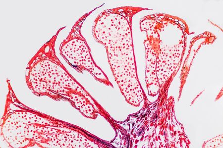
Tongue with taste buds Papilla across 100x
Коллекция по умолчанию
Коллекция по умолчанию
Создать новую
A microscopic view of tissue showing pink-stained cells and structures, indicative of biological samples, possibly related to histology or pathology
Коллекция по умолчанию
Коллекция по умолчанию
Создать новую
Abstract nature background- reproductive system flower. Ovary and ovule- plant cell.
Коллекция по умолчанию
Коллекция по умолчанию
Создать новую
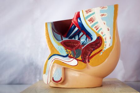
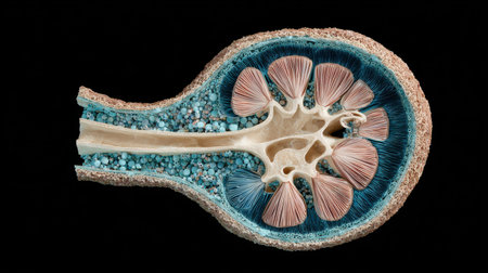
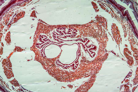
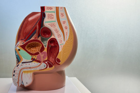
Male reproductive system model in half-cut perspective for education.
Коллекция по умолчанию
Коллекция по умолчанию
Создать новую

Prostate cancer, light micrograph, photo under microscope
Коллекция по умолчанию
Коллекция по умолчанию
Создать новую
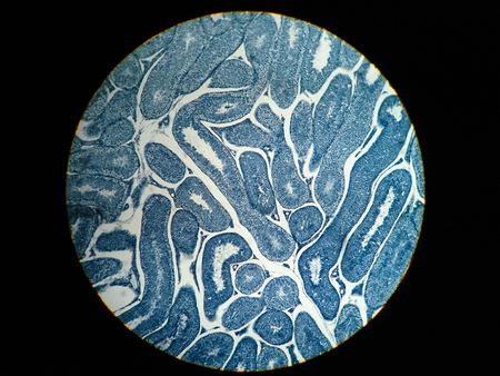
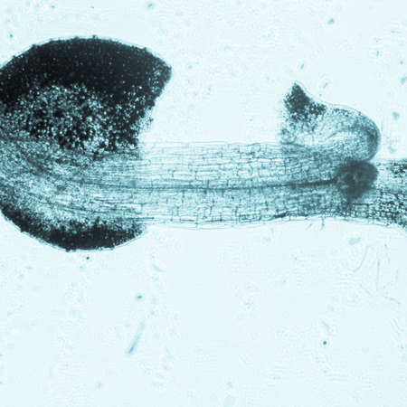
Earthworm histology cross section 10th segment 100x
Коллекция по умолчанию
Коллекция по умолчанию
Создать новую
Slime molds, as a group, are polyphyletic under the microscope for education.
Коллекция по умолчанию
Коллекция по умолчанию
Создать новую
Tissue of Stomach Human under the microscope in Lab.
Коллекция по умолчанию
Коллекция по умолчанию
Создать новую
Photomicrograph showing histological features of benign prostatic hyperplasia. Enlarged prostate gland with nodular proliferation of glandular and stromal components. High-resolution histology image.
Коллекция по умолчанию
Коллекция по умолчанию
Создать новую

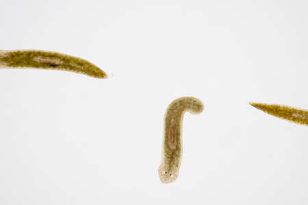
Planaria flatworm, under microscope view.(Soft focus)
Коллекция по умолчанию
Коллекция по умолчанию
Создать новую
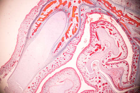
Histological Uterus human, Uterine tube human, Placenta human and Umbilical cord Human under the microscope for education.
Коллекция по умолчанию
Коллекция по умолчанию
Создать новую
Histology of human tissue, show tracheitis as seen under the microscope
Коллекция по умолчанию
Коллекция по умолчанию
Создать новую
Columnar epithelium of human gall bladder under the microscope in Lab.
Коллекция по умолчанию
Коллекция по умолчанию
Создать новую
Earthworm histology cross section 10th segment 100x
Коллекция по умолчанию
Коллекция по умолчанию
Создать новую

Papillary thyroid carcinoma, light micrograph, photo under microscope. The most common type of thyroid cancer
Коллекция по умолчанию
Коллекция по умолчанию
Создать новую

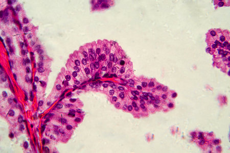
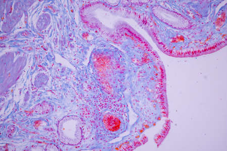
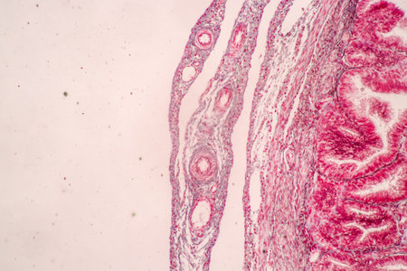
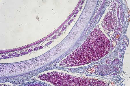
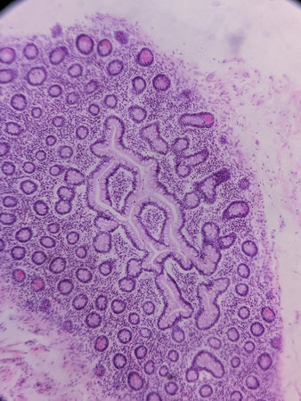
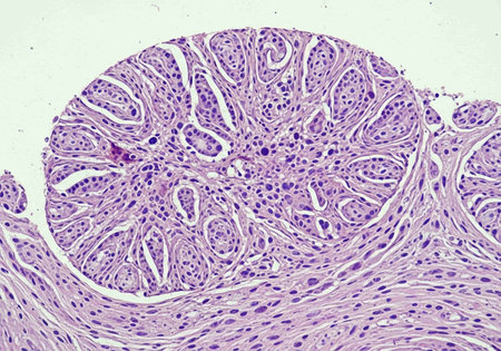
Education of Uterine tube under the microscope in Lab.
Коллекция по умолчанию
Коллекция по умолчанию
Создать новую
Ovarian cyst, light micrograph, photo under microscope
Коллекция по умолчанию
Коллекция по умолчанию
Создать новую
science botany micrograph plant arabidopsis thaliana root tissue micro
Коллекция по умолчанию
Коллекция по умолчанию
Создать новую

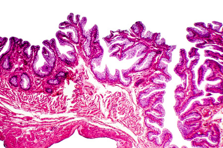
Tissue of Small intestine (Duodenum) and Vermiform appendix Human under the microscope in Lab.
Коллекция по умолчанию
Коллекция по умолчанию
Создать новую
Photomicrograph showing histological features of benign prostatic hyperplasia. Enlarged prostate gland with nodular proliferation of glandular and stromal components.
Коллекция по умолчанию
Коллекция по умолчанию
Создать новую
Diagram showing the structure of the blood vessels in the human body
Коллекция по умолчанию
Коллекция по умолчанию
Создать новую
Microscope view of Obelia hydroid, a marine and sometimes freshwater animal.
Коллекция по умолчанию
Коллекция по умолчанию
Создать новую
Detailed observation of a human arterys cross section showcasing colorful layers and cellular structures in a laboratory setting.
Коллекция по умолчанию
Коллекция по умолчанию
Создать новую
Cross-section through the lichen symbiote body 100x
Коллекция по умолчанию
Коллекция по умолчанию
Создать новую
Tongue Tissue with taste buds across 200x
Коллекция по умолчанию
Коллекция по умолчанию
Создать новую
Blue paint strokes on white canvas create abstract design with two elongated shapes and textured brush marks showing paint application technique and color contrast
Коллекция по умолчанию
Коллекция по умолчанию
Создать новую
3d rendered illustration of the human appendix
Коллекция по умолчанию
Коллекция по умолчанию
Создать новую
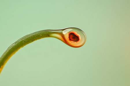
Up-close perspective highlights a curved green plant stem culminating in a rounded, vivid red seed encased in transparent gel, set against a pastel background.
Коллекция по умолчанию
Коллекция по умолчанию
Создать новую
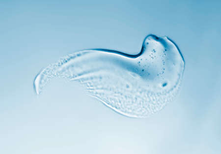
Transparent gel with bubbles close-up. Smear of face gel cream. The texture of gel cream. A sample of a cosmetic product. Antibacterial gel.
Коллекция по умолчанию
Коллекция по умолчанию
Создать новую
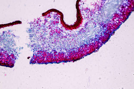
Cross-section leaf Plant of under the microscope for classroom education.
Коллекция по умолчанию
Коллекция по умолчанию
Создать новую
The study of tissue samples of Trachea of Cat, Epididymis, Prostate, Uterus with embryo of rat and Mammary gland cow under the microscope in Lab.
Коллекция по умолчанию
Коллекция по умолчанию
Создать новую

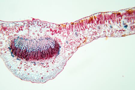
Horse chestnut with flower bud in cross section 100x
Коллекция по умолчанию
Коллекция по умолчанию
Создать новую

Histological Spermatic cord human, Seminal vesicle human, Prostate human and Human chromosomes under the microscope for education.
Коллекция по умолчанию
Коллекция по умолчанию
Создать новую
Flower with pollen along 100x
Коллекция по умолчанию
Коллекция по умолчанию
Создать новую
Cell Gene Microscopic Series
Коллекция по умолчанию
Коллекция по умолчанию
Создать новую
squamous cell
Коллекция по умолчанию
Коллекция по умолчанию
Создать новую
Planarian parasite (flatworm) under microscope view.
Коллекция по умолчанию
Коллекция по умолчанию
Создать новую
Histopathology of human under microscope view for education in laboratory.
Коллекция по умолчанию
Коллекция по умолчанию
Создать новую
Light micrograph of human pancreas, light micrograph, photo under microscope
Коллекция по умолчанию
Коллекция по умолчанию
Создать новую
Pancreas cancer cell under microscope view for medical education.
Коллекция по умолчанию
Коллекция по умолчанию
Создать новую

cells biology- endosperm weevil rye
Коллекция по умолчанию
Коллекция по умолчанию
Создать новую
Histopathology of Human liver under microscope view, light micrograph
Коллекция по умолчанию
Коллекция по умолчанию
Создать новую

Condyloma acuminatum, also known as genital warts. Light micrograph, photo under microscope
Коллекция по умолчанию
Коллекция по умолчанию
Создать новую
Anatomical Model of Human Respiratory and Digestive Systems for education and demonstrations
Коллекция по умолчанию
Коллекция по умолчанию
Создать новую
Moniliformis dubius in the Intestine of rat, intermediate host
Коллекция по умолчанию
Коллекция по умолчанию
Создать новую
This visual representation reveals a cross-section of a plant stem, displaying the intricate structures essential for plant physiology.
Коллекция по умолчанию
Коллекция по умолчанию
Создать новую

Johannes berry fruit cross 100x
Коллекция по умолчанию
Коллекция по умолчанию
Создать новую
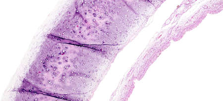
science micrograph of bone cell osteocyte
Коллекция по умолчанию
Коллекция по умолчанию
Создать новую
Taste buds in foliate tongue papillae. Many of them show the taste or gustatory pore. Hematoxylin & eosin stain.
Коллекция по умолчанию
Коллекция по умолчанию
Создать новую
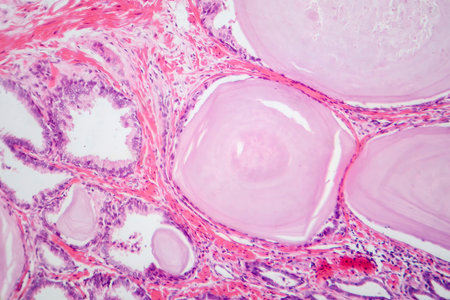
Benign prostatic hyperplasia. Micrograph shows dilated glands, papillary projections inside the lumen of the glands, cystic dilatation with accumulation of secretory material. Photo under microscope
Коллекция по умолчанию
Коллекция по умолчанию
Создать новую
Juicy Algae Extract in Nature's Garden
Коллекция по умолчанию
Коллекция по умолчанию
Создать новую

Photomicrograph, cross section of Hirudo medicinalis, the medicinal leech. Focus = the center crop. Uneven focus elsewhere due to speciman thickness. 14MP camera and microscope. Isolated.
Коллекция по умолчанию
Коллекция по умолчанию
Создать новую
Blue paint strokes on white background show two elongated shapes with different widths and heights, creating textured patterns for artistic applications
Коллекция по умолчанию
Коллекция по умолчанию
Создать новую
Prostate cancer, light micrograph, photo under microscope
Коллекция по умолчанию
Коллекция по умолчанию
Создать новую
Legion-Media
Создайте свои проекты на основе качественных стоковых фотографий и видео.


Copyright © Legion-Media.