
tongue with ulcers of adult man.Cracks in the tongue of a young man.man with halitosis for candida albicans on tongue.
Коллекция по умолчанию
Коллекция по умолчанию
Создать новую
Tongue Tissue with taste buds across 200x
Коллекция по умолчанию
Коллекция по умолчанию
Создать новую
Columnar epithelium of human gall bladder under the microscope in Lab.
Коллекция по умолчанию
Коллекция по умолчанию
Создать новую
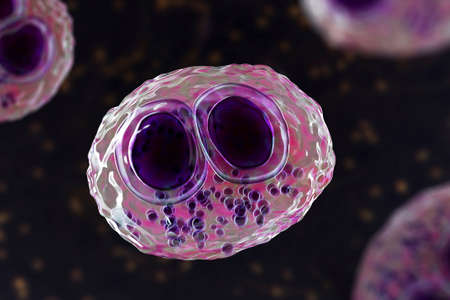
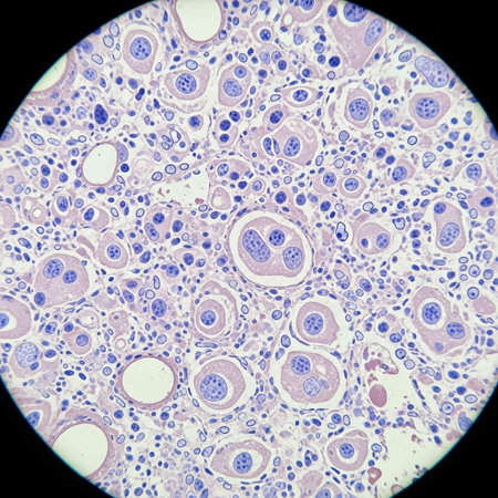
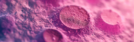
Cytomegalovirus CMV in a human cell, owl's eye inclusion in nucleus, multinucleated cell, 3D illustration. It is herpes virus, causes diseases in fetus, organ transplant patients, HIV infected people
Коллекция по умолчанию
Коллекция по умолчанию
Создать новую
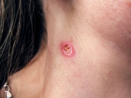
A wound on a woman’s neck after burning a wart, close-up. Round painful hole in human skin, purulent wound
Коллекция по умолчанию
Коллекция по умолчанию
Создать новую

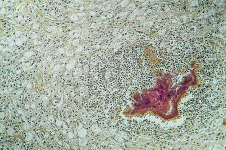
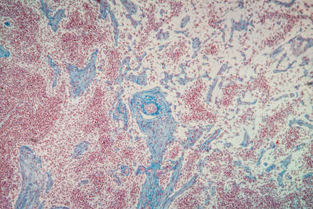
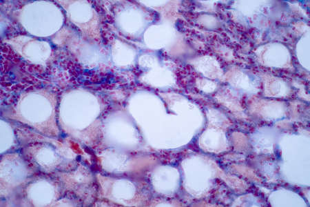
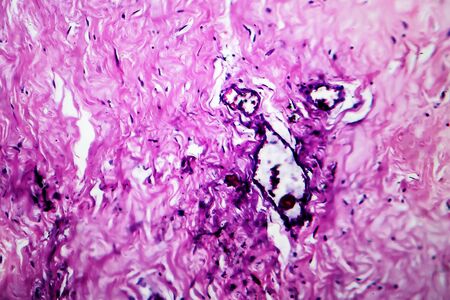
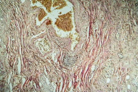
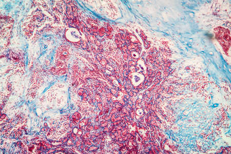
Thyroid follicular carcinoma, light micrograph, photo under microscope
Коллекция по умолчанию
Коллекция по умолчанию
Создать новую
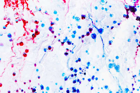
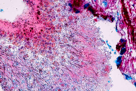
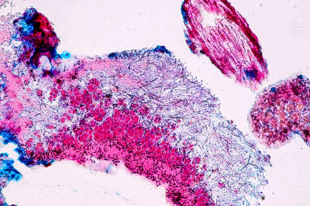
Actinomyces in the jaw diseased tissue 200x
Коллекция по умолчанию
Коллекция по умолчанию
Создать новую
Chaos ink texture background, ink in water pattern frost. Crystal winter design
Коллекция по умолчанию
Коллекция по умолчанию
Создать новую
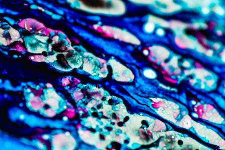
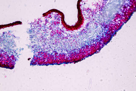
Characteristics of Lichen, hyphae and Symbiotic algae under the microscope for education.
Коллекция по умолчанию
Коллекция по умолчанию
Создать новую

Bladder cancer, light micrograph, photo under microscope
Коллекция по умолчанию
Коллекция по умолчанию
Создать новую
Characteristics of Lichen, hyphae and Symbiotic algae under the microscope for education.
Коллекция по умолчанию
Коллекция по умолчанию
Создать новую
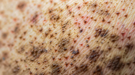
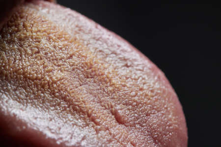
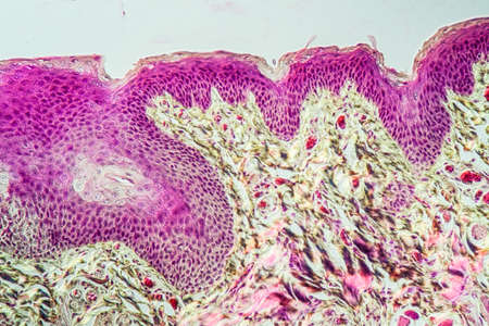
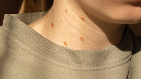
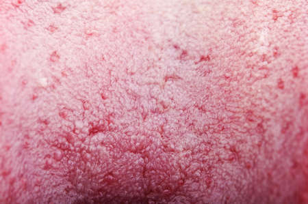
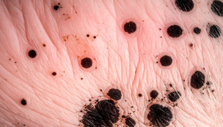
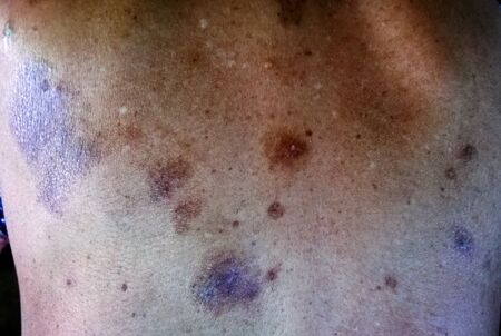
The image shows a close up of skin with a textured pattern. The skin is light brown and has many small, brown spots and a few red marks. The surface is textured with small bumps and ridges.
Коллекция по умолчанию
Коллекция по умолчанию
Создать новую

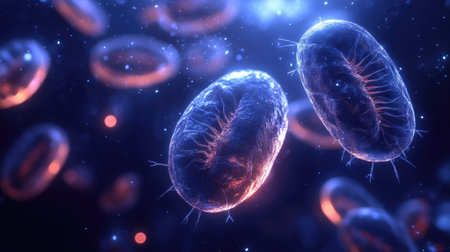
Blood vessel with flowing blood cells, 3D illustration. Small blood vessels, capillaries
Коллекция по умолчанию
Коллекция по умолчанию
Создать новую
Histopathology of human liver under microscope view for medical education.
Коллекция по умолчанию
Коллекция по умолчанию
Создать новую

Itch mites under the skin cross-section 100x
Коллекция по умолчанию
Коллекция по умолчанию
Создать новую

Tongue with taste buds Papilla across 100x
Коллекция по умолчанию
Коллекция по умолчанию
Создать новую
Lymph node tissue under the microscope 100x
Коллекция по умолчанию
Коллекция по умолчанию
Создать новую
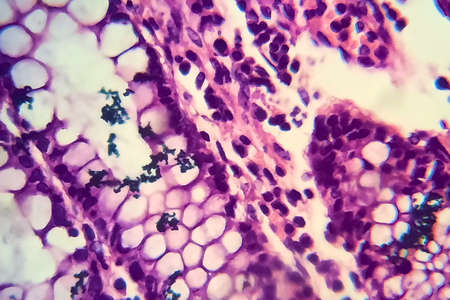
Bacillary dysentery, light micrograph, photo under microscope showing presence of bacteria and accumulation of inflammatory cells in intestinal epithelium
Коллекция по умолчанию
Коллекция по умолчанию
Создать новую

Condyloma acuminatum, also known as genital warts. Light micrograph, photo under microscope
Коллекция по умолчанию
Коллекция по умолчанию
Создать новую

Characteristics of Lichen, hyphae and Symbiotic algae under the microscope for education.
Коллекция по умолчанию
Коллекция по умолчанию
Создать новую

old woman skin
Коллекция по умолчанию
Коллекция по умолчанию
Создать новую

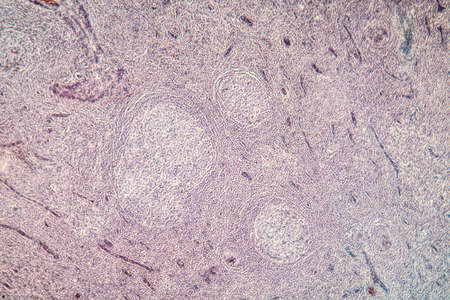
Ovarian cancer, light micrograph, photo under microscope. Photograph shows a fragment of a cancerous tumor in the female ovary. Selective focus
Коллекция по умолчанию
Коллекция по умолчанию
Создать новую

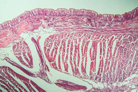
Stomach tissue under the microscope 100x
Коллекция по умолчанию
Коллекция по умолчанию
Создать новую
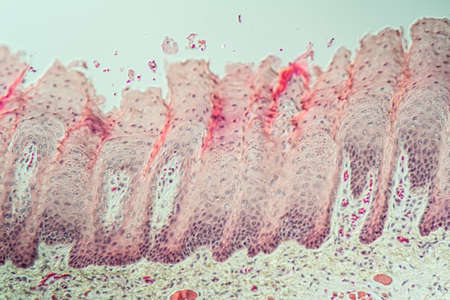
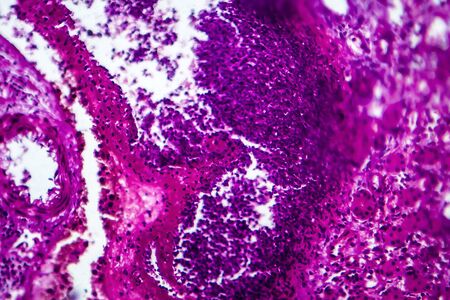
Human lung pathology under light microscope, The lungs is organs of the respiratory system in humans. Human pathology education. Haematoxylin and eosin staining technique slide.
Коллекция по умолчанию
Коллекция по умолчанию
Создать новую
Tongue Tissue with taste buds across 200x
Коллекция по умолчанию
Коллекция по умолчанию
Создать новую
Tissue of Stomach Human under the microscope in Lab.
Коллекция по умолчанию
Коллекция по умолчанию
Создать новую
Lungworm under the microscope 100x
Коллекция по умолчанию
Коллекция по умолчанию
Создать новую

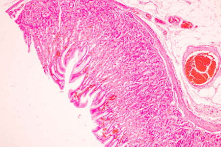
Scalp and hair follicles of human under the microscope in Lab.
Коллекция по умолчанию
Коллекция по умолчанию
Создать новую

Dry Man Hand With Skin Peeling Off
Коллекция по умолчанию
Коллекция по умолчанию
Создать новую
Molluscum contagiosum virus, 3D illustration. A virus from Poxvirus family, causes skin infection with numerous small raised lesions
Коллекция по умолчанию
Коллекция по умолчанию
Создать новую
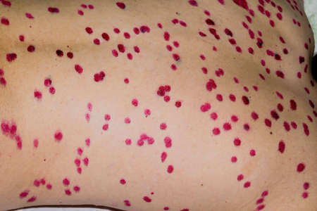
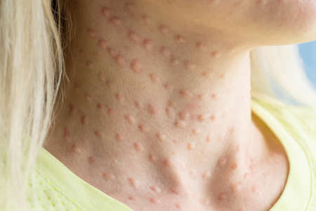
Papules on the skin of a patient with chickenpox close-up. Varicella Zoster virus.
Коллекция по умолчанию
Коллекция по умолчанию
Создать новую

parasite eggs.
Коллекция по умолчанию
Коллекция по умолчанию
Создать новую

a close up of the inside of a breast
Коллекция по умолчанию
Коллекция по умолчанию
Создать новую
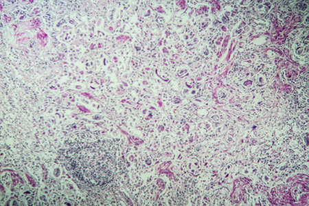
AIDS with fungi 200x infected tissue
Коллекция по умолчанию
Коллекция по умолчанию
Создать новую
Microbes of different shapes, 3D illustration. Group of microorganisms. Digital art. Picture render by neural network.
Коллекция по умолчанию
Коллекция по умолчанию
Создать новую
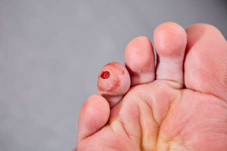
Laser removal of wart from the toe of foot. Cut out verruca.
Коллекция по умолчанию
Коллекция по умолчанию
Создать новую
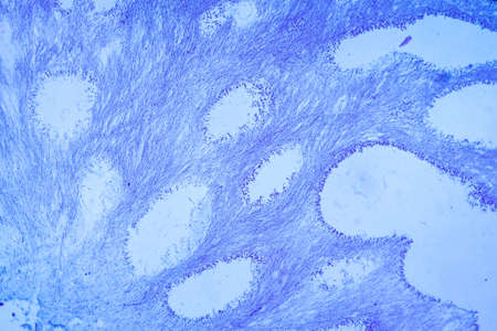
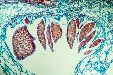
Anatomy and Histological Ovary, Testis and Sperm human cells under microscope.
Коллекция по умолчанию
Коллекция по умолчанию
Создать новую
Blue and pink refill ink spilled onto the white washbasin and the ink mixed into abstract blobs and patterns.
Коллекция по умолчанию
Коллекция по умолчанию
Создать новую

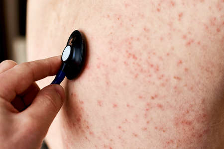
Doctor hands in gloves examining red rash on patient skin closeup
Коллекция по умолчанию
Коллекция по умолчанию
Создать новую

Human hyaline cartilage bone under microscope view for education pathology. Human tissue.
Коллекция по умолчанию
Коллекция по умолчанию
Создать новую

Parasitic protozoans Toxoplasma gondii, the causative agent of toxoplasmosis in tachyzoite stage, 3D illustration
Коллекция по умолчанию
Коллекция по умолчанию
Создать новую
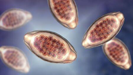
Eggs of parasitic roundworm Trichuris trichiura, or whipworm, the causative agent of trichuriasis, disease of a human large intestine, 3D illustration
Коллекция по умолчанию
Коллекция по умолчанию
Создать новую
Breast fibroadenosis, light micrograph, photo under microscope. Common benign hyperplastic process involving breast glands
Коллекция по умолчанию
Коллекция по умолчанию
Создать новую

Allergy concept. Young woman with pimples on the chest, closeup
Коллекция по умолчанию
Коллекция по умолчанию
Создать новую
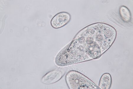
Paramecium caudatum is a genus of unicellular ciliated protozoan and Bacterium under the microscope.
Коллекция по умолчанию
Коллекция по умолчанию
Создать новую
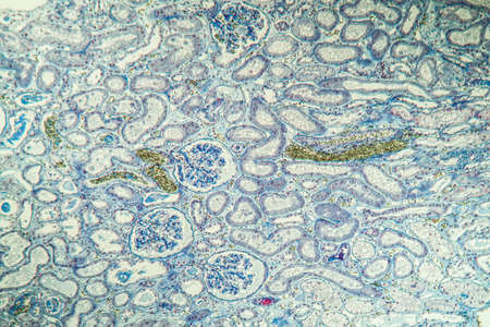
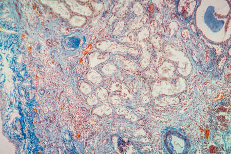
science medical anthropotomy physiology microscopic section of human kidney tissue background
Коллекция по умолчанию
Коллекция по умолчанию
Создать новую
Tissue of Stomach Human under the microscope in Lab.
Коллекция по умолчанию
Коллекция по умолчанию
Создать новую
Histopathology of acute nephritis, light micrograph, photo under microscope
Коллекция по умолчанию
Коллекция по умолчанию
Создать новую
Moniliformis dubius in the Intestine of rat, intermediate host
Коллекция по умолчанию
Коллекция по умолчанию
Создать новую
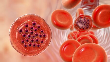
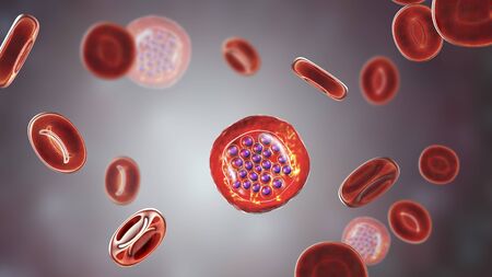
The malaria-infected red blood cells. 3D illustration showing parasite Plasmodium falciparum in schizont stage inside red blood cells, the causative agent of tropical malaria
Коллекция по умолчанию
Коллекция по умолчанию
Создать новую

Closeup of a man protruding tongue.
Коллекция по умолчанию
Коллекция по умолчанию
Создать новую
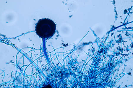
Aspergillus niger and Aspergillus oryzae (mold) under microscope for Microbiology in Lab.
Коллекция по умолчанию
Коллекция по умолчанию
Создать новую
water flowing over boulders, rocks, and stones
Коллекция по умолчанию
Коллекция по умолчанию
Создать новую

Tooth development from human under microscope view for education.
Коллекция по умолчанию
Коллекция по умолчанию
Создать новую
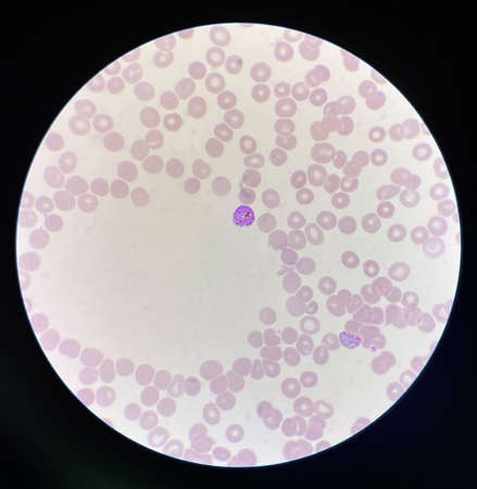
Malaria blood parasite infected red blood cells laboratory background.
Коллекция по умолчанию
Коллекция по умолчанию
Создать новую
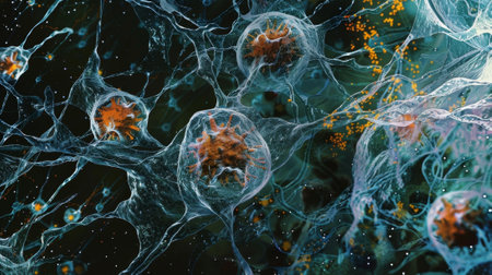
The activation of TLRs leading to the production of proinflammatory cytokines as seen in a micrograph of immune cells
Коллекция по умолчанию
Коллекция по умолчанию
Создать новую
Fibrin deposits in the kidney, microscopy 100x
Коллекция по умолчанию
Коллекция по умолчанию
Создать новую

Apple pollen from a blossom in spring under the microscope
Коллекция по умолчанию
Коллекция по умолчанию
Создать новую

Real biorevitalization of the skin on a white background. Traces of injections of biorevitalization on the face of a woman. Traces of biorevitalization needles.
Коллекция по умолчанию
Коллекция по умолчанию
Создать новую

Skin lesions on the leg. Symptoms characteristic of the elderly begin with a red rash in a small circle and spread to a wider area. Large scabs on the legs. Elderly care. Consequences of diabetes
Коллекция по умолчанию
Коллекция по умолчанию
Создать новую
Drops of blood on a white background
Коллекция по умолчанию
Коллекция по умолчанию
Создать новую
destructive mushroom in wood fabric 100x
Коллекция по умолчанию
Коллекция по умолчанию
Создать новую
Palatal tonsils transverse 100x under a microscope
Коллекция по умолчанию
Коллекция по умолчанию
Создать новую
Allergic rash on the skin. Woman with dermatology problem on back skin
Коллекция по умолчанию
Коллекция по умолчанию
Создать новую
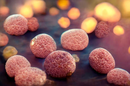
An intricate digital representation of bacteria displaying vibrant colors and glowing elements, highlighting the fascinating world of microscopic life in a stunning abstract setting.
Коллекция по умолчанию
Коллекция по умолчанию
Создать новую
Birthmarks on skin Close up detail of the bare skin Sun Exposure effect on skin, Health Effects of UV Radiation Woman with birthmarks Pigmentation and lot of birthmarks.
Коллекция по умолчанию
Коллекция по умолчанию
Создать новую
Abstract ink design template mixed texture background. Blue abstract texture. Multicolored liquid marble pattern. Fluid Art
Коллекция по умолчанию
Коллекция по умолчанию
Создать новую
Papules after beauty injections on the face. Closeup photo
Коллекция по умолчанию
Коллекция по умолчанию
Создать новую
Microscopic view of human cells under microscope.
Коллекция по умолчанию
Коллекция по умолчанию
Создать новую

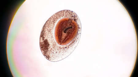
Opisthorchis viverrini egg in stool under microscopic
Коллекция по умолчанию
Коллекция по умолчанию
Создать новую

Ringworm on the body. It is caused by a fungus,High Dynamic Range tone
Коллекция по умолчанию
Коллекция по умолчанию
Создать новую
closeup of tongue structure background
Коллекция по умолчанию
Коллекция по умолчанию
Создать новую

Histopathology of human under microscope view for education in laboratory.
Коллекция по умолчанию
Коллекция по умолчанию
Создать новую

Tongue Tissue with taste buds across 100x
Коллекция по умолчанию
Коллекция по умолчанию
Создать новую
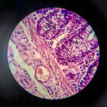
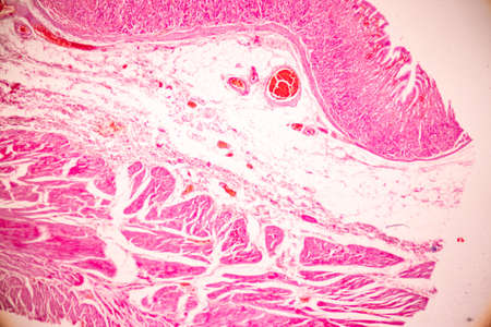
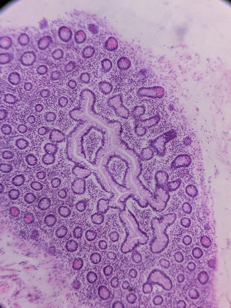
Colon tissue with diverticulum 100x
Коллекция по умолчанию
Коллекция по умолчанию
Создать новую
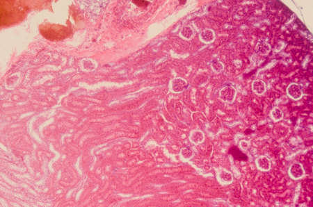
Atrophy kidney tissue under the microscope 100x
Коллекция по умолчанию
Коллекция по умолчанию
Создать новую
A detailed macro view captures the varied landscape of human skin, highlighting multiple nevi and moles. This image serves as a powerful visual for dermatology, skin cancer awareness, and the importance of regular health screenings.
Коллекция по умолчанию
Коллекция по умолчанию
Создать новую
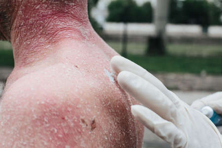
A dermatologist wearing gloves examines the skin of a sick patient. Examination and diagnosis of skin diseases-allergies, psoriasis, eczema, dermatitis. The doctor applies the cream to the skin
Коллекция по умолчанию
Коллекция по умолчанию
Создать новую
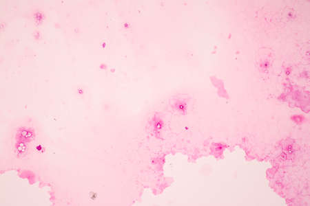
Abstract marbling floral pattern for fabric, tile design. background texture
Коллекция по умолчанию
Коллекция по умолчанию
Создать новую

Skin damaged by insect bites. Close up. Skin with many red spot and scar from sand fly bites
Коллекция по умолчанию
Коллекция по умолчанию
Создать новую
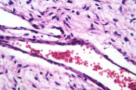
A small blood vessel with red blood cells in neurofibroma tissue sample, light photomicrograph.
Коллекция по умолчанию
Коллекция по умолчанию
Создать новую
Characteristics of Lichen, hyphae and Symbiotic algae under the microscope for education.
Коллекция по умолчанию
Коллекция по умолчанию
Создать новую

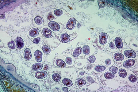
Egg of Ascaris lumbricoides (roundworm) in human stool, analyze by microscope, 400x
Коллекция по умолчанию
Коллекция по умолчанию
Создать новую
Dry scars on the back of an old woman, tan separately on a white background, damage caused by severe itchy dermatitis, 10 March 2020
Коллекция по умолчанию
Коллекция по умолчанию
Создать новую
Earthworm histology cross section 10th segment 100x
Коллекция по умолчанию
Коллекция по умолчанию
Создать новую
Worms in infected liver 100x
Коллекция по умолчанию
Коллекция по умолчанию
Создать новую
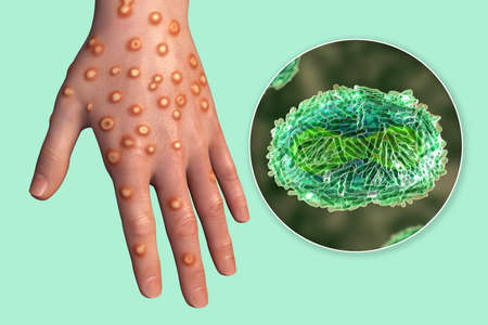
Hand of a patient with monkeypox infection, 3D illustration. Monkeypox is a zoonotic virus from Poxviridae family, causes monkeypox, a pox-like disease
Коллекция по умолчанию
Коллекция по умолчанию
Создать новую
Fibroepithelium Diseased tissue 100x
Коллекция по умолчанию
Коллекция по умолчанию
Создать новую

Trichinella spiralis larvae in muscle tissue under the microscope. Trichinella spiralis is a nematode parasite responsible for trichosis and affecting mammals.
Коллекция по умолчанию
Коллекция по умолчанию
Создать новую

Histopathology of alcoholic hepatitis, light micrograph, photo under microscope. High magnification
Коллекция по умолчанию
Коллекция по умолчанию
Создать новую
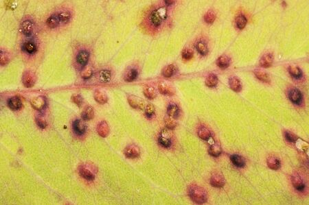
Parasitization of a green leaf by fungi Puccinia commonly called plant rust orange color natural lighting
Коллекция по умолчанию
Коллекция по умолчанию
Создать новую
The malaria-infected red blood cells. 3D illustration showing malaria parasite Plasmodium falciparum in schizont stage inside red blood cells, the causative agent of tropical malaria
Коллекция по умолчанию
Коллекция по умолчанию
Создать новую

Air and oil bubbles inside water base form patterns
Коллекция по умолчанию
Коллекция по умолчанию
Создать новую
Nestwurz orchid root cross 100x
Коллекция по умолчанию
Коллекция по умолчанию
Создать новую
Bacillary dysentery, light micrograph, photo under microscope showing presence of bacteria and accumulation of inflammatory cells in intestinal epithelium
Коллекция по умолчанию
Коллекция по умолчанию
Создать новую

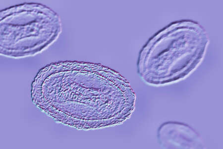
Ascaris lumbricoides, a large roundworm, fertilized egg, 3D illustration
Коллекция по умолчанию
Коллекция по умолчанию
Создать новую
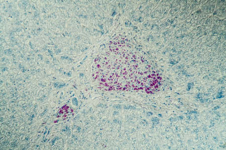
Pancreas cancer cell under microscope view for medical education.
Коллекция по умолчанию
Коллекция по умолчанию
Создать новую
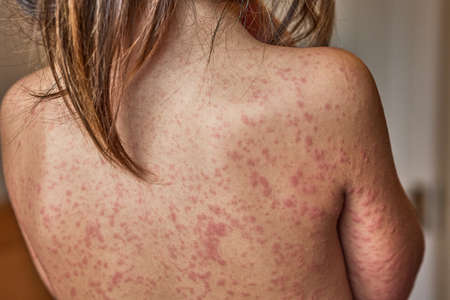
allergic rash on the body of the patient. 5 year old girl.
Коллекция по умолчанию
Коллекция по умолчанию
Создать новую
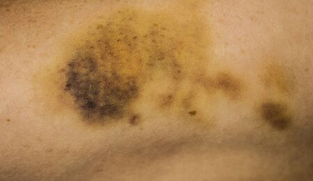
Violence victim with a bruise on her arm
Коллекция по умолчанию
Коллекция по умолчанию
Создать новую
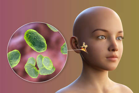
Haemophilus influenzae bacterium as a cause of otitis media. 3D illustration showing purulent inflammation of the middle ear in a girl and close-up view of Haemophilus bacteria
Коллекция по умолчанию
Коллекция по умолчанию
Создать новую
Legion-Media
Создайте свои проекты на основе качественных стоковых фотографий и видео.


Copyright © Legion-Media.