
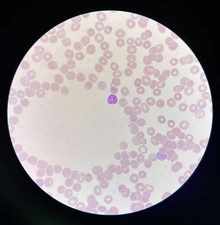
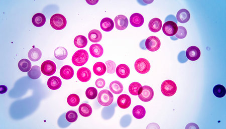
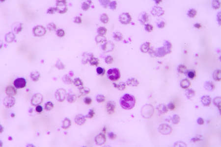
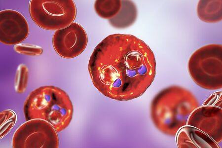
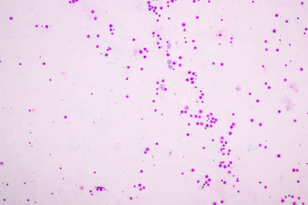
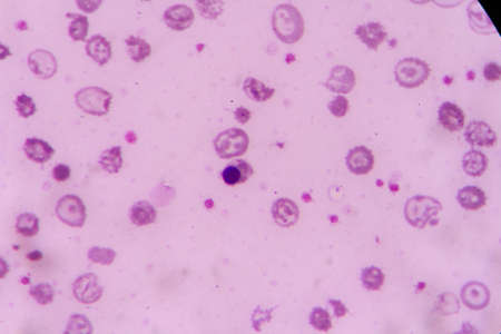
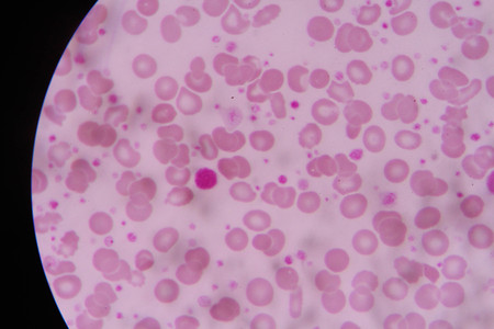
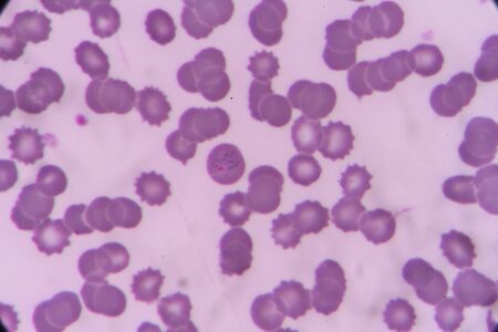
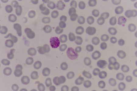
Malaria blood parasite infected red blood cells laboratory background.
Коллекция по умолчанию
Коллекция по умолчанию
Создать новую
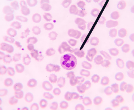
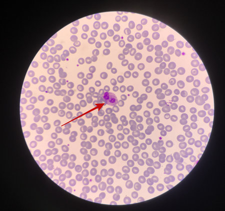
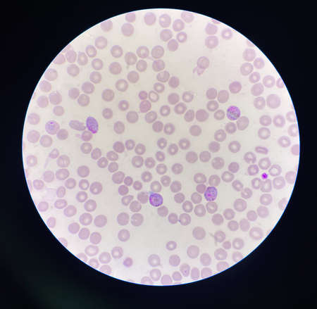
Neutrophil show in blood smear CBC test find with microscope.
Коллекция по умолчанию
Коллекция по умолчанию
Создать новую
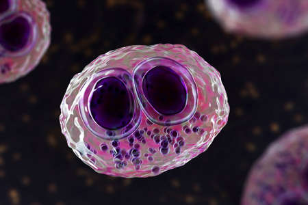
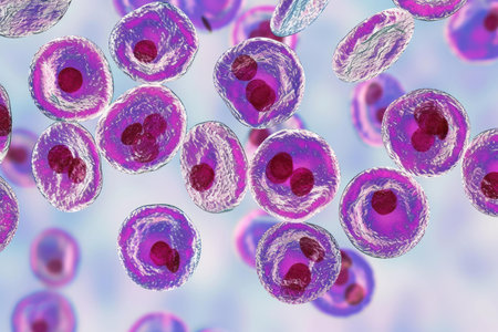
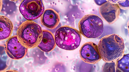
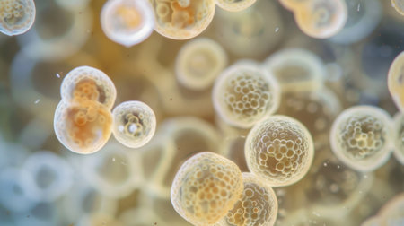
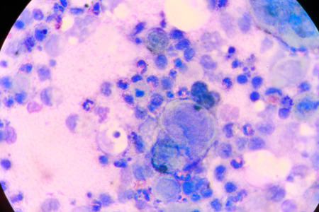
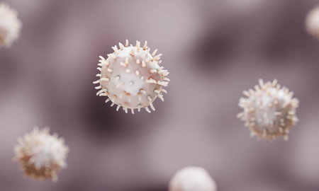
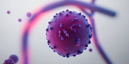
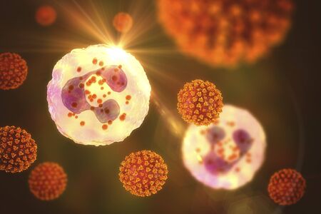
Cytomegalovirus CMV in a human cell, owl's eye inclusion in nucleus, multinucleated cell, 3D illustration. It is herpes virus, causes diseases in fetus, organ transplant patients, HIV infected people
Коллекция по умолчанию
Коллекция по умолчанию
Создать новую

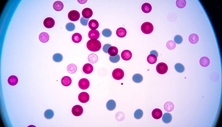
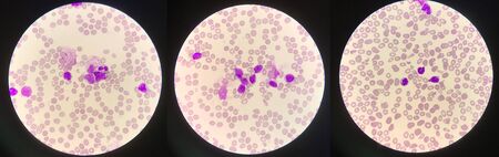
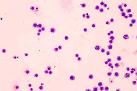
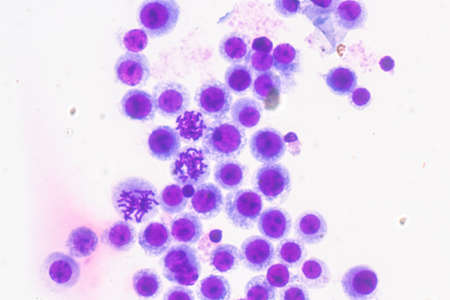
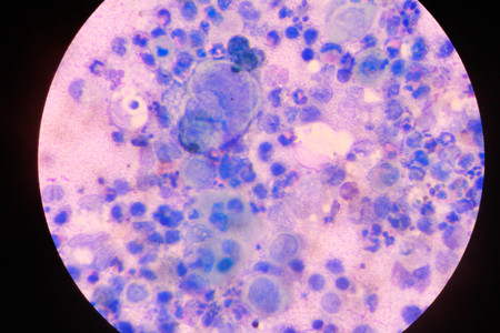
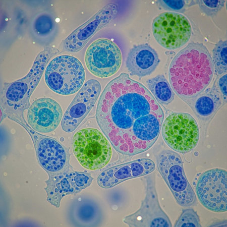
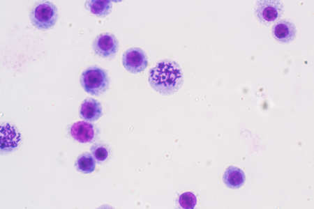
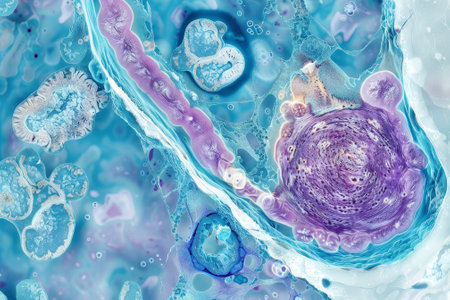
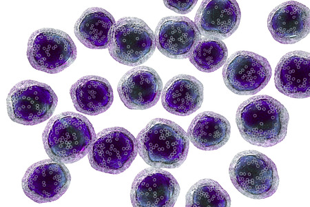
Picture of acute lymphocytic leukemia or ALL cells in blood smear, analyze by microscope, 400x
Коллекция по умолчанию
Коллекция по умолчанию
Создать новую
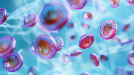
Blood smear with red blood cells in human body, medical background.
Коллекция по умолчанию
Коллекция по умолчанию
Создать новую
Microscope with metal lens at laboratory. Medical equipment.
Коллекция по умолчанию
Коллекция по умолчанию
Создать новую
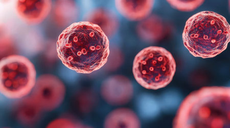
Immature white blood cells in leukemia.Science concept.
Коллекция по умолчанию
Коллекция по умолчанию
Создать новую
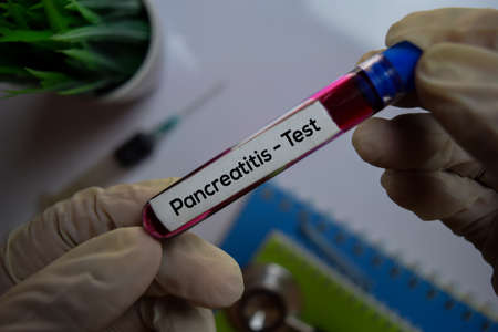
Pancreatitis - Test with blood sample. Top view isolated on office desk. Healthcare/Medical concept
Коллекция по умолчанию
Коллекция по умолчанию
Создать новую
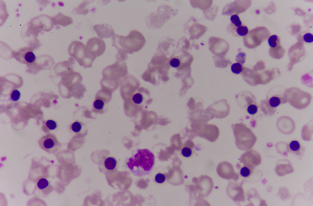
nucleated red cell
Коллекция по умолчанию
Коллекция по умолчанию
Создать новую

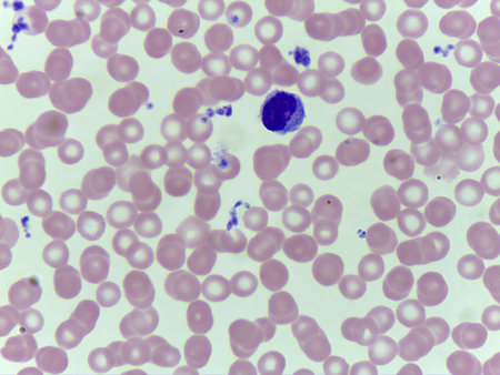
neutrophils. blood smear is often used as a follow-up test to abnormal results on a complete blood count (CBC) to evaluate the different types of blood cells.
Коллекция по умолчанию
Коллекция по умолчанию
Создать новую

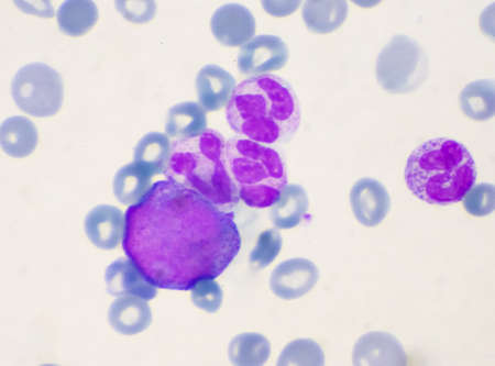
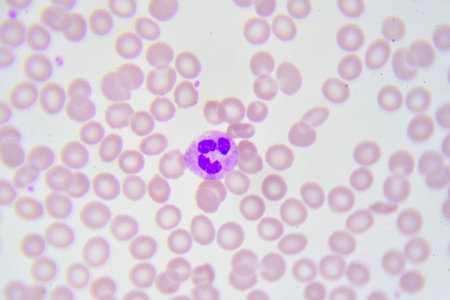
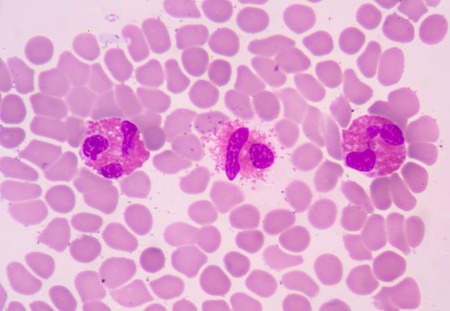
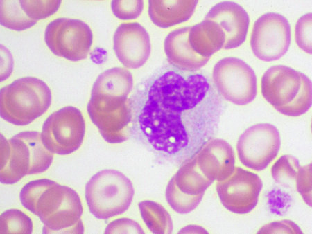
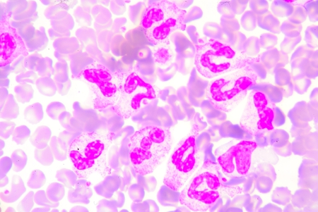
Blood smear showing, in the center, three neutrophil with hypersegmented nucleus. These cells appear in pathological situations such as megaloblastic anemias. Wright stain.
Коллекция по умолчанию
Коллекция по умолчанию
Создать новую
Blood cells in human body under microscope view for education in laboratory.
Коллекция по умолчанию
Коллекция по умолчанию
Создать новую
Chromosomes Human under the microscope for education.
Коллекция по умолчанию
Коллекция по умолчанию
Создать новую
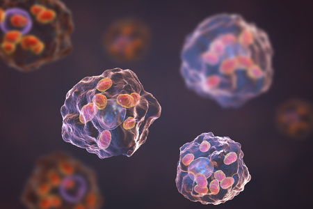
Macrophages infected by Leishmania amastigotes, 3D illustration
Коллекция по умолчанию
Коллекция по умолчанию
Создать новую
Chromosomes Human under the microscope for education.
Коллекция по умолчанию
Коллекция по умолчанию
Создать новую
nucleated red cell
Коллекция по умолчанию
Коллекция по умолчанию
Создать новую

Chronic myeloid leukemia cells or CML, analyze by microscope, original magnification 1000x
Коллекция по умолчанию
Коллекция по умолчанию
Создать новую
Microscopic close-up of vibrant stained human cells on a blue backdrop
Коллекция по умолчанию
Коллекция по умолчанию
Создать новую
Chromosomes Human under the microscope for education.
Коллекция по умолчанию
Коллекция по умолчанию
Создать новую
Immature cells in myeloid serie myelocyte metamyelocyte.
Коллекция по умолчанию
Коллекция по умолчанию
Создать новую
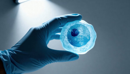
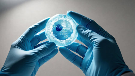
Gloved hand holds a translucent cell model under focused laboratory light with visible nucleus and organelle structures, suggesting scientific research and educational demonstration, with empty background space available for text
Коллекция по умолчанию
Коллекция по умолчанию
Создать новую
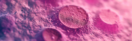
The malaria-infected red blood cells. 3D illustration showing ring-form trophozoites of malaria parasite Plasmodium falciparum inside red blood cells, the causative agent of tropical malaria
Коллекция по умолчанию
Коллекция по умолчанию
Создать новую
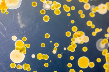
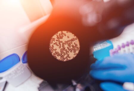
Backgrounds of Characteristics and Different shaped Colony of Bacteria and Mold growing on agar plates from Soil samples for education in Microbiology laboratory.
Коллекция по умолчанию
Коллекция по умолчанию
Создать новую

Gloved hands hold a translucent cell culture sample with visible cellular structures and bubbles under focused clinical light, indicating laboratory research and microscopy, with available space for text on a neutral background
Коллекция по умолчанию
Коллекция по умолчанию
Создать новую
Anatomy and Histological Ovary, Testis and Sperm human cells under microscope.
Коллекция по умолчанию
Коллекция по умолчанию
Создать новую
multinucleated giant
Коллекция по умолчанию
Коллекция по умолчанию
Создать новую

Meningococcal meningitis, cerebrospinal fluid smear containing neutrophils with and without bacteria Neisseria meningitidis
Коллекция по умолчанию
Коллекция по умолчанию
Создать новую

Neutrophil cell (white blood cell) in peripheral blood smear
Коллекция по умолчанию
Коллекция по умолчанию
Создать новую
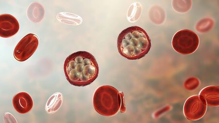
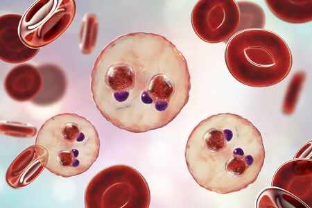
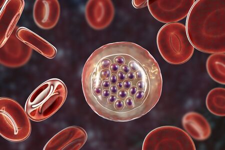
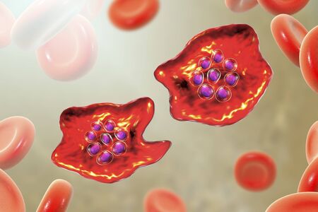
Red blood cells infected with malaria parasite, 3D illustration showing Plasmodium parasites inside red blood cells in the stage of schizont
Коллекция по умолчанию
Коллекция по умолчанию
Создать новую
Red blood cells infected with malaria parasite Plasmodium vivax, schizont stage, 3D illustration
Коллекция по умолчанию
Коллекция по умолчанию
Создать новую
Microscopic View Rendered Image of Abnormal, Diseased Cells in Biology and Medicine Illustration
Коллекция по умолчанию
Коллекция по умолчанию
Создать новую
Blood smear showing white and red blood cells
Коллекция по умолчанию
Коллекция по умолчанию
Создать новую
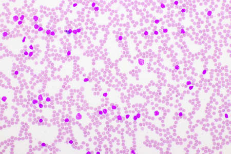
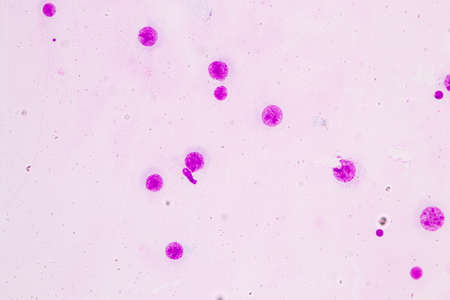
Picture of acute lymphocytic leukemia or ALL cells in blood smear, analyze by microscope, 400x
Коллекция по умолчанию
Коллекция по умолчанию
Создать новую
abstract acrylic watercolor paint brush stroke texture isolated on white background for logo and banner. design, creative, and illustration.
Коллекция по умолчанию
Коллекция по умолчанию
Создать новую
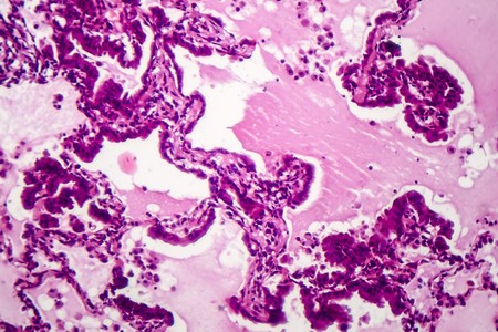
Lung adenocarcinoma, light micrograph, photo under microscope
Коллекция по умолчанию
Коллекция по умолчанию
Создать новую
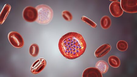
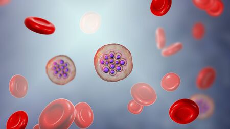
The malaria-infected red blood cells. 3D illustration showing malaria parasite Plasmodium falciparum in schizont stage inside red blood cells, the causative agent of tropical malaria
Коллекция по умолчанию
Коллекция по умолчанию
Создать новую
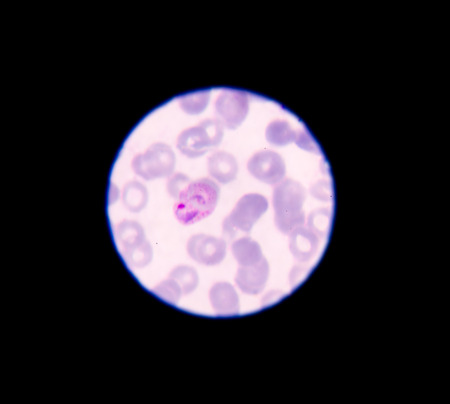
Malaria parasite in blood smear, gemetocyte stage
Коллекция по умолчанию
Коллекция по умолчанию
Создать новую
The malaria-infected red blood cells. 3D illustration showing ring-form trophozoites of malaria parasite Plasmodium falciparum inside red blood cells, the causative agent of tropical malaria
Коллекция по умолчанию
Коллекция по умолчанию
Создать новую
blood films for Malaria parasite
Коллекция по умолчанию
Коллекция по умолчанию
Создать новую
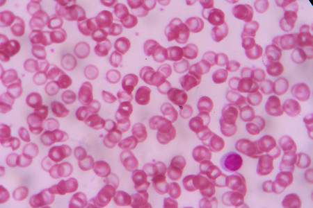
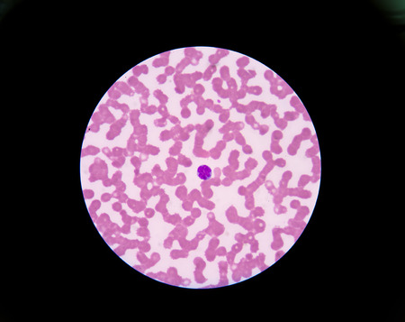
Abnormal red blood cells in Blood smear Thalassemia patient.
Коллекция по умолчанию
Коллекция по умолчанию
Создать новую
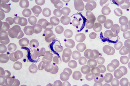
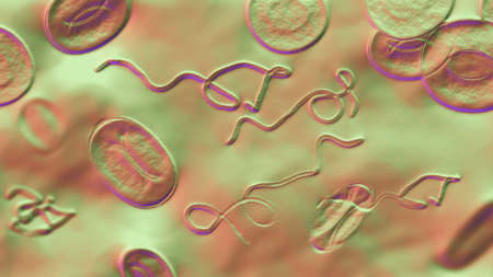
Trypanosoma gambiense blood smear viewed under a microscope at 1250 power.
Коллекция по умолчанию
Коллекция по умолчанию
Создать новую
complete blood count
Коллекция по умолчанию
Коллекция по умолчанию
Создать новую
Testicular seminoma, light micrograph, photo under microscope. A most common germ cell tumor of the testis
Коллекция по умолчанию
Коллекция по умолчанию
Создать новую
Neutrophil cell
Коллекция по умолчанию
Коллекция по умолчанию
Создать новую
Chromosomes Human under the microscope for education.
Коллекция по умолчанию
Коллекция по умолчанию
Создать новую
Red blood cells infected with malaria parasite
Коллекция по умолчанию
Коллекция по умолчанию
Создать новую
Microscopic view of cells, bacteria and viruses. Pathogens and microscopic organisms. Vivid biomedical backdrop. Banner. Concept of microbiology, immunology, health research, infection
Коллекция по умолчанию
Коллекция по умолчанию
Создать новую
nucleated red cell
Коллекция по умолчанию
Коллекция по умолчанию
Создать новую

Thyroid follicular carcinoma, light micrograph, photo under microscope
Коллекция по умолчанию
Коллекция по умолчанию
Создать новую

Apple pollen from a blossom in spring under the microscope
Коллекция по умолчанию
Коллекция по умолчанию
Создать новую
Numerous tiny white bubbles float gracefully on the surface of the water. The bubbles appear to be delicate and light, moving with the gentle current in a mesmerizing manner.
Коллекция по умолчанию
Коллекция по умолчанию
Создать новую
The malaria-infected red blood cells. 3D illustration showing malaria parasite Plasmodium falciparum in schizont stage inside red blood cells, the causative agent of tropical malaria
Коллекция по умолчанию
Коллекция по умолчанию
Создать новую
Cell division under microscope view. Microscopic view of cell.
Коллекция по умолчанию
Коллекция по умолчанию
Создать новую
complete blood count
Коллекция по умолчанию
Коллекция по умолчанию
Создать новую
Red arrow showing neutrophil with toxic granule active PMN.
Коллекция по умолчанию
Коллекция по умолчанию
Создать новую
Gloved hands hold a translucent cell culture sample with visible cellular structures and bubbles under focused clinical light, indicating laboratory research and microscopy, with available space for text on a neutral background
Коллекция по умолчанию
Коллекция по умолчанию
Создать новую

Blood vessel with flowing blood cells, 3D illustration. Small blood vessels, capillaries
Коллекция по умолчанию
Коллекция по умолчанию
Создать новую
Anatomy and Histological Ovary, Testis and Sperm human cells under microscope.
Коллекция по умолчанию
Коллекция по умолчанию
Создать новую
Chromosomes Human under the microscope for education.
Коллекция по умолчанию
Коллекция по умолчанию
Создать новую

Malaria parasites in red blood cells under the microscope 400x
Коллекция по умолчанию
Коллекция по умолчанию
Создать новую

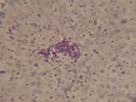
Histopathology of cirrhosis in blood smear, photo under microscope.
Коллекция по умолчанию
Коллекция по умолчанию
Создать новую
multinucleated giant
Коллекция по умолчанию
Коллекция по умолчанию
Создать новую
Creative colorful sand flows on blue background. Place for text or product promotion. Copy space. High quality photo
Коллекция по умолчанию
Коллекция по умолчанию
Создать новую
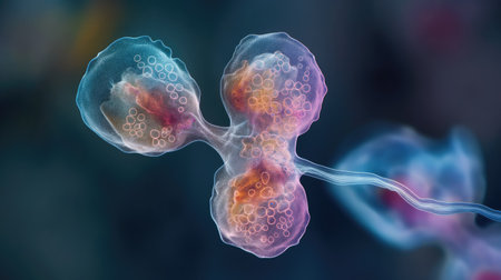
Cell division process, micro
Коллекция по умолчанию
Коллекция по умолчанию
Создать новую

plasmodium
Коллекция по умолчанию
Коллекция по умолчанию
Создать новую

Bacteria Neisseria gonorrhoeae inside phagocytes, gonoccoccus, diplococci which cause sexually transmitted infection gonorrhoea. 3D illustration. Incomplete phagocytosis
Коллекция по умолчанию
Коллекция по умолчанию
Создать новую
Nanoparticles Functionalization Therapeutics, Nanoparticles application in bioiechnology illustration
Коллекция по умолчанию
Коллекция по умолчанию
Создать новую

Backgrounds of Characteristics and Different shaped Colony of Bacteria and Mold growing on agar plates from Soil samples for education in Microbiology laboratory.
Коллекция по умолчанию
Коллекция по умолчанию
Создать новую
Malaria blood parasite infected red blood cells laboratory background.
Коллекция по умолчанию
Коллекция по умолчанию
Создать новую
The malaria-infected red blood cell. 3D illustration showing malaria parasite Plasmodium ovale in the stage of schizont
Коллекция по умолчанию
Коллекция по умолчанию
Создать новую
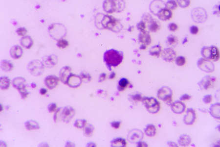
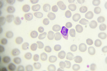
blood smear is often used as a follow-up test to abnormal results on a complete blood count (CBC) to evaluate the different types of blood cells.Atypical lymphocyte.
Коллекция по умолчанию
Коллекция по умолчанию
Создать новую

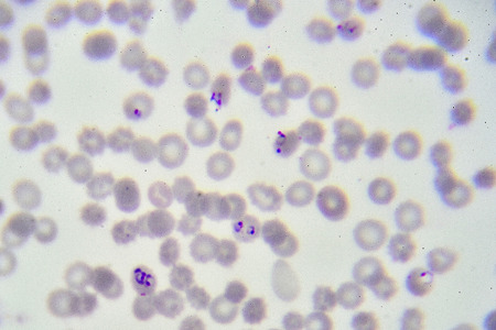
malaria parasite plasmodium falciparum on a thick blood smear reading under a microscope
Коллекция по умолчанию
Коллекция по умолчанию
Создать новую
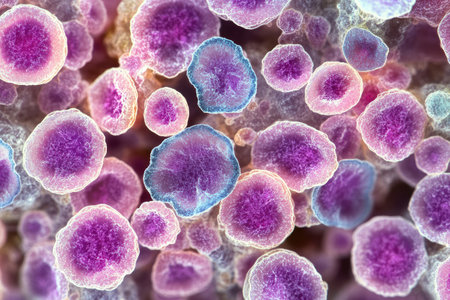
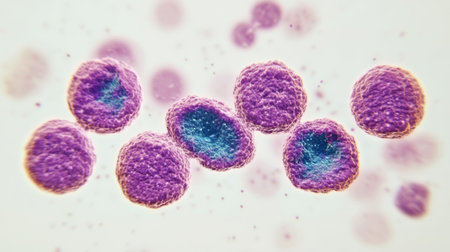
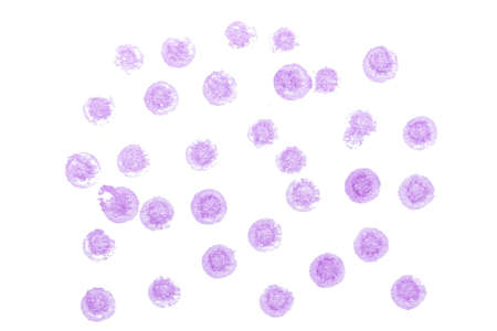
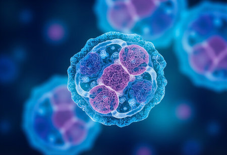
Close-up of a Cluster of Purple and Blue Cells
Коллекция по умолчанию
Коллекция по умолчанию
Создать новую
Coronavirus outbreak. Pathogen affecting the respiratory tract. COVID-19 infection. Concept of pandemic, viral infection. Coronavirus inside a human. Human cells, virus infects cells. 3D illustration
Коллекция по умолчанию
Коллекция по умолчанию
Создать новую
plasmodium
Коллекция по умолчанию
Коллекция по умолчанию
Создать новую
White blood cells of a human, Eosinophil photomicrograph panorama as seen under the microscope
Коллекция по умолчанию
Коллекция по умолчанию
Создать новую
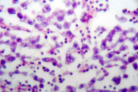
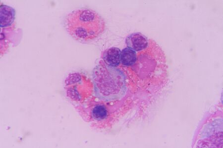
Abnomal cells look like malignant cells from specimen plural fluid.
Коллекция по умолчанию
Коллекция по умолчанию
Создать новую
Chaos ink texture background, ink in water pattern frost. Crystal winter design
Коллекция по умолчанию
Коллекция по умолчанию
Создать новую
Blood smear with white blood cells and red blood cells. Medical background.
Коллекция по умолчанию
Коллекция по умолчанию
Создать новую

Fungi Paracoccidioides lutzii, a dimorphic fungus that causes paracoccidioidomycosis found in Brazil and Ecuador. Scientific 3D illustration showing characteristic morphology of the fungus
Коллекция по умолчанию
Коллекция по умолчанию
Создать новую
Eosinophil
Коллекция по умолчанию
Коллекция по умолчанию
Создать новую
Virus cells, 3D illustration. Viruses and bacteria in human body. Viruses in infected organism.
Коллекция по умолчанию
Коллекция по умолчанию
Создать новую

Polycythemia vera, a rare slow-growing blood cancer with an increase in the number of red blood cells in the body, 3D illustration showing abundant erythrocytes inside blood vessel
Коллекция по умолчанию
Коллекция по умолчанию
Создать новую

Ascaris lumbricoides, a large roundworm, fertilized egg, 3D illustration
Коллекция по умолчанию
Коллекция по умолчанию
Создать новую
Borrelia bacteria in blood, 3D illustration. The causative agent of Lyme disease and relapsing fever. Borrelia recurrentis, B. burgdorferi in blood smear under microscope
Коллекция по умолчанию
Коллекция по умолчанию
Создать новую
Staphylococcus aureus bacteria under the microscope
Коллекция по умолчанию
Коллекция по умолчанию
Создать новую
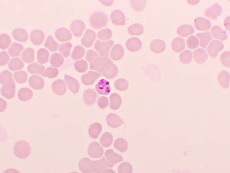
Malaria parasite in red blood cells, ring form stage of Plasmodium falciparum, original magnification 1000x
Коллекция по умолчанию
Коллекция по умолчанию
Создать новую
Neutrophils are a type of phagocyte and are normally found in the bloodstream.
Коллекция по умолчанию
Коллекция по умолчанию
Создать новую

The malaria-infected red blood cell. 3D illustration showing malaria parasite Plasmodium ovale in the stage of schizont
Коллекция по умолчанию
Коллекция по умолчанию
Создать новую
AI Generated. Microscopic View of a Cell Under Observation
Коллекция по умолчанию
Коллекция по умолчанию
Создать новую
blood cells with microscope.
Коллекция по умолчанию
Коллекция по умолчанию
Создать новую
Microscopic view of human cells under microscope.
Коллекция по умолчанию
Коллекция по умолчанию
Создать новую
SARS-CoV-2 viruses activating neutrophil, conceptual 3D illustration. Overactive neutrophils in COVID-19 are associated with hyperinflammation that drives lung and multi-organ damage
Коллекция по умолчанию
Коллекция по умолчанию
Создать новую
A microscopic image shows a cluster of pink and orange cells, with light refracting off their surfaces. The cells are suspended in a blue liquid, with a fine network of pale blue lines extending throughout the background.
Коллекция по умолчанию
Коллекция по умолчанию
Создать новую
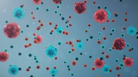
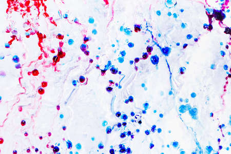
This macro image showcases red and blue spherical cells suspended in an abstract scientific background, ideal for themes of biology, health, and microscopic research.
Коллекция по умолчанию
Коллекция по умолчанию
Создать новую
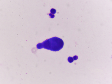
A colorful image of a cell with a purple and blue blob in the center. The image is abstract and has a mood of curiosity and wonder
Коллекция по умолчанию
Коллекция по умолчанию
Создать новую

Pichia is a genus of yeasts in the family Saccharomycetaceae under the microscope for education.
Коллекция по умолчанию
Коллекция по умолчанию
Создать новую
Backgrounds of Characteristics and Different shaped Colony of Bacteria and Mold growing on agar plates from Soil samples for education in Microbiology laboratory.
Коллекция по умолчанию
Коллекция по умолчанию
Создать новую

Photomicrograph of canine eosinphil
Коллекция по умолчанию
Коллекция по умолчанию
Создать новую
Burkitts lymphoma cells, a cancer of the lymphatic system, monoclonal B-cell tumor, 3D illustration
Коллекция по умолчанию
Коллекция по умолчанию
Создать новую
Legion-Media
Создайте свои проекты на основе качественных стоковых фотографий и видео.


Copyright © Legion-Media.