

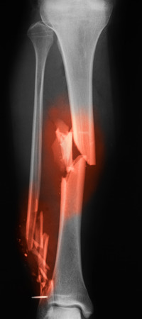
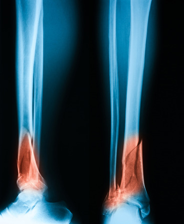
lower leg x-ray of a 48 year old female with a spiral fracture of the distal tibia
Коллекция по умолчанию
Коллекция по умолчанию
Создать новую
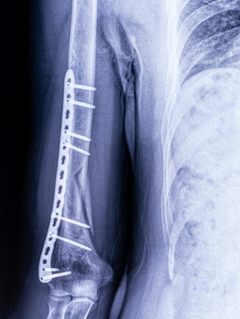
X-ray image of osteosynthesis of a humerus fracture in a man close-up
Коллекция по умолчанию
Коллекция по умолчанию
Создать новую
Arthrosis. Yellow warning tapes
Коллекция по умолчанию
Коллекция по умолчанию
Создать новую
Коллекция по умолчанию
Коллекция по умолчанию
Создать новую

X-ray of both human legs (broken legs)
Коллекция по умолчанию
Коллекция по умолчанию
Создать новую
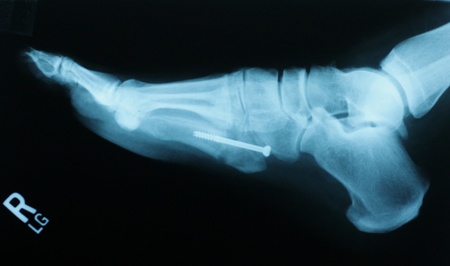
X-ray Foot Ankle Calcaneus
Коллекция по умолчанию
Коллекция по умолчанию
Создать новую
Close up of beaters with dripping cake mix vertical
Коллекция по умолчанию
Коллекция по умолчанию
Создать новую

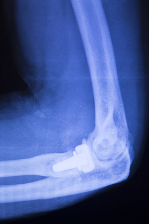
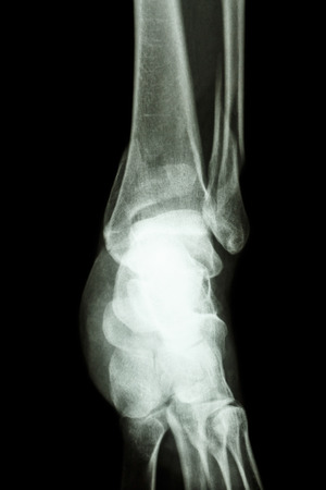
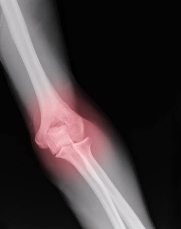
Elbow joint tennis elbow inury x-ray test scan result before traumatology and orthopedics surgery.
Коллекция по умолчанию
Коллекция по умолчанию
Создать новую

image of x-ray Osteoarthritis knee
Коллекция по умолчанию
Коллекция по умолчанию
Создать новую
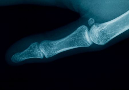
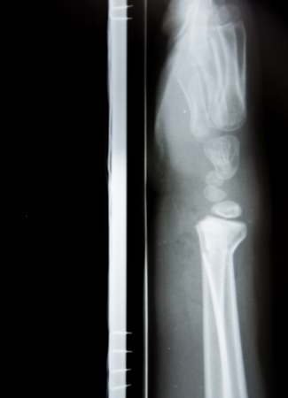
radiography of a middle aged woman finger
Коллекция по умолчанию
Коллекция по умолчанию
Создать новую

X-rays image of leg fracture patients
Коллекция по умолчанию
Коллекция по умолчанию
Создать новую

X-ray of a broken arm in a gypsum. The doctor shows the patient the location of the injury. A picture of a human skeleton.
Коллекция по умолчанию
Коллекция по умолчанию
Создать новую

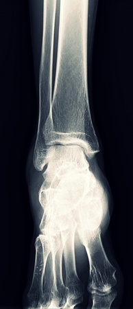
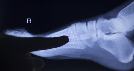
X-ray medical picture - Human foot
Коллекция по умолчанию
Коллекция по умолчанию
Создать новую

X-Ray Image Of Human Chest for a medical diagnosis
Коллекция по умолчанию
Коллекция по умолчанию
Создать новую
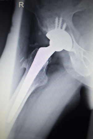
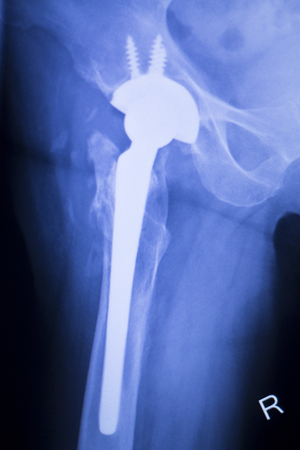
Hip joint replacement orthopedic titanium metal Traaumatology ball and socket implant x-ray image of old age patient.
Коллекция по умолчанию
Коллекция по умолчанию
Создать новую

human back bone model with old color style
Коллекция по умолчанию
Коллекция по умолчанию
Создать новую

Dead mosquito with blood crushed in a hand
Коллекция по умолчанию
Коллекция по умолчанию
Создать новую
X-ray scan image of hip joints with orthopedic hip joint replacement implant head and screws in human skeleton in blue gray tones. Scanned in orthopedics traumatology surgery hospital clinic.
Коллекция по умолчанию
Коллекция по умолчанию
Создать новую
X-ray image of broken leg. AP view
Коллекция по умолчанию
Коллекция по умолчанию
Создать новую

X-ray scan image of hip joints with orthopedic hip joint replacement implant head and screws in human skeleton in blue gray tones. Scanned in orthopedics traumatology surgery hospital clinic.
Коллекция по умолчанию
Коллекция по умолчанию
Создать новую

x-ray of human leg (broken leg)
Коллекция по умолчанию
Коллекция по умолчанию
Создать новую
Cocktail style drink full of fruit
Коллекция по умолчанию
Коллекция по умолчанию
Создать новую
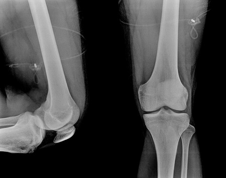
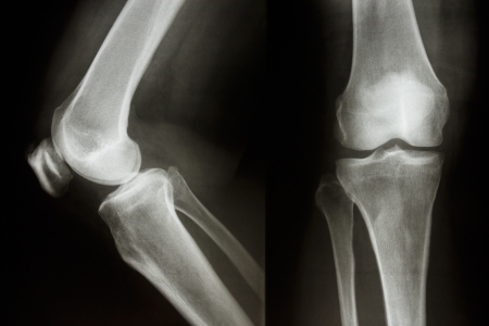
X-Ray picture of knees front and side view
Коллекция по умолчанию
Коллекция по умолчанию
Создать новую
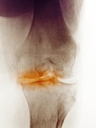
X-ray of the knee of a 83 year old woman showing degenerative arthritis. This woman was scheduled for a knee replacement.
Коллекция по умолчанию
Коллекция по умолчанию
Создать новую
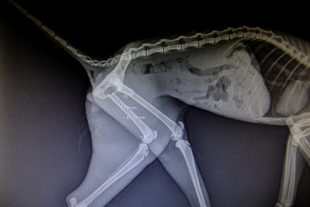
X-ray images of wild animals used by veterinarians to diagnose, treat diseases and illnesses
Коллекция по умолчанию
Коллекция по умолчанию
Создать новую

X-ray of the leg
Коллекция по умолчанию
Коллекция по умолчанию
Создать новую

Surgical suture
Коллекция по умолчанию
Коллекция по умолчанию
Создать новую

Side view of human knee-joint with kneecap on X-ray image
Коллекция по умолчанию
Коллекция по умолчанию
Создать новую
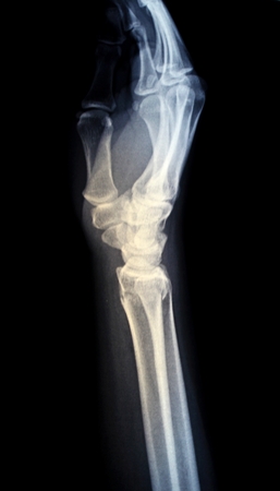
X-Ray image of human hands
Коллекция по умолчанию
Коллекция по умолчанию
Создать новую
film x-ray ankle show fracture distal tibia and fibula leg
Коллекция по умолчанию
Коллекция по умолчанию
Создать новую

X-ray scan image of hip joints with orthopedic hip joint replacement implant head and screws in human skeleton in blue gray tones. Scanned in orthopedics traumatology surgery hospital clinic.
Коллекция по умолчанию
Коллекция по умолчанию
Создать новую
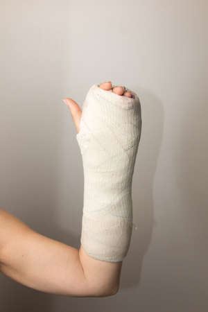
Lady with a Broken Hand and Wrist wrapped in a cast
Коллекция по умолчанию
Коллекция по умолчанию
Создать новую
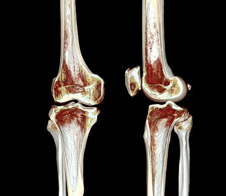
A half of CT Scan of knee 3D rendering image isolated on black background for diagnosis knee fracture.
Коллекция по умолчанию
Коллекция по умолчанию
Создать новую

Medicine bandage on human injury hand after accident with chainsaw
Коллекция по умолчанию
Коллекция по умолчанию
Создать новую

X-ray image of portal vein after Doctor doing ERCP and laparoscopic cholecystectomy inside modern operating room.
Коллекция по умолчанию
Коллекция по умолчанию
Создать новую

hand x-ray
Коллекция по умолчанию
Коллекция по умолчанию
Создать новую

Osteoarthritis Knee ( OA Knee ) ( Film x-ray both knee with arthritis of knee joint : narrow knee joint space ) ( Medical and Science background )
Коллекция по умолчанию
Коллекция по умолчанию
Создать новую
Close up of a painful knee joint against a blurred hospital background highlighting skin texture
Коллекция по умолчанию
Коллекция по умолчанию
Создать новую
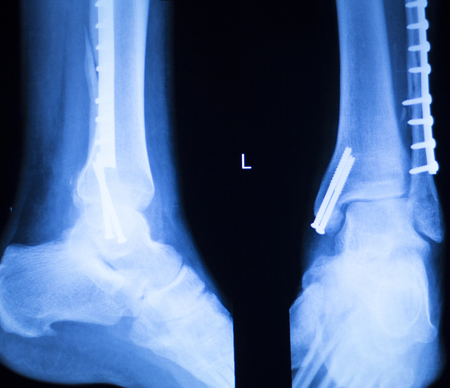
Foot ankle and shin orthopedics x-ray scan results showing Traumatology plate and screw titanium implants.
Коллекция по умолчанию
Коллекция по умолчанию
Создать новую
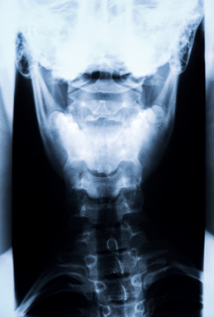
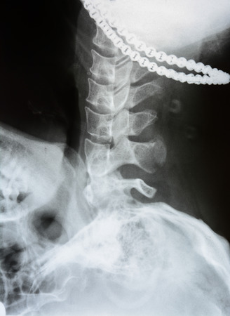
X-Ray photo of neck and skull.
Коллекция по умолчанию
Коллекция по умолчанию
Создать новую
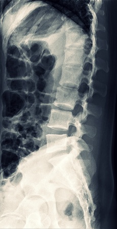
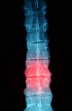
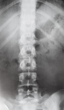
X-Ray Lumbar Spine
Коллекция по умолчанию
Коллекция по умолчанию
Создать новую
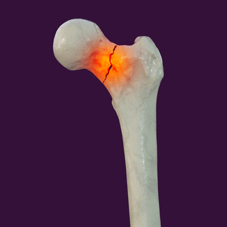
A fracture of the femur neck, a common type of hip fracture that typically occurs in older adults and can lead to mobility issues and other complications, isolated on white background, 3D illustration
Коллекция по умолчанию
Коллекция по умолчанию
Создать новую
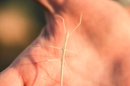
Stick insect on a person's hand
Коллекция по умолчанию
Коллекция по умолчанию
Создать новую

CT Scan ankle joint with 3d rendering of calcaneus bone showing Calcaneus (Heel Bone) Fractures.
Коллекция по умолчанию
Коллекция по умолчанию
Создать новую

An image of different man body parts back arm leg
Коллекция по умолчанию
Коллекция по умолчанию
Создать новую

Vintage style of white sand crystal and wooden hourglass with warm sunlight in soft-focus in the background, time concept.
Коллекция по умолчанию
Коллекция по умолчанию
Создать новую

Femoral artery angiogram or angiography at lower extremity area.
Коллекция по умолчанию
Коллекция по умолчанию
Создать новую
X ray fracture with fixation screws, broken leg
Коллекция по умолчанию
Коллекция по умолчанию
Создать новую
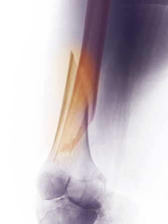
X-ray of the femur of a 60 year old female who fell and fractured her distal femur
Коллекция по умолчанию
Коллекция по умолчанию
Создать новую

x-ray of human muscle
Коллекция по умолчанию
Коллекция по умолчанию
Создать новую
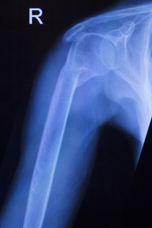
Arm X RAY
Коллекция по умолчанию
Коллекция по умолчанию
Создать новую
X-ray image of knee joint, AP view.
Коллекция по умолчанию
Коллекция по умолчанию
Создать новую
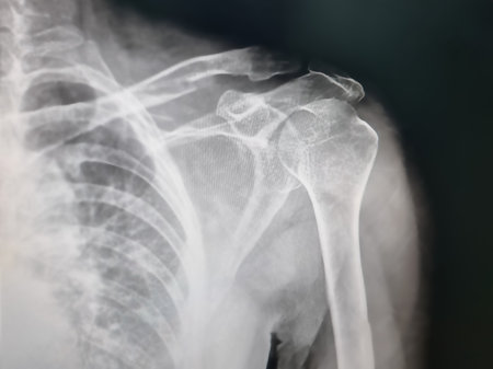
Shoulder joint injury xray traumatology and orthopedics test medical scan used to diagnose sports injuries in patient.
Коллекция по умолчанию
Коллекция по умолчанию
Создать новую
Front view of human spine in torso on X-ray image
Коллекция по умолчанию
Коллекция по умолчанию
Создать новую
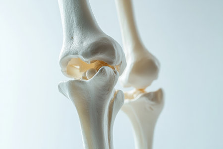
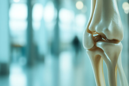
A translucent close-up of the knee joint in a human skeleton model, showing its complex structure in a sleek and minimalistic design, set on a crisp white background.
Коллекция по умолчанию
Коллекция по умолчанию
Создать новую

x-ray of legs. Blurry Traumatology orthopedic surgery hospital operating room for the legs operation. Medical health and Education concept.
Коллекция по умолчанию
Коллекция по умолчанию
Создать новую
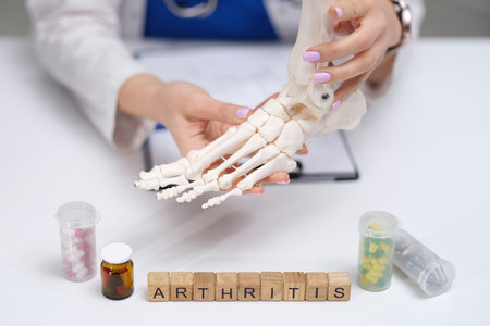
A Comprehensive Overview of Arthritis Including Its Anatomy, Symptoms, and Treatment Options
Коллекция по умолчанию
Коллекция по умолчанию
Создать новую
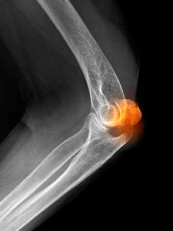
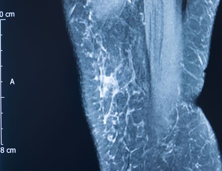
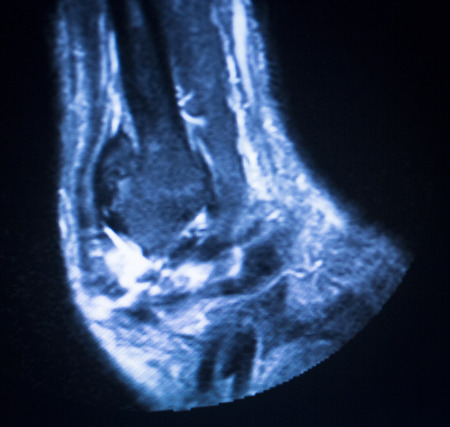
x-ray of an olecranon fracture
Коллекция по умолчанию
Коллекция по умолчанию
Создать новую

Arthritic hip xray test scan orthopedic and Traumatology results showing titanium hip replacement plate and screws surgical implant.
Коллекция по умолчанию
Коллекция по умолчанию
Создать новую
Knee sports injury mri mcl grade 2 tear magnetic resonance imaging orthopedic traumatology scan.
Коллекция по умолчанию
Коллекция по умолчанию
Создать новую
Knee joint xray test scan results of patient with arthritis and joints pain in knees on screen with surgeon.
Коллекция по умолчанию
Коллекция по умолчанию
Создать новую
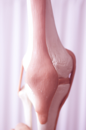
Human knee joint meniscus medical teaching model showing bones and anterior cruciate ligaments tendons.
Коллекция по умолчанию
Коллекция по умолчанию
Создать новую
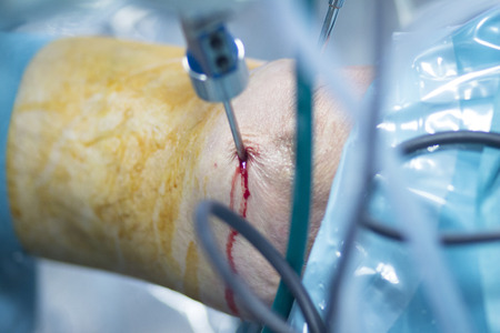
Orthopedic surgery meniscus operation hospital emergency operating room photo.
Коллекция по умолчанию
Коллекция по умолчанию
Создать новую
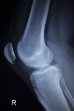
X-ray image of normal old age Knee
Коллекция по умолчанию
Коллекция по умолчанию
Создать новую
arm bone x-ray images blur and noise
Коллекция по умолчанию
Коллекция по умолчанию
Создать новую
Patient foot, leg and ankle in electro physiotherapy electrical impulse stimulation rehabiliation treatment from injury in hospital clinic with electrical stimulus attached with plaster.
Коллекция по умолчанию
Коллекция по умолчанию
Создать новую

X-ray
Коллекция по умолчанию
Коллекция по умолчанию
Создать новую

Radiology x-ray photograph of human hip
Коллекция по умолчанию
Коллекция по умолчанию
Создать новую
Hip joint replacement xray showing ball and socket joint's titanium screw implant in medical orthpodedics scan.
Коллекция по умолчанию
Коллекция по умолчанию
Создать новую
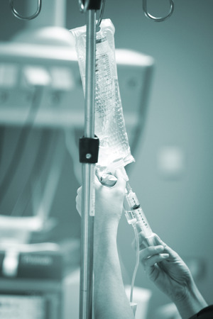
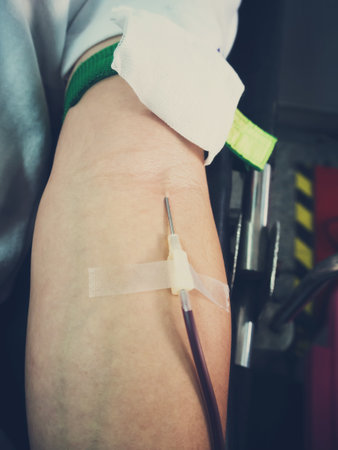
Hospital surgery emergency operating room surgical liquid drip equipment photo.
Коллекция по умолчанию
Коллекция по умолчанию
Создать новую
Jones Fracture
Коллекция по умолчанию
Коллекция по умолчанию
Создать новую

X-ray orthopedic medical CAT scan of painful neck injury in Traumatology hospital clinic.
Коллекция по умолчанию
Коллекция по умолчанию
Создать новую
Foot toes joint xray test scan results of patient with arthritis and joints pain in feet.
Коллекция по умолчанию
Коллекция по умолчанию
Создать новую
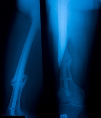
X-ray image of leg, AP and lateral view, Showing tibia and fibula fracture
Коллекция по умолчанию
Коллекция по умолчанию
Создать новую
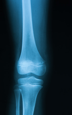
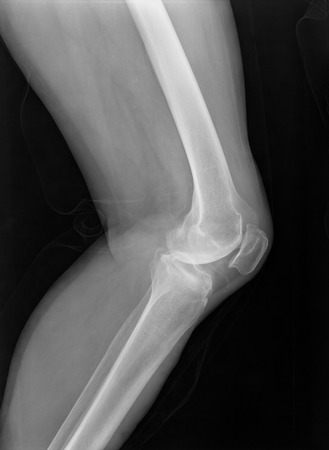
Film X-ray show normal knee joint AP Lateral
Коллекция по умолчанию
Коллекция по умолчанию
Создать новую
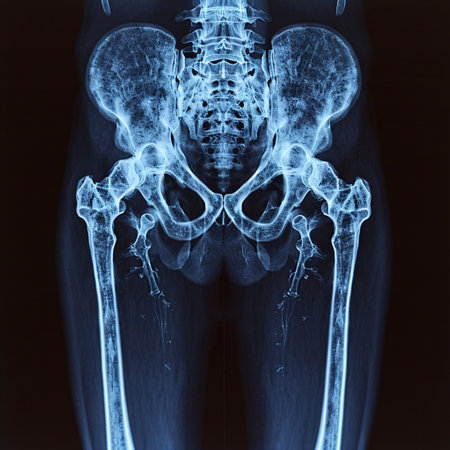
X-ray image of the pelvis and hip of a patient
Коллекция по умолчанию
Коллекция по умолчанию
Создать новую
Knee joint xray test scan results of patient with arthritis and joints pain in knees on screen with surgeon.
Коллекция по умолчанию
Коллекция по умолчанию
Создать новую
Blood Donors Making Donation In Hospital, focus at center
Коллекция по умолчанию
Коллекция по умолчанию
Создать новую
MRI magnetic resonance imaging medical scan test results showing ligaments, cartilege and cross section of bones in human skeleton of ankle.
Коллекция по умолчанию
Коллекция по умолчанию
Создать новую
x-ray image
Коллекция по умолчанию
Коллекция по умолчанию
Создать новую
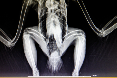
lateral x-ray film of bird
Коллекция по умолчанию
Коллекция по умолчанию
Создать новую

normal foot x-ray of a 17 year old woman
Коллекция по умолчанию
Коллекция по умолчанию
Создать новую
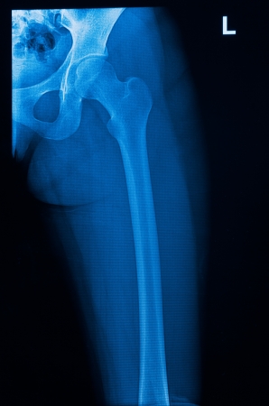
Hip x-ray of human
Коллекция по умолчанию
Коллекция по умолчанию
Создать новую

3d rendered illustration of an arthritic elbow joint
Коллекция по умолчанию
Коллекция по умолчанию
Создать новую
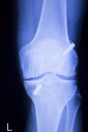
Knee joint implant screw xray showing in medical orthpodedic traumatology scan.
Коллекция по умолчанию
Коллекция по умолчанию
Создать новую
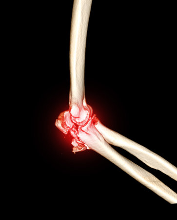
Computed Tomography Volume Rendering examination of elbow joint 3D rendering in patient fracture elbow joint.
Коллекция по умолчанию
Коллекция по умолчанию
Создать новую

Orthopedics knee joint meniscus, ligament, tendon and cartilage injury titanium modern metal implant X-ray scan.
Коллекция по умолчанию
Коллекция по умолчанию
Создать новую
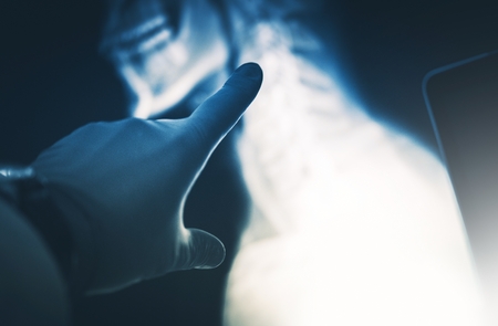
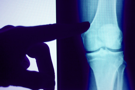
Xray Scan Examination. Doctor Hand in Safety Gloves Exposing the Problem.
Коллекция по умолчанию
Коллекция по умолчанию
Создать новую

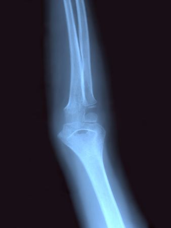
x-ray of an elbow dislocation
Коллекция по умолчанию
Коллекция по умолчанию
Создать новую

X-ray film
Коллекция по умолчанию
Коллекция по умолчанию
Создать новую
X-ray orthopedic medical CAT scan of painful knee meniscus injury leg in traumatology hospital clinic.
Коллекция по умолчанию
Коллекция по умолчанию
Создать новую
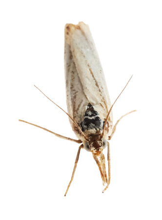
Single chrysoteuchia culmella moth isolated on white
Коллекция по умолчанию
Коллекция по умолчанию
Создать новую
x-ray of the neck
Коллекция по умолчанию
Коллекция по умолчанию
Создать новую
xray of an arm with elbow joint visible and red pain circle
Коллекция по умолчанию
Коллекция по умолчанию
Создать новую

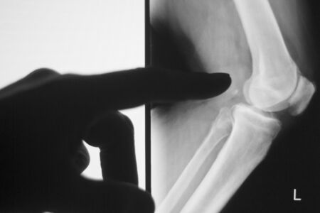
x-ray of an injured elbow
Коллекция по умолчанию
Коллекция по умолчанию
Создать новую
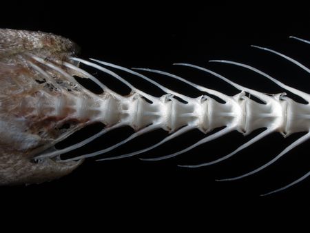
Sea fish white bone close-up
Коллекция по умолчанию
Коллекция по умолчанию
Создать новую

Upper part of broken human thigh with steel screw x-ray
Коллекция по умолчанию
Коллекция по умолчанию
Создать новую

X-rays image of leg fracture patients
Коллекция по умолчанию
Коллекция по умолчанию
Создать новую

X-Ray photo of neck and skull.
Коллекция по умолчанию
Коллекция по умолчанию
Создать новую

Operation for cesarean section with new born infant in operating theater
Коллекция по умолчанию
Коллекция по умолчанию
Создать новую
Legion-Media
Создайте свои проекты на основе качественных стоковых фотографий и видео.


Copyright © Legion-Media.