
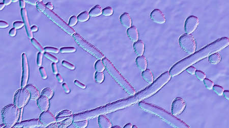
Microscopic fungi Trichosporon, 3D illustration shows septate hyphae, pseudohyphae, blastoconidia singly or in short chains, arthroconidia. Cause white piedra, superficial and invasive infections
Коллекция по умолчанию
Коллекция по умолчанию
Создать новую
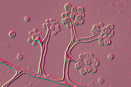
Microscopic fungi Cunninghamella, scientific 3D illustration. Pathogenic fungi from the order Mucorales, cause sinopulmonary and disseminated infections, one of the causative agents of mucormycosis
Коллекция по умолчанию
Коллекция по умолчанию
Создать новую
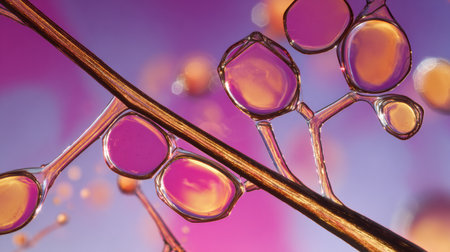
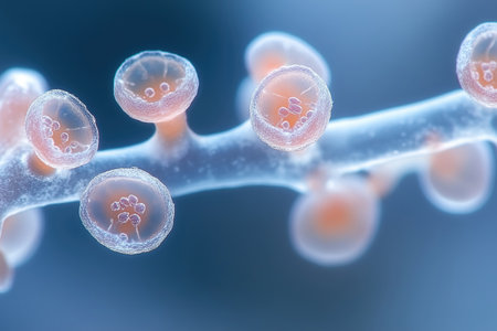
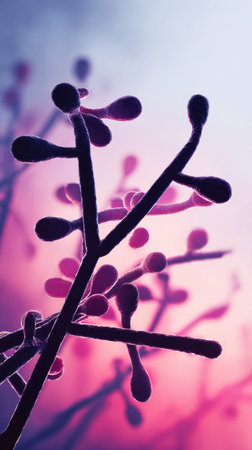
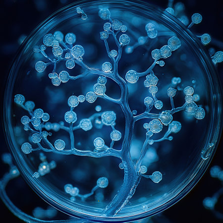
Translucent bubbles cluster around a branch, illuminated against a vibrant purple backdrop, showing intricate structures and captivating light reflections.
Коллекция по умолчанию
Коллекция по умолчанию
Создать новую
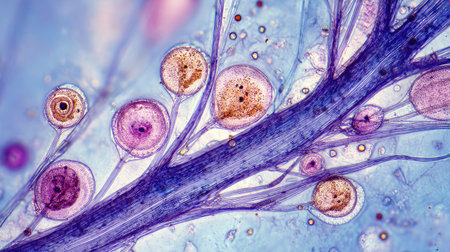
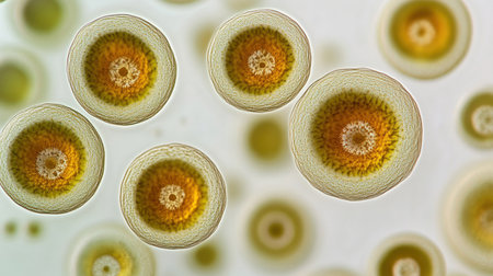
Aspergillus niger and Aspergillus oryzae (mold) under microscope for Microbiology in Lab.
Коллекция по умолчанию
Коллекция по умолчанию
Создать новую

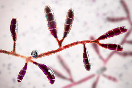
Fungi Trichophyton mentagrophytes, 3D illustration showing macroconidia, branched conidiophores bearing spherical conidia, septate and spiral hyphae. Causes ringworm, hair and nail infections
Коллекция по умолчанию
Коллекция по умолчанию
Создать новую
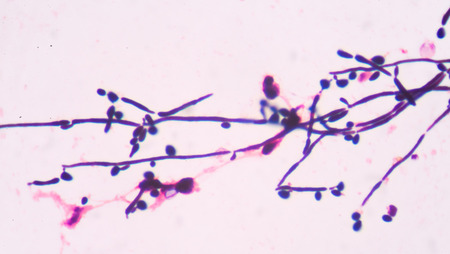
Branching budding yeast cells with pseudohyphae in urine gram stain fine with microscope.
Коллекция по умолчанию
Коллекция по умолчанию
Создать новую
Clusters of bacteria can be observed rejecting antibiotics, showing their resistance mechanisms in a laboratory setting during an examination.
Коллекция по умолчанию
Коллекция по умолчанию
Создать новую
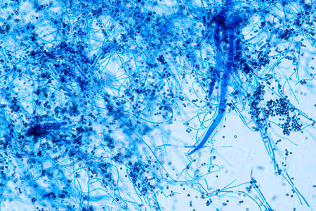
Explore a stunning microscopic view featuring colorful cells and thin filaments against a vibrant blue background, showcasing scientific beauty in detail.
Коллекция по умолчанию
Коллекция по умолчанию
Создать новую
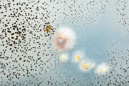
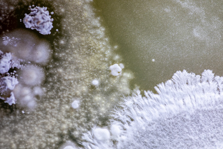
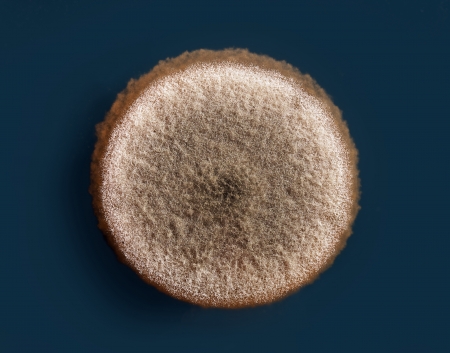
Petri dish with mold
Коллекция по умолчанию
Коллекция по умолчанию
Создать новую
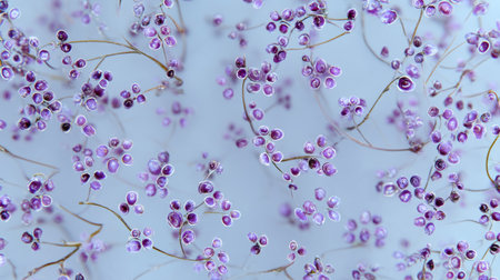
This image captures delicate purple flowers with fine stems set against a soft blue background, evoking a tranquil and ethereal feeling perfect for decor.
Коллекция по умолчанию
Коллекция по умолчанию
Создать новую
Aspergillus niger and Aspergillus oryzae (mold) under microscope for Microbiology in Lab.
Коллекция по умолчанию
Коллекция по умолчанию
Создать новую
Mould fungi Madurella, 3D illustration. The microscopic fungus that causes black-grain mycetoma, or maduromycosis, an infection of human extremities and nervous system found in tropical areas
Коллекция по умолчанию
Коллекция по умолчанию
Создать новую

Microscopic fungi Microsporum audouinii, 3D illustration. Anthropophilic dermatophyte fungus, causes infections of scalp (tinea capitis), body skin (tinea corporis) mainly in children
Коллекция по умолчанию
Коллекция по умолчанию
Создать новую

Pathogen, Bacteria and bacterium cells in microscopic as a medical illustration of bacterial disease infection in a human body
Коллекция по умолчанию
Коллекция по умолчанию
Создать новую

Candida tropicalis yeasts, microscopic fungi that cause infections in immunocompromised patients. Scientific 3D illustration showing pseudohyphae and blastoconidia formed singly or in small groups
Коллекция по умолчанию
Коллекция по умолчанию
Создать новую
Candida kefyr yeasts, formely Candida pseudotropicalis, microscopic fungi, scientific illustration. Causes invasive candidiasis and candidemia in patients with hematologic malignancies
Коллекция по умолчанию
Коллекция по умолчанию
Создать новую
Microscopic fungi Epidermophyton floccosum, scientific 3D illustration. A filamentous fungus, causes skin and nail infections, such as athlete's foot, tinea cruris, tinea corporis and onychomycosis
Коллекция по умолчанию
Коллекция по умолчанию
Создать новую
Microscopic View of Septate Dematiaceous Fungal Hyphae and Possible Chlamydospores
Коллекция по умолчанию
Коллекция по умолчанию
Создать новую
Fungi Trichophyton mentagrophytes, 3D illustration showing macroconidia, branched conidiophores bearing spherical conidia, septate and spiral hyphae. Causes ringworm, hair and nail infections
Коллекция по умолчанию
Коллекция по умолчанию
Создать новую
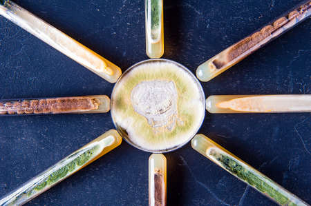
Slant tubes and petri dish with various mold strain cultures including neurospora intermedia, aspergillus niger and trichoderma
Коллекция по умолчанию
Коллекция по умолчанию
Создать новую
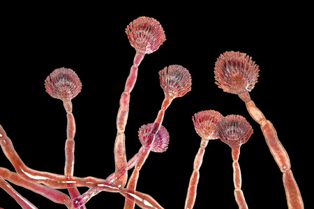
sporangia of a microscopic organism, microbiology concept
Коллекция по умолчанию
Коллекция по умолчанию
Создать новую
microscope slide with detailed view of plant stem, complete with cells and minutiae, created with generative ai
Коллекция по умолчанию
Коллекция по умолчанию
Создать новую

Aspergillus (mold) for Microbiology in Lab.
Коллекция по умолчанию
Коллекция по умолчанию
Создать новую
An enchanting close-up view of underwater flora illuminated by soft light, showcasing bubbles and vibrant colors in a tranquil aquatic environment.
Коллекция по умолчанию
Коллекция по умолчанию
Создать новую

Apple pollen from a blossom in spring under the microscope
Коллекция по умолчанию
Коллекция по умолчанию
Создать новую
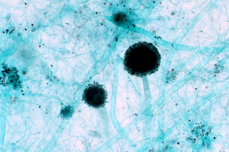
Microscopic Visualization of Darkly Pigmented Fungal Conidia and Hyphal Structures in a Clear Wet Mount Specimen.
Коллекция по умолчанию
Коллекция по умолчанию
Создать новую
A microscope slide containing a sample of plankton viewed under high magnification to study its composition
Коллекция по умолчанию
Коллекция по умолчанию
Создать новую
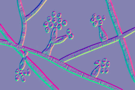
A captivating close-up of a delicate branch-like structure against a gentle blue bokeh background, ideal for highlighting themes of nature and abstract beauty.
Коллекция по умолчанию
Коллекция по умолчанию
Создать новую
Microscopic fungi Microsporum audouinii, 3D illustration. Anthropophilic dermatophyte fungus, causes infections of scalp (tinea capitis), body skin (tinea corporis) mainly in children
Коллекция по умолчанию
Коллекция по умолчанию
Создать новую
Aspergillus niger and Aspergillus oryzae (mold) under microscope for Microbiology in Lab.
Коллекция по умолчанию
Коллекция по умолчанию
Создать новую
Characteristics of fungi living in wood as a group, are polyphyletic under the microscope for education.
Коллекция по умолчанию
Коллекция по умолчанию
Создать новую
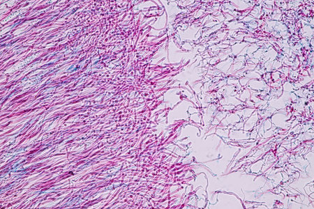
Characteristics of Lichen, hyphae and Symbiotic algae under the microscope for education.
Коллекция по умолчанию
Коллекция по умолчанию
Создать новую

Backgrounds of Characteristics and Different shaped Colony of Bacteria and Mold growing on agar plates from Soil samples for education in Microbiology laboratory.
Коллекция по умолчанию
Коллекция по умолчанию
Создать новую
Backgrounds of Characteristics and Different shaped Colony of Bacteria and Mold growing on agar plates from Soil samples for education in Microbiology laboratory.
Коллекция по умолчанию
Коллекция по умолчанию
Создать новую
microbiology background made of fungi colonies. Surface of agar petri dish.
Коллекция по умолчанию
Коллекция по умолчанию
Создать новую

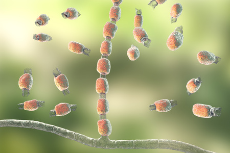
Fungus Trichophyton rubrum, 3D illustration showing macroconidium, microconidia and septate hyphae. Infects skin and nails causing dermatophytosis, especially on feet (tinea pedis), and onychomycosis
Коллекция по умолчанию
Коллекция по умолчанию
Создать новую

Pichia is a genus of yeasts in the family Saccharomycetaceae under the microscope for education.
Коллекция по умолчанию
Коллекция по умолчанию
Создать новую
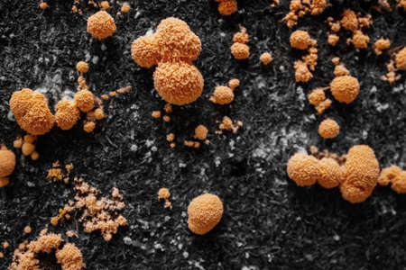
The Orange Peel Fungus (Aleuria aurantia) - edible
Коллекция по умолчанию
Коллекция по умолчанию
Создать новую

Euglena is a genus of single-celled flagellate Eukaryotes under microscopic view for education.
Коллекция по умолчанию
Коллекция по умолчанию
Создать новую

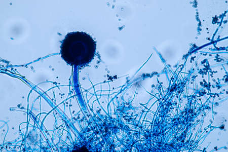
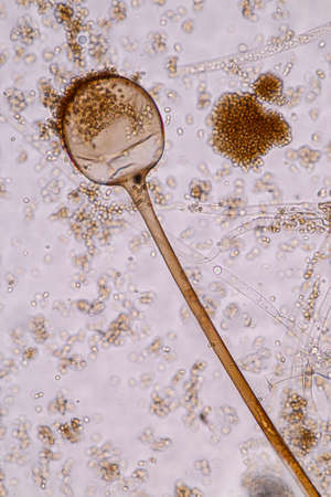
Microscope view of mold sporangia.
Коллекция по умолчанию
Коллекция по умолчанию
Создать новую
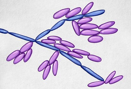
Fungi Trichophyton mentagrophytes, 3D illustration showing branched conidiophores bearing spherical microconidia. Causes skin infection (ringworm), hair and nail infections
Коллекция по умолчанию
Коллекция по умолчанию
Создать новую
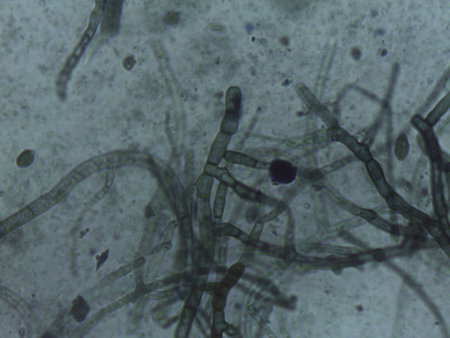
Microscopic View of Septate Dematiaceous Fungal Hyphae and Possible Chlamydospores
Коллекция по умолчанию
Коллекция по умолчанию
Создать новую
Characteristics of Rhizopus is a genus of common saprophytic fungi on Slide under the microscope for education.
Коллекция по умолчанию
Коллекция по умолчанию
Создать новую
Microscopic View of Septate Dematiaceous Fungal Hyphae and Possible Chlamydospores
Коллекция по умолчанию
Коллекция по умолчанию
Создать новую
Backgrounds Colony Characteristics of Rhizopus (bread mold) is a genus of common saprophytic fungi, Rhizopus (bread mold) under the microscope.(soft focus and have Grain/Noise)
Коллекция по умолчанию
Коллекция по умолчанию
Создать новую
Characteristics of Lichen, hyphae and Symbiotic algae under the microscope for education.
Коллекция по умолчанию
Коллекция по умолчанию
Создать новую
Fusarium oxysporum mycelium
Коллекция по умолчанию
Коллекция по умолчанию
Создать новую
Microscopic View of Septate Dematiaceous Fungal Hyphae and Possible Chlamydospores
Коллекция по умолчанию
Коллекция по умолчанию
Создать новую
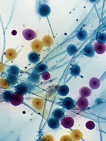
Beautiful microworld, microbes of different shapes, 3D illustration.
Коллекция по умолчанию
Коллекция по умолчанию
Создать новую
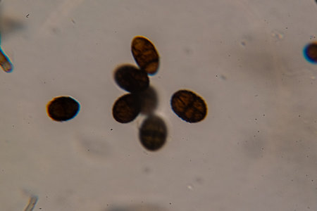
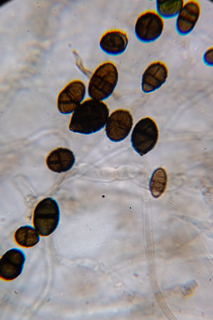
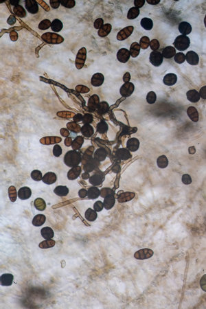
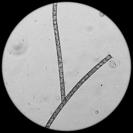
Dematiaceous Fungal Spores Exhibiting Transverse and Longitudinal Septation, Likely of the Genus Alternaria.
Коллекция по умолчанию
Коллекция по умолчанию
Создать новую
macro of green fungi on petri dish.
Коллекция по умолчанию
Коллекция по умолчанию
Создать новую
Dangerous head mold with hyphae and mold sponsors under the microscope 200x
Коллекция по умолчанию
Коллекция по умолчанию
Создать новую

Fungus Trichophyton rubrum, 3D illustration showing macroconidia, microconidia and septate hyphae. Infects skin and nails causing dermatophytosis, especially on feet (tinea pedis), and onychomycosis
Коллекция по умолчанию
Коллекция по умолчанию
Создать новую

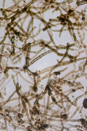
fungal hyphae and soil fungi in a soil sample, showing the living soil form a farm
Коллекция по умолчанию
Коллекция по умолчанию
Создать новую
Detailed image of rod-shaped bacteria under a high-powered microscope, showing cell structure
Коллекция по умолчанию
Коллекция по умолчанию
Создать новую
Mold on pork caused by moisture and mold it for a cause the diarrhea. Abstract blur background.
Коллекция по умолчанию
Коллекция по умолчанию
Создать новую
close-up of vibrant bacteria colonies on a petri dish, created with generative ai
Коллекция по умолчанию
Коллекция по умолчанию
Создать новую

Backgrounds Colony Characteristics of Rhizopus (bread mold) is a genus of common saprophytic fungi, Rhizopus (bread mold) under the microscope.
Коллекция по умолчанию
Коллекция по умолчанию
Создать новую

budding yeast cells with pseudohyphae in urine gram stain fine with microscope.
Коллекция по умолчанию
Коллекция по умолчанию
Создать новую
Colorful cells reveal stunning patterns, showcasing the beauty of microscopic life.
Коллекция по умолчанию
Коллекция по умолчанию
Создать новую
Colony characteristic of Actinomyces, Bacteria, yeast and Mold on selective media from soil samples for study in laboratory microbiology.
Коллекция по умолчанию
Коллекция по умолчанию
Создать новую
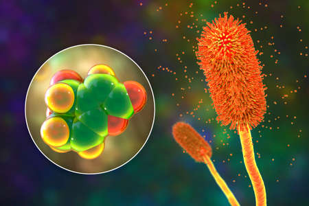
Fungus Aspergillus flavus, 3D illustration. It is the major producer of aflatoxin in crops, a potent carcinogen
Коллекция по умолчанию
Коллекция по умолчанию
Создать новую

Opisthorchis viverrini, common name Southeast Asian liver fluke, is a trematode parasite.
Коллекция по умолчанию
Коллекция по умолчанию
Создать новую
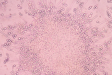
Aspergillus (mold) under the light microscopic view for microbiology education.
Коллекция по умолчанию
Коллекция по умолчанию
Создать новую
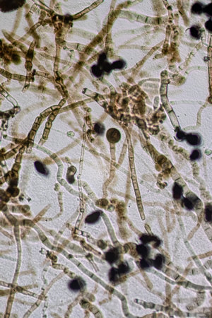
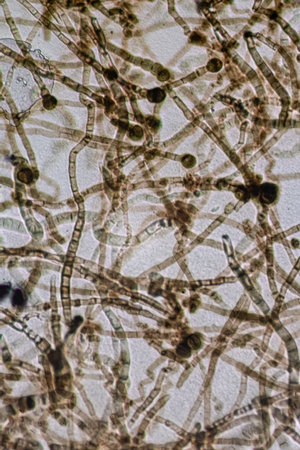
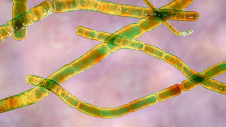
Micrograph showing intertwined septate and dematiaceous fungal hyphae in a dense network.
Коллекция по умолчанию
Коллекция по умолчанию
Создать новую
Microscopic mold fungi Curvularia, scientific 3D illustration. Curvularia causes keratitis, chronic allergic sinusitis, onychomycosis, mycetoma, is commonly found in soil and plants
Коллекция по умолчанию
Коллекция по умолчанию
Создать новую

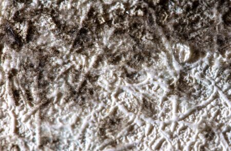
Backgrounds of Colony Characteristics of Mold under the microscope.
Коллекция по умолчанию
Коллекция по умолчанию
Создать новую
Microscopic Visualization of Darkly Pigmented Fungal Conidia and Hyphal Structures in a Clear Wet Mount Specimen.
Коллекция по умолчанию
Коллекция по умолчанию
Создать новую

Budding yeast cells with pseudohyphae Gram stain method
Коллекция по умолчанию
Коллекция по умолчанию
Создать новую
Nocardia bacteria, 3D illustration. Nocardia are rod-shaped Gram-positive bacteria that cause pulmonary infection nocardiosis, tropical infection of skin and bones mycetoma, nocardiosis in animals
Коллекция по умолчанию
Коллекция по умолчанию
Создать новую
Aspergillus clavatus mold fungi and molecule of patulin toxin, 3D illustration. A mycotoxin produced by mold fungi Aspergillus, Penicillium and Byssochlamys, found in rotting apples and other foods
Коллекция по умолчанию
Коллекция по умолчанию
Создать новую

Whooping cough bacteria Bordetella pertussis in respiratory tract, 3D illustration showing cilia of respiratory epithelium and bacteria
Коллекция по умолчанию
Коллекция по умолчанию
Создать новую
Bacterial colony under the microscope
Коллекция по умолчанию
Коллекция по умолчанию
Создать новую
Colony characteristic of Actinomyces, Bacteria, yeast and Mold on selective media from soil samples for study in laboratory microbiology.
Коллекция по умолчанию
Коллекция по умолчанию
Создать новую
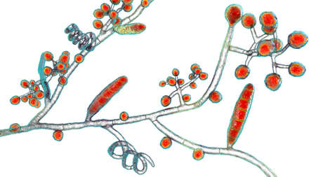
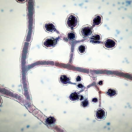
Fungi Coccidioides immitis, saprophytic stage, 3D illustration showing fungal arthroconidia. Pathogenic fungi that reside in soil and can cause infection coccidioidomycosis, or Valley fever
Коллекция по умолчанию
Коллекция по умолчанию
Создать новую
Closeup of Mold. Yellow Mold Growing on background. Microbiology life
Коллекция по умолчанию
Коллекция по умолчанию
Создать новую
Image of data processing over dna strand on black background. Science, medicine and data processing concept digitally generated image.
Коллекция по умолчанию
Коллекция по умолчанию
Создать новую
gram stian was show gram positive bacilli.
Коллекция по умолчанию
Коллекция по умолчанию
Создать новую
Mold growing in yellow, orange, red, white and blue showing signs of multiplication and reproduction
Коллекция по умолчанию
Коллекция по умолчанию
Создать новую
Magnified view of diverse microbes shows various shapes and sizes against a colorful background. This observation highlights the complexity of microbial ecosystems.
Коллекция по умолчанию
Коллекция по умолчанию
Создать новую

Aspergillus niger and Aspergillus oryzae (mold) under microscope for Microbiology in Lab.
Коллекция по умолчанию
Коллекция по умолчанию
Создать новую
Microscopic View of Septate Dematiaceous Fungal Hyphae and Possible Chlamydospores
Коллекция по умолчанию
Коллекция по умолчанию
Создать новую
Candida tropicalis yeasts, microscopic fungi that cause infections in immunocompromised patients. Scientific 3D illustration showing pseudohyphae and blastoconidia formed singly or in small groups
Коллекция по умолчанию
Коллекция по умолчанию
Создать новую
Rhizopus is a genus of common saprophytic fungi. Rhizopus bread mold under the microscope.
Коллекция по умолчанию
Коллекция по умолчанию
Создать новую
Botulinum therapy techniques utilizing plant extracts for medical treatments are highlighted with delicate lighting and focus on the floral elements.
Коллекция по умолчанию
Коллекция по умолчанию
Создать новую
Picture of fungus and red blood cells in hemoculture tube, analyze by microscope 400x
Коллекция по умолчанию
Коллекция по умолчанию
Создать новую
Branching budding yeast cells with pseudohyphae in urine sample fine with microscope.
Коллекция по умолчанию
Коллекция по умолчанию
Создать новую
Mold on the wallpaper on the wall in the room. Macro
Коллекция по умолчанию
Коллекция по умолчанию
Создать новую
water algae cell macro, micrograph.
Коллекция по умолчанию
Коллекция по умолчанию
Создать новую
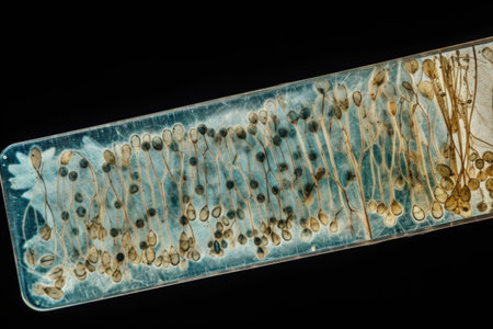
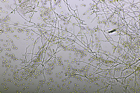
Microscopic view of intricate branching structures inside a petri dish, showcasing a captivating biological composition.
Коллекция по умолчанию
Коллекция по умолчанию
Создать новую
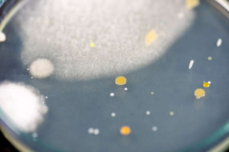
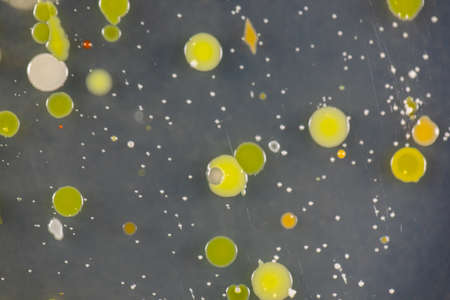
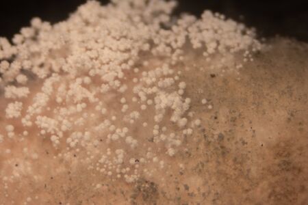
a mold colony on an agar plate
Коллекция по умолчанию
Коллекция по умолчанию
Создать новую
Host cells with spores (mold) are inside wood under the microscope for education.
Коллекция по умолчанию
Коллекция по умолчанию
Создать новую

Microscopic fungi Trichosporon, 3D illustration shows septate hyphae, pseudohyphae, blastoconidia singly or in short chains, arthroconidia. Cause white piedra, superficial and invasive infections
Коллекция по умолчанию
Коллекция по умолчанию
Создать новую
Multi-colored spots and lines. Blurred defocused background for web design
Коллекция по умолчанию
Коллекция по умолчанию
Создать новую
Backgrounds of Characteristics and Different shaped Colony of Bacteria and Mold growing on agar plates from Soil samples for education in Microbiology laboratory.
Коллекция по умолчанию
Коллекция по умолчанию
Создать новую
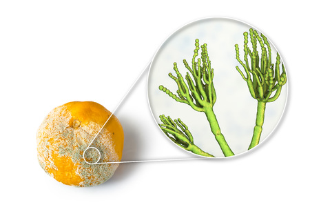
A mandarin with mold. Photo and 3D illustration of microscopic fungi Penicillium which cause food spoilage and produce antibiotic penicillin
Коллекция по умолчанию
Коллекция по умолчанию
Создать новую
Backgrounds of Characteristics and Different shaped Colony of Bacteria and Mold growing on agar plates from Soil samples for education in Microbiology laboratory.
Коллекция по умолчанию
Коллекция по умолчанию
Создать новую

Budding yeast cells with pseudohyphae in urine sample finding with microscope 100X
Коллекция по умолчанию
Коллекция по умолчанию
Создать новую
Aspergillus (mold) for Microbiology in Lab.
Коллекция по умолчанию
Коллекция по умолчанию
Создать новую
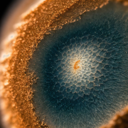
Macro image of the structure of the spiral section of an agate
Коллекция по умолчанию
Коллекция по умолчанию
Создать новую
Legion-Media
Создайте свои проекты на основе качественных стоковых фотографий и видео.


Copyright © Legion-Media.