

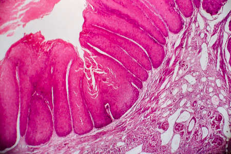
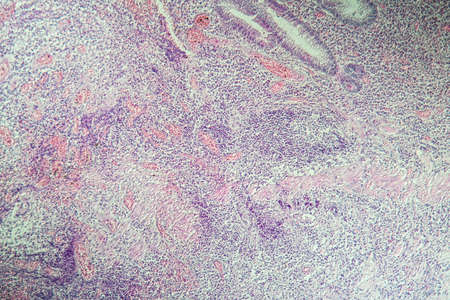
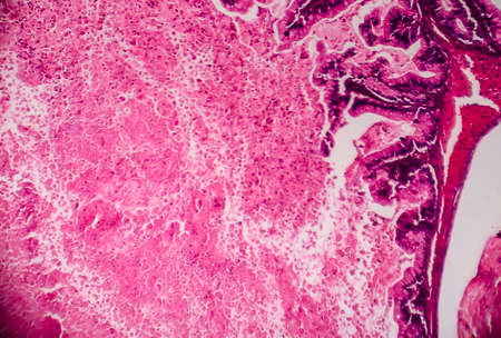
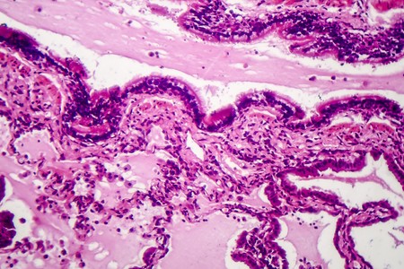
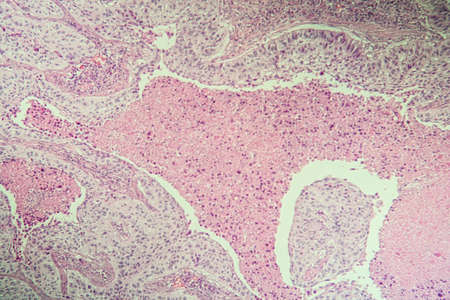
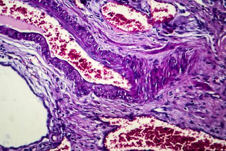
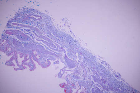
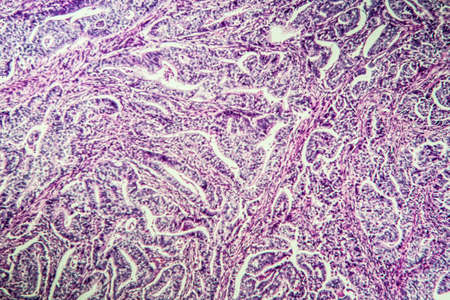
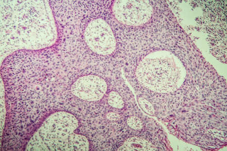
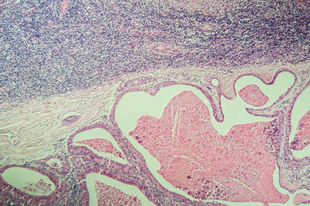
Ovarian cancer, light micrograph, photo under microscope. Photograph shows a fragment of a cancerous tumor in the female ovary. Selective focus
Коллекция по умолчанию
Коллекция по умолчанию
Создать новую
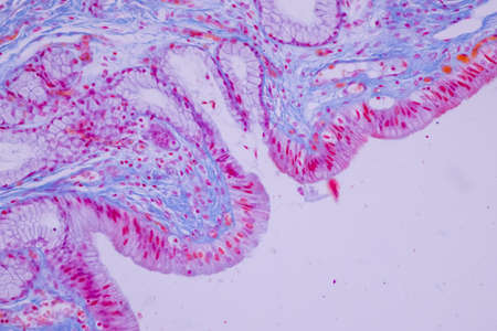
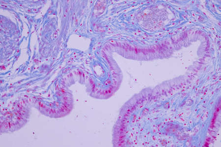
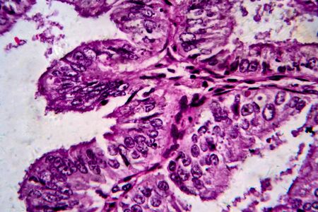
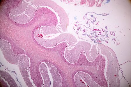
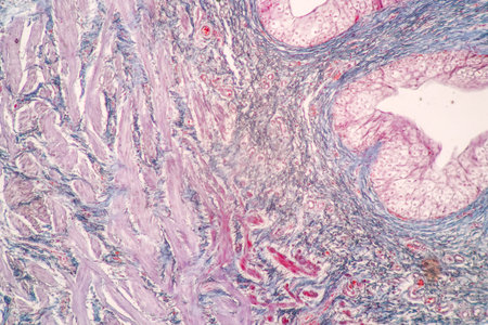
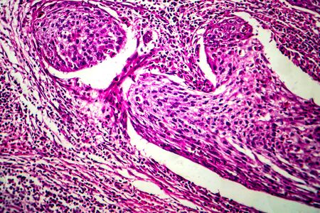
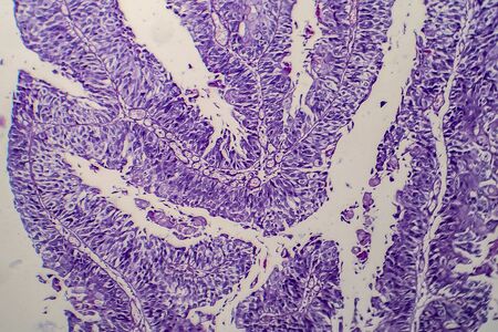
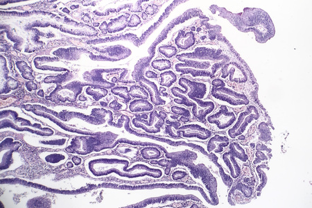
Condyloma acuminatum, also known as genital warts. Light micrograph, photo under microscope
Коллекция по умолчанию
Коллекция по умолчанию
Создать новую
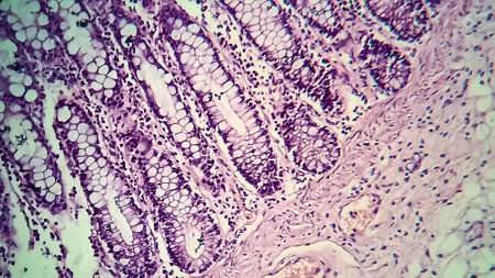
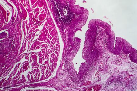
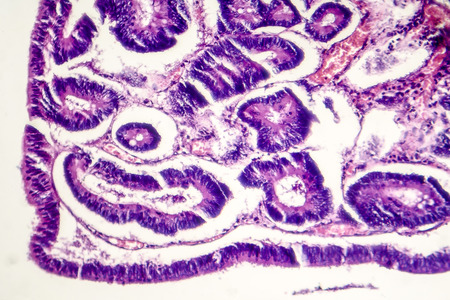
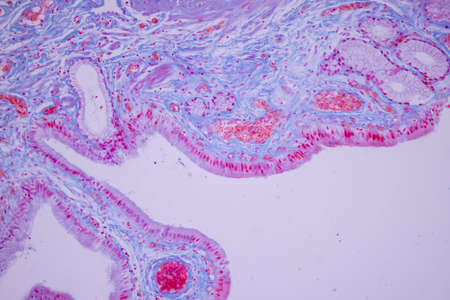
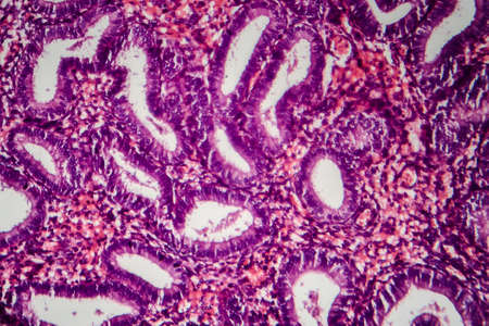
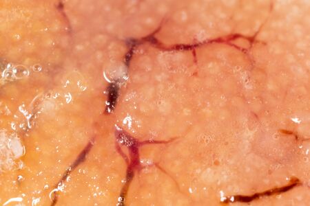
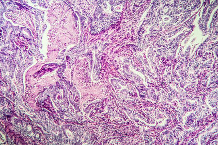
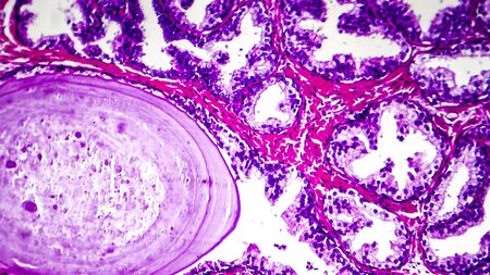
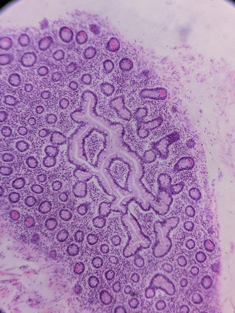
Columnar epithelium of human gall bladder under the microscope in Lab.
Коллекция по умолчанию
Коллекция по умолчанию
Создать новую
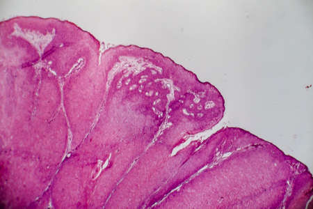
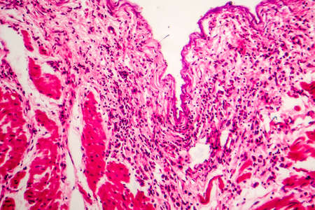
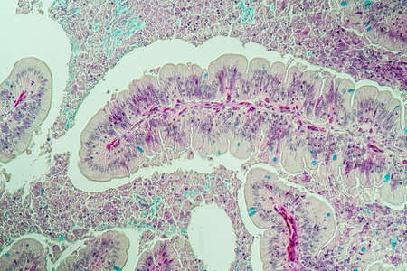
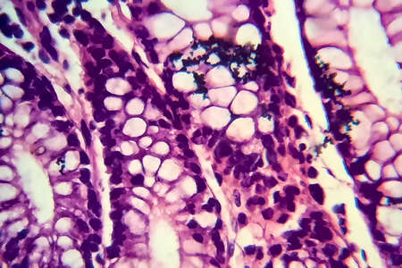
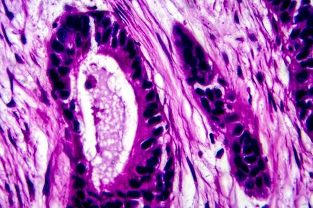
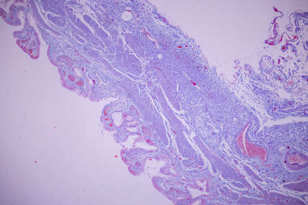
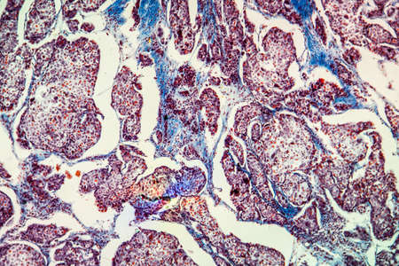
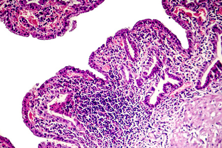
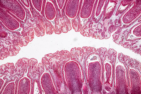
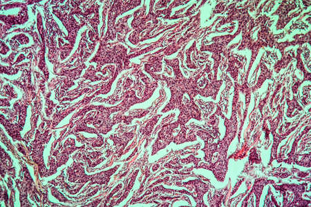
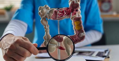
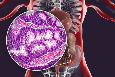
Bacillary dysentery, light micrograph, photo under microscope showing presence of bacteria and accumulation of inflammatory cells in intestinal epithelium
Коллекция по умолчанию
Коллекция по умолчанию
Создать новую

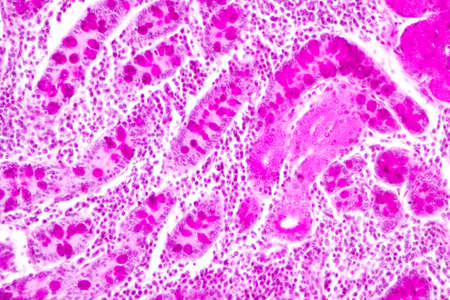
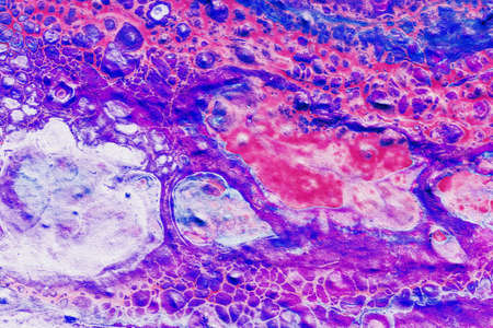
Abstract science background- pyloric division of the stomach of the dog. Cell biology
Коллекция по умолчанию
Коллекция по умолчанию
Создать новую

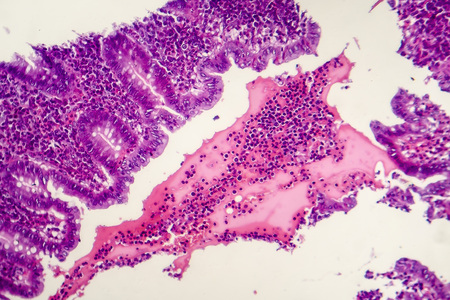
Stomach tissue under the microscope 100x
Коллекция по умолчанию
Коллекция по умолчанию
Создать новую

Endometriosis, a disorder in which cells similar to those in the endometrium grow outside the uterus. Light micrograph, photo under microscope
Коллекция по умолчанию
Коллекция по умолчанию
Создать новую
Condyloma acuminatum, also known as genital warts. Light micrograph, photo under microscope
Коллекция по умолчанию
Коллекция по умолчанию
Создать новую
Esophageal squamous cell carcinoma, light micrograph, photo under microscope
Коллекция по умолчанию
Коллекция по умолчанию
Создать новую
Stomach tissue under the microscope 100x
Коллекция по умолчанию
Коллекция по умолчанию
Создать новую
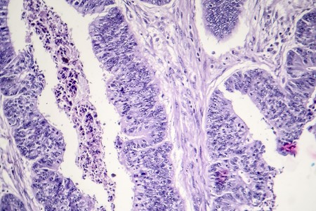
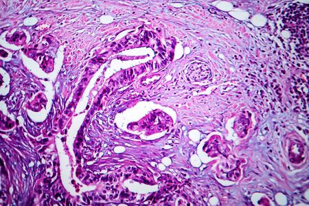
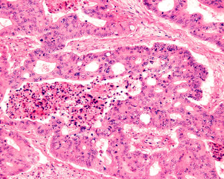
Differentiated intestinal adenocarcinoma, light micrograph, photo under microscope
Коллекция по умолчанию
Коллекция по умолчанию
Создать новую

Colon polyp, one of the largest polyps
Коллекция по умолчанию
Коллекция по умолчанию
Создать новую

Columnar epithelium of human gall bladder under the microscope in Lab.
Коллекция по умолчанию
Коллекция по умолчанию
Создать новую
Colon carcinoma arising from adenoma, 100x
Коллекция по умолчанию
Коллекция по умолчанию
Создать новую
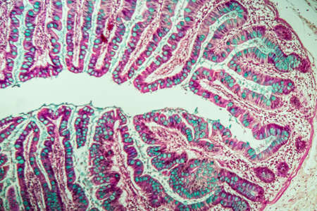
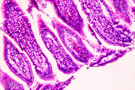
Small intestine with villi under the microscope 100x
Коллекция по умолчанию
Коллекция по умолчанию
Создать новую

Endometriosis, a disorder in which cells similar to those in the endometrium grow outside the uterus. Light micrograph, photo under microscope
Коллекция по умолчанию
Коллекция по умолчанию
Создать новую

Histopathology of interstitial nephritis, light micrograph, photo under microscope
Коллекция по умолчанию
Коллекция по умолчанию
Создать новую
Bacillary dysentery, light micrograph, photo under microscope showing presence of bacteria and accumulation of inflammatory cells in intestinal epithelium
Коллекция по умолчанию
Коллекция по умолчанию
Создать новую

Histopathology of intestinal adenoma, light micrograph, photo under microscope
Коллекция по умолчанию
Коллекция по умолчанию
Создать новую
Education anatomy and Histological sample of Human under the microscope.
Коллекция по умолчанию
Коллекция по умолчанию
Создать новую

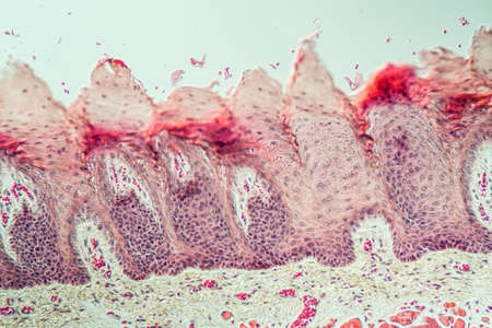
micrograph of medical science stratified squamous epithelium tissue cell
Коллекция по умолчанию
Коллекция по умолчанию
Создать новую
Papillary serous ovarian adenocarcinoma, cancer of ovary, light micrograph, photo under microscope
Коллекция по умолчанию
Коллекция по умолчанию
Создать новую

Cross section of human skin under microscope view for education in laboratory.
Коллекция по умолчанию
Коллекция по умолчанию
Создать новую
Colon inflammation in Crohn's disease 100x
Коллекция по умолчанию
Коллекция по умолчанию
Создать новую
Columnar epithelium of human gall bladder under the microscope in Lab.
Коллекция по умолчанию
Коллекция по умолчанию
Создать новую
Bladder cancer, light micrograph, photo under microscope
Коллекция по умолчанию
Коллекция по умолчанию
Создать новую
Signet ring cell carcinoma of the stomach, light micrograph, photo under microscope
Коллекция по умолчанию
Коллекция по умолчанию
Создать новую
Ovarian cancer, light micrograph, photo under microscope. Photograph shows a fragment of a cancerous tumor in the female ovary. Selective focus
Коллекция по умолчанию
Коллекция по умолчанию
Создать новую
Endometrial adenocarcinoma, light micrograph, photo under microscope
Коллекция по умолчанию
Коллекция по умолчанию
Создать новую
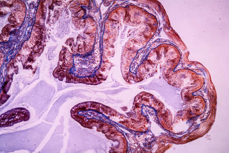
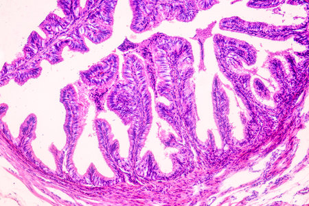
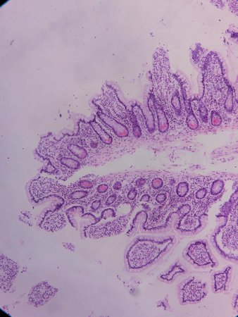
Intestinal polypoid adenoma, light micrograph, photo under microscope
Коллекция по умолчанию
Коллекция по умолчанию
Создать новую
Slow worm histology bowel transverse 100x
Коллекция по умолчанию
Коллекция по умолчанию
Создать новую

Ovarian cancer, light micrograph, photo under microscope. Photograph shows a fragment of a cancerous tumor in the female ovary. Selective focus
Коллекция по умолчанию
Коллекция по умолчанию
Создать новую
Columnar epithelium of human gall bladder under the microscope in Lab.
Коллекция по умолчанию
Коллекция по умолчанию
Создать новую
Bacillary dysentery, light micrograph, photo under microscope showing presence of bacteria and accumulation of inflammatory cells in intestinal epithelium
Коллекция по умолчанию
Коллекция по умолчанию
Создать новую
Tissue of Small intestine (Duodenum) and Vermiform appendix Human under the microscope in Lab.
Коллекция по умолчанию
Коллекция по умолчанию
Создать новую
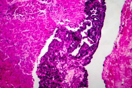
Suppurative appendicitis, light micrograph, photo under microscope showing neutrophilic infiltrates of the appendix wall and lumen
Коллекция по умолчанию
Коллекция по умолчанию
Создать новую
Breast cancer, light micrograph, photo under microscope
Коллекция по умолчанию
Коллекция по умолчанию
Создать новую

Education anatomy and Histological sample Spinal cord Tissue under the microscope.
Коллекция по умолчанию
Коллекция по умолчанию
Создать новую
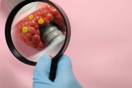
Endocrinologist looking at model of thyroid gland through magnifying glass on pink background, closeup. Space for text
Коллекция по умолчанию
Коллекция по умолчанию
Создать новую
Tissue of Stomach Human under the microscope in Lab.
Коллекция по умолчанию
Коллекция по умолчанию
Создать новую
Histopathology of intestinal adenoma, light micrograph, photo under microscope
Коллекция по умолчанию
Коллекция по умолчанию
Создать новую
Tongue Tissue with taste buds across 200x
Коллекция по умолчанию
Коллекция по умолчанию
Создать новую
Chronic cholecystitis, light micrograph, photo under microscope showing fibrosis and muscular hypertrophy of gallbladder wall, entrapped epithelial crypts, foamy macrophages
Коллекция по умолчанию
Коллекция по умолчанию
Создать новую
Histopathology of human liver under microscope view for medical education.
Коллекция по умолчанию
Коллекция по умолчанию
Создать новую
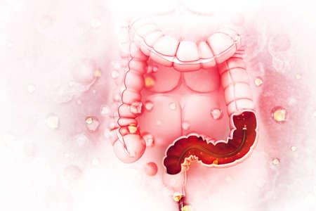
Doctor examines digestive system model and ultrasound image. Stomach intestine ulcers cyst and oncology and examination
Коллекция по умолчанию
Коллекция по умолчанию
Создать новую

Villous colon adenocarcinoma, light micrograph, photo under microscope
Коллекция по умолчанию
Коллекция по умолчанию
Создать новую
Lung adenocarcinoma, light micrograph, photo under microscope
Коллекция по умолчанию
Коллекция по умолчанию
Создать новую
Cerebellum and Nerve human under the microscope for education in Lab.
Коллекция по умолчанию
Коллекция по умолчанию
Создать новую
Endometriosis, a disorder in which cells similar to those in the endometrium grow outside the uterus. Light micrograph, photo under microscope
Коллекция по умолчанию
Коллекция по умолчанию
Создать новую
Uterine cancer, light micrograph, photo under microscope
Коллекция по умолчанию
Коллекция по умолчанию
Создать новую
Squamous cell carcinoma diseased tissue under the microscope 100x
Коллекция по умолчанию
Коллекция по умолчанию
Создать новую
Colonoscopy on scientific background. 3d illustration
Коллекция по умолчанию
Коллекция по умолчанию
Создать новую
Education anatomy and Histological sample of Human under the microscope.
Коллекция по умолчанию
Коллекция по умолчанию
Создать новую
Extreme Close up of microscopic kidney Bowman's Capsule and Glomerulus
Коллекция по умолчанию
Коллекция по умолчанию
Создать новую

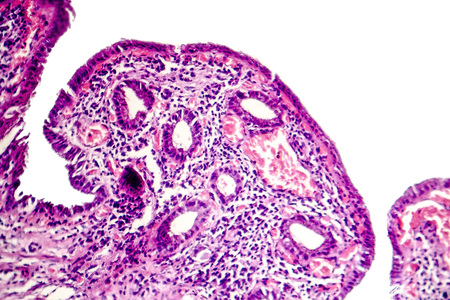
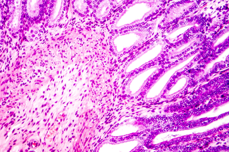
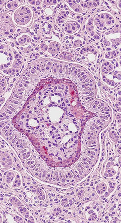
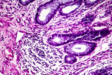
Histopathology of chronic atrophic gastritis, light micrograph, photo under microscope
Коллекция по умолчанию
Коллекция по умолчанию
Создать новую
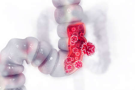
Colon cancer. Cancer attacking cell. Colon disease concept. 3d illustration
Коллекция по умолчанию
Коллекция по умолчанию
Создать новую
Columnar epithelium of human gall bladder under the microscope in Lab.
Коллекция по умолчанию
Коллекция по умолчанию
Создать новую
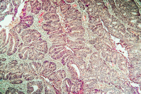
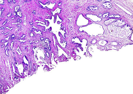
Large-bowel adenocarcinoma. Cancer cells arranged in cords or strands with empty central spaces remembering the normal crypts of the colon mucosa.
Коллекция по умолчанию
Коллекция по умолчанию
Создать новую
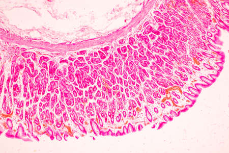
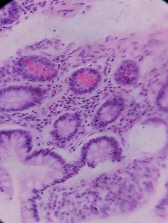
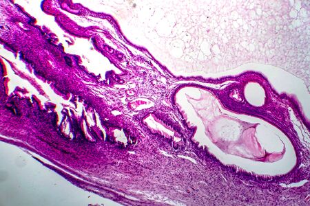
Histology of human stomach, fundic region. Light micrograph, isolated on white background, hematoxylin and eosin staining
Коллекция по умолчанию
Коллекция по умолчанию
Создать новую

Columnar epithelium of human gall bladder under the microscope in Lab.
Коллекция по умолчанию
Коллекция по умолчанию
Создать новую

Villous colon adenocarcinoma, light micrograph, photo under microscope. High magnification
Коллекция по умолчанию
Коллекция по умолчанию
Создать новую
Chronic nephritis, light micrograph, photo under microscope
Коллекция по умолчанию
Коллекция по умолчанию
Создать новую
Histological Uterus human, Uterine tube human, Placenta human and Umbilical cord Human under the microscope for education.
Коллекция по умолчанию
Коллекция по умолчанию
Создать новую
fish caviar as a background. macro
Коллекция по умолчанию
Коллекция по умолчанию
Создать новую
Basal cell cancer Diseased tissue 100x
Коллекция по умолчанию
Коллекция по умолчанию
Создать новую
Histopathology of cholera under microscope view for education.
Коллекция по умолчанию
Коллекция по умолчанию
Создать новую
Painting acrylic paint- abstract drawing. Texture background
Коллекция по умолчанию
Коллекция по умолчанию
Создать новую
Chronic cholecystitis, light micrograph, photo under microscope showing fibrosis and muscular hypertrophy of gallbladder wall, entrapped epithelial crypts, foamy macrophages
Коллекция по умолчанию
Коллекция по умолчанию
Создать новую
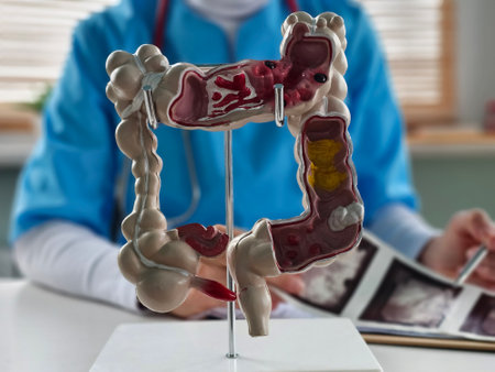
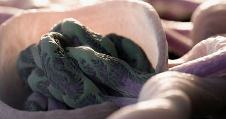
Detailed anatomical model of the human digestive system displayed during a medical consultation in a clinic
Коллекция по умолчанию
Коллекция по умолчанию
Создать новую
Columnar epithelium of human gall bladder under the microscope in Lab.
Коллекция по умолчанию
Коллекция по умолчанию
Создать новую

Ovarian cancer, light micrograph, photo under microscope. Photograph shows a fragment of a cancerous tumor in the female ovary. Selective focus
Коллекция по умолчанию
Коллекция по умолчанию
Создать новую
Squamous cell carcinoma of the uterus, light micrograph, photo under microscope
Коллекция по умолчанию
Коллекция по умолчанию
Создать новую

intestinal constipation. Bowel disorder characterized by difficulty in excreting faeces. Laxatives resolve the disorder. 3D rendering
Коллекция по умолчанию
Коллекция по умолчанию
Создать новую

Chronic pyelonephritis, light micrograph, photo under microscope. High magnification
Коллекция по умолчанию
Коллекция по умолчанию
Создать новую
Gastric carcinoma in tissue section 100x
Коллекция по умолчанию
Коллекция по умолчанию
Создать новую
Pathology and Histology Tissue of Mammals under microscope.
Коллекция по умолчанию
Коллекция по умолчанию
Создать новую
Gastric carcinoma in tissue section 100x
Коллекция по умолчанию
Коллекция по умолчанию
Создать новую
Bladder transitional cell carcinoma, light micrograph, photo under microscope
Коллекция по умолчанию
Коллекция по умолчанию
Создать новую
Histopathology of prostate gland hyperplasia, light micrograph, photo under microscope
Коллекция по умолчанию
Коллекция по умолчанию
Создать новую
Breast cancer of the woman diseased tissue 100x
Коллекция по умолчанию
Коллекция по умолчанию
Создать новую

Colon cancer. Colon disease concept
Коллекция по умолчанию
Коллекция по умолчанию
Создать новую
Bowen's Disease Tumor under the microscope 100x
Коллекция по умолчанию
Коллекция по умолчанию
Создать новую
Histopathology of fibroids, light micrograph, photo under microscope
Коллекция по умолчанию
Коллекция по умолчанию
Создать новую
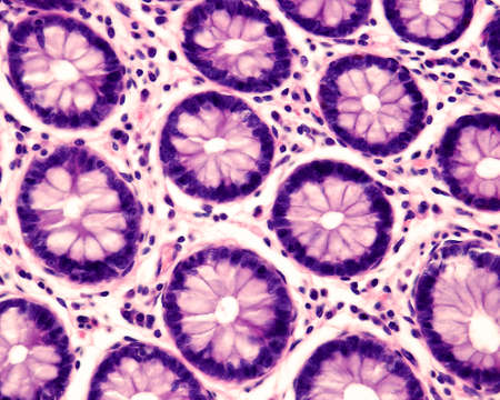
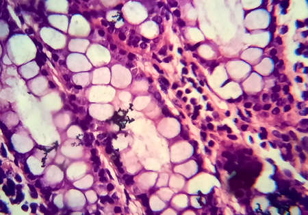
Cross section of intestinal glands (crypts of Lieberkühn) showing mucous goblet cells. Human colon.
Коллекция по умолчанию
Коллекция по умолчанию
Создать новую
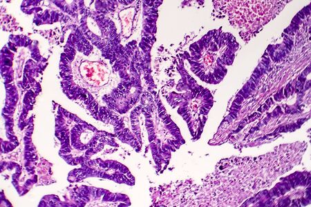
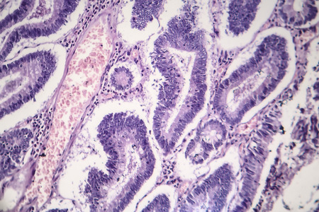
Well-differentiated intestinal adenocarcinoma, light micrograph, photo under microscope
Коллекция по умолчанию
Коллекция по умолчанию
Создать новую

Papillary thyroid carcinoma, light micrograph, photo under microscope. The most common type of thyroid cancer
Коллекция по умолчанию
Коллекция по умолчанию
Создать новую
Salivary gland swollen diseased tissue under the microscope 100x
Коллекция по умолчанию
Коллекция по умолчанию
Создать новую

Histopathology of intestinal adenoma, light micrograph, photo under microscope
Коллекция по умолчанию
Коллекция по умолчанию
Создать новую
Histopathology of intestinal adenoma, light micrograph, photo under microscope
Коллекция по умолчанию
Коллекция по умолчанию
Создать новую
Pancreas cancer cell under microscope view for medical education.
Коллекция по умолчанию
Коллекция по умолчанию
Создать новую
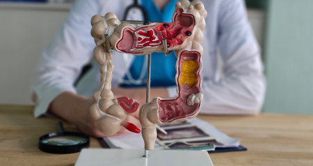
Doctor explains model of intestine and appendicitis discusses anatomy concept
Коллекция по умолчанию
Коллекция по умолчанию
Создать новую
Prostate cancer of a human, highly detailed segment of panorama. Photomicrograph as seen under the microscope, 10x zoom.
Коллекция по умолчанию
Коллекция по умолчанию
Создать новую
Ovarian mucinous cystadenoma, a benign tumor of ovary, light micrograph, photo under microscope
Коллекция по умолчанию
Коллекция по умолчанию
Создать новую

Section of a dog ciliated epithelium under the microscope.
Коллекция по умолчанию
Коллекция по умолчанию
Создать новую
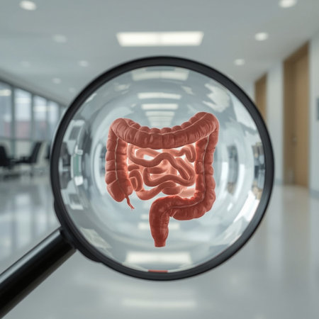
Magnifying glass enlarging large intestine inside hospital building. 3D rendering
Коллекция по умолчанию
Коллекция по умолчанию
Создать новую
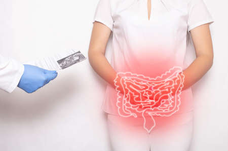
The doctor holds the results of the examination of the female patient on a white background. Bowel inflammation and disease concept, abdominal pain, dolichosigma
Коллекция по умолчанию
Коллекция по умолчанию
Создать новую

Histopathology of human under microscope view for education in laboratory.
Коллекция по умолчанию
Коллекция по умолчанию
Создать новую
Colon cancer, 3D illustration and photo under microscope. Light micrograph showing colon adenocarcinoma
Коллекция по умолчанию
Коллекция по умолчанию
Создать новую

Smokers lung, histopathology, light micrograph showing accumulation of carbon particles in lung tissue
Коллекция по умолчанию
Коллекция по умолчанию
Создать новую

Colon cancer. Cancer attacking cell. Colon disease concept. 3d illustration
Коллекция по умолчанию
Коллекция по умолчанию
Создать новую
Legion-Media
Создайте свои проекты на основе качественных стоковых фотографий и видео.


Copyright © Legion-Media.