

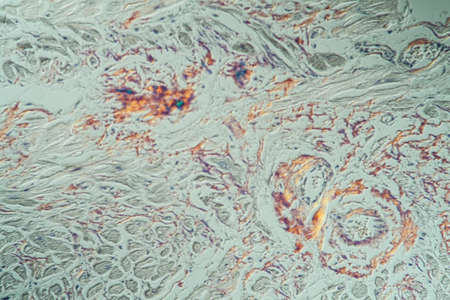
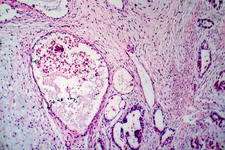
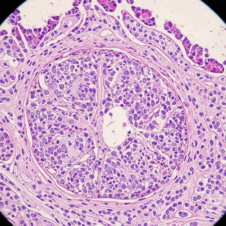
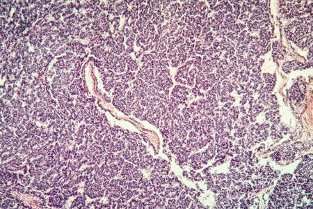
Thyroid follicular carcinoma, light micrograph, photo under microscope
Коллекция по умолчанию
Коллекция по умолчанию
Создать новую

Ovarian cancer, light micrograph, photo under microscope. Photograph shows a fragment of a cancerous tumor in the female ovary. Selective focus
Коллекция по умолчанию
Коллекция по умолчанию
Создать новую
Close up of patients wound and medical workers hand in sterile gloves doing abdominoplasty surgery. Plastic surgeon and assistant using medical instruments. Concept of medicine, abdominoplasty.
Коллекция по умолчанию
Коллекция по умолчанию
Создать новую
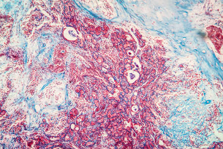
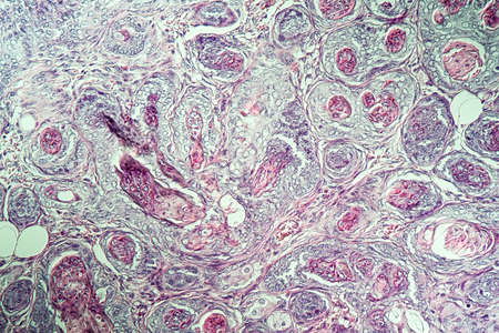
Fibroepithelium Diseased tissue 100x
Коллекция по умолчанию
Коллекция по умолчанию
Создать новую

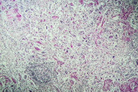
diseased liver with cirrhosis 100x under the microscope
Коллекция по умолчанию
Коллекция по умолчанию
Создать новую

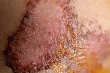
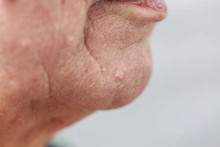
Freshly healed scar on the neck after thyroid surgery.
Коллекция по умолчанию
Коллекция по умолчанию
Создать новую

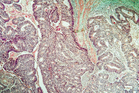
Papillary thyroid carcinoma, light micrograph, photo under microscope. The most common type of thyroid cancer
Коллекция по умолчанию
Коллекция по умолчанию
Создать новую
Squamous cell carcinoma of the uterus, light micrograph, photo under microscope
Коллекция по умолчанию
Коллекция по умолчанию
Создать новую

Wound from laser on a face from dermatologist
Коллекция по умолчанию
Коллекция по умолчанию
Создать новую
Colon tissue with diverticulum 100x
Коллекция по умолчанию
Коллекция по умолчанию
Создать новую
hearth with amyloid deposits of sick tissue under the microscope 200x
Коллекция по умолчанию
Коллекция по умолчанию
Создать новую
Metastases tumor diseased tissue 100x
Коллекция по умолчанию
Коллекция по умолчанию
Создать новую
Colon carcinoma arising from adenoma, 100x
Коллекция по умолчанию
Коллекция по умолчанию
Создать новую
Papillary thyroid carcinoma, light micrograph, photo under microscope. The most common type of thyroid cancer
Коллекция по умолчанию
Коллекция по умолчанию
Создать новую
Close-up Medical Image: Lipoma Growth on Scalp - Documentary Photography Style
Коллекция по умолчанию
Коллекция по умолчанию
Создать новую
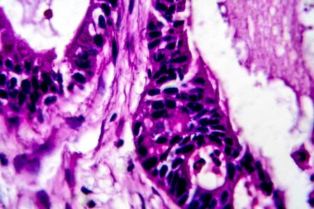
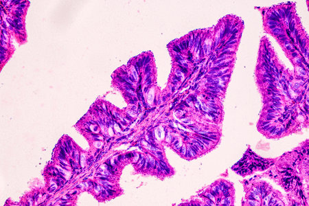
Columnar epithelium of human gall bladder under the microscope in Lab.
Коллекция по умолчанию
Коллекция по умолчанию
Создать новую
Breast cancer, light micrograph, photo under microscope
Коллекция по умолчанию
Коллекция по умолчанию
Создать новую
Tongue Tissue with taste buds across 200x
Коллекция по умолчанию
Коллекция по умолчанию
Создать новую
Light micrograph of teratoma, a tumor made up of several different types of tissue, such as hair, teeth, muscle, or bone. Teratoma is typically found in the ovary, testicle, or coccyx
Коллекция по умолчанию
Коллекция по умолчанию
Создать новую
Carcinoma in guinea pigs, tissue 100x
Коллекция по умолчанию
Коллекция по умолчанию
Создать новую

Stomach tissue under the microscope 100x
Коллекция по умолчанию
Коллекция по умолчанию
Создать новую

Papillary thyroid carcinoma, light micrograph, photo under microscope. The most common type of thyroid cancer
Коллекция по умолчанию
Коллекция по умолчанию
Создать новую
Lungworm under the microscope 100x
Коллекция по умолчанию
Коллекция по умолчанию
Создать новую
Closeup injured, Fresh wounds from accident.
Коллекция по умолчанию
Коллекция по умолчанию
Создать новую
Wound on the knee on the child leg. A Scar on the skin of the child.
Коллекция по умолчанию
Коллекция по умолчанию
Создать новую
Bacillary dysentery, light micrograph, photo under microscope showing presence of bacteria and accumulation of inflammatory cells in intestinal epithelium
Коллекция по умолчанию
Коллекция по умолчанию
Создать новую
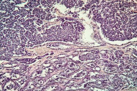
Cancer swollen Parotid diseased tissue 100x
Коллекция по умолчанию
Коллекция по умолчанию
Создать новую
Ovarian cancer, light micrograph, photo under microscope. Photograph shows a fragment of a cancerous tumor in the female ovary. Selective focus
Коллекция по умолчанию
Коллекция по умолчанию
Создать новую
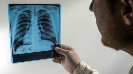
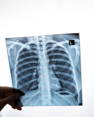
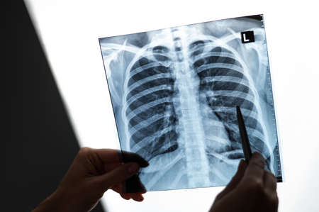
X-ray picture in the hands of a doctor on a white background, a medical worker looks at a picture of an X-ray of the human lungs.
Коллекция по умолчанию
Коллекция по умолчанию
Создать новую

Characteristics of Lichen, hyphae and Symbiotic algae under the microscope for education.
Коллекция по умолчанию
Коллекция по умолчанию
Создать новую
Colon inflammation in Crohn's disease 100x
Коллекция по умолчанию
Коллекция по умолчанию
Создать новую
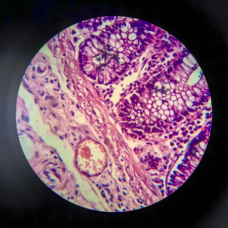
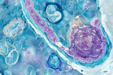
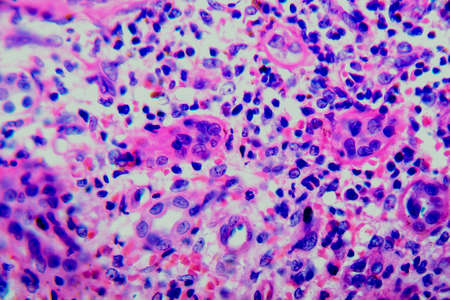
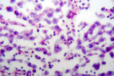
This detailed microscopic image showcases various cellular structures, highlighted in striking purple tones. The intricate patterns and textures reveal the complexity of biological tissues, making it a valuable resource for educational and scientific purposes
Коллекция по умолчанию
Коллекция по умолчанию
Создать новую
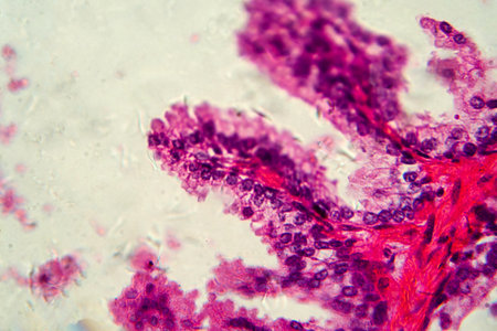
A closeup of glandular epithelium highlighting its specialized cells for secretion and absorption
Коллекция по умолчанию
Коллекция по умолчанию
Создать новую
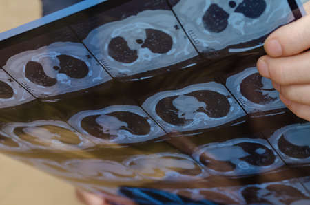
A man's hand holds the CT scan. Transverse view or axial plain of CT chest showing normal study of heart, lungs, spine, rib, other. Selective focuse
Коллекция по умолчанию
Коллекция по умолчанию
Создать новую
Goiter colloid goiter disease 100x
Коллекция по умолчанию
Коллекция по умолчанию
Создать новую

Details of senior woman face. Elderly pensioner female, dermal fibroma close up.
Коллекция по умолчанию
Коллекция по умолчанию
Создать новую
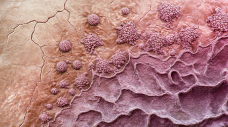
3D illustration of a close-up of skin cancer like the malign melanoma inflaming surrounding tissue.
Коллекция по умолчанию
Коллекция по умолчанию
Создать новую
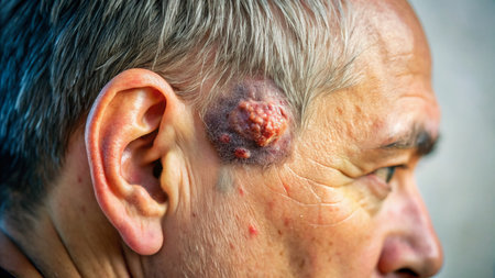
Basal cell cancer Diseased tissue 100x
Коллекция по умолчанию
Коллекция по умолчанию
Создать новую

Condyloma acuminatum, also known as genital warts. Light micrograph, photo under microscope
Коллекция по умолчанию
Коллекция по умолчанию
Создать новую
Histopathology of cirrhosis, light micrograph, photo under microscope
Коллекция по умолчанию
Коллекция по умолчанию
Создать новую
Uterine cancer, light micrograph, photo under microscope
Коллекция по умолчанию
Коллекция по умолчанию
Создать новую
Details of senior woman face. Elderly pensioner female, chin and lips close up.
Коллекция по умолчанию
Коллекция по умолчанию
Создать новую
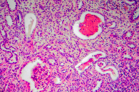
Chronic pyelonephritis, light micrograph, photo under microscope
Коллекция по умолчанию
Коллекция по умолчанию
Создать новую
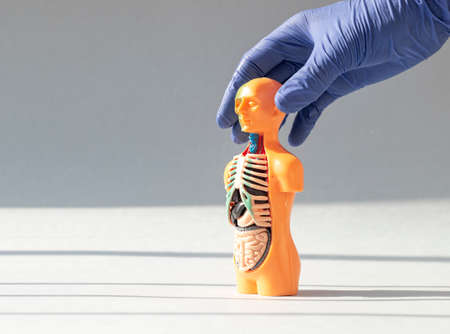
3d human model with inner organs. Medical anatomical concept. Anatomy banner with copy space for text.
Коллекция по умолчанию
Коллекция по умолчанию
Создать новую
A colorful image of a cell with a purple and blue blob in the center. The image is abstract and has a mood of curiosity and wonder
Коллекция по умолчанию
Коллекция по умолчанию
Создать новую
Horizontal view on open stomach during an operation
Коллекция по умолчанию
Коллекция по умолчанию
Создать новую
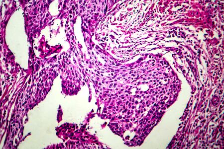
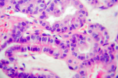
Photomicrograph showing histological features of benign prostatic hyperplasia. Enlarged prostate gland with nodular proliferation of glandular and stromal components. High-resolution histology image.
Коллекция по умолчанию
Коллекция по умолчанию
Создать новую
a pimple on the skin of a light-skinned man. a pimple on the skin of a light-skinned man.
Коллекция по умолчанию
Коллекция по умолчанию
Создать новую
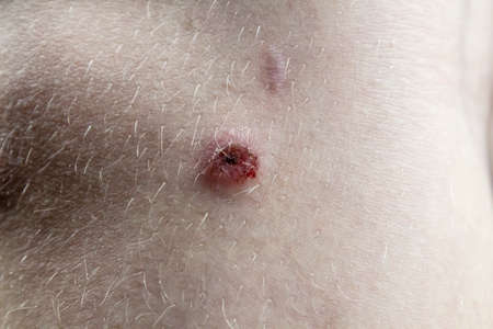
A wound on a woman’s neck after burning a wart, close-up. Round painful hole in human skin, purulent wound
Коллекция по умолчанию
Коллекция по умолчанию
Создать новую

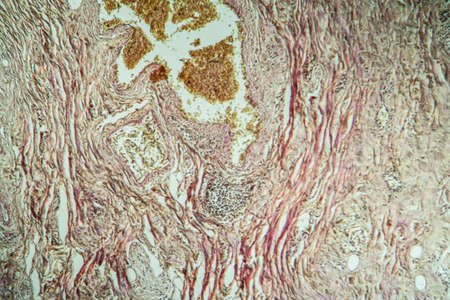
Hemosiderosis liver 200x under a microscope
Коллекция по умолчанию
Коллекция по умолчанию
Создать новую
Laser removal of wart from the toe of foot. Cut out verruca.
Коллекция по умолчанию
Коллекция по умолчанию
Создать новую
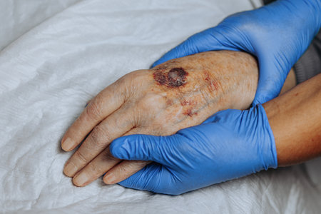
Senior adult male with stitches in the cut after surgery for removal of basal cell carcinoma caused by sun damage
Коллекция по умолчанию
Коллекция по умолчанию
Создать новую

Black tongue. A man shows the consequences of an injury, bite or burn of the tongue. Part is damaged. Treatment of internal injuries
Коллекция по умолчанию
Коллекция по умолчанию
Создать новую
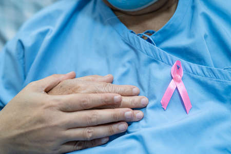
Breast cancer, pink ribbon at Asian senior lady patient for supporting awareness.
Коллекция по умолчанию
Коллекция по умолчанию
Создать новую
Actinomyces in the jaw diseased tissue 200x
Коллекция по умолчанию
Коллекция по умолчанию
Создать новую

Breast ductal carcinoma, light micrograph, photo under microscope
Коллекция по умолчанию
Коллекция по умолчанию
Создать новую
close up of red cabbage leaf with water droplets, abstract background
Коллекция по умолчанию
Коллекция по умолчанию
Создать новую
removal of benign tumor on the dogs paw by surgery
Коллекция по умолчанию
Коллекция по умолчанию
Создать новую
Doctor showing x-ray of patients lungs.
Коллекция по умолчанию
Коллекция по умолчанию
Создать новую

Skin disease. Closeup brown mole on caucasian human body.
Коллекция по умолчанию
Коллекция по умолчанию
Создать новую
Breast cancer, light micrograph, photo under microscope
Коллекция по умолчанию
Коллекция по умолчанию
Создать новую
Uterine cancer, light micrograph, photo under microscope
Коллекция по умолчанию
Коллекция по умолчанию
Создать новую
Atrophy kidney tissue under the microscope 100x
Коллекция по умолчанию
Коллекция по умолчанию
Создать новую
Metastases tumor diseased tissue 100x
Коллекция по умолчанию
Коллекция по умолчанию
Создать новую
Doctor showing x-ray of patients lungs.
Коллекция по умолчанию
Коллекция по умолчанию
Создать новую
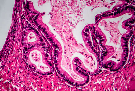
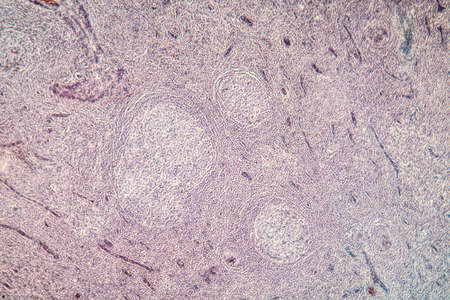
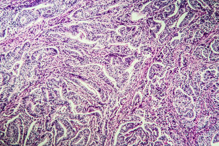
Education anatomy and Histological sample of Human under the microscope.
Коллекция по умолчанию
Коллекция по умолчанию
Создать новую

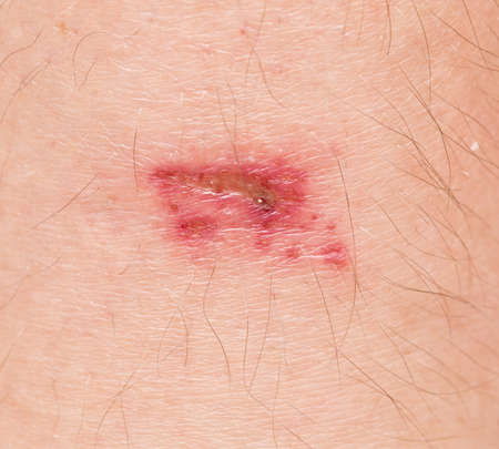
Scab wound infected on man arm closeup
Коллекция по умолчанию
Коллекция по умолчанию
Создать новую
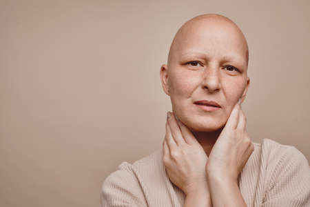
Minimal head and shoulders portrait of bald woman looking at camera while posing against beige background in studio, alopecia and cancer awareness, copy space
Коллекция по умолчанию
Коллекция по умолчанию
Создать новую
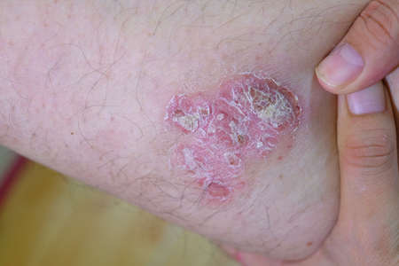
Psoriasis
Коллекция по умолчанию
Коллекция по умолчанию
Создать новую
Chronic pyelonephritis, light micrograph, photo under microscope
Коллекция по умолчанию
Коллекция по умолчанию
Создать новую
Lung adenocarcinoma, light micrograph, photo under microscope
Коллекция по умолчанию
Коллекция по умолчанию
Создать новую
Palatal tonsils transverse 100x under a microscope
Коллекция по умолчанию
Коллекция по умолчанию
Создать новую

Close up of hydrogen peroxide in a wound on human skin. Macro
Коллекция по умолчанию
Коллекция по умолчанию
Создать новую
A bruise on the hand of an elderly person. Known as senile purpura. Caused by the fragility of the skin and blood vessels in old age. Elderly care
Коллекция по умолчанию
Коллекция по умолчанию
Создать новую
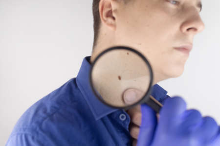
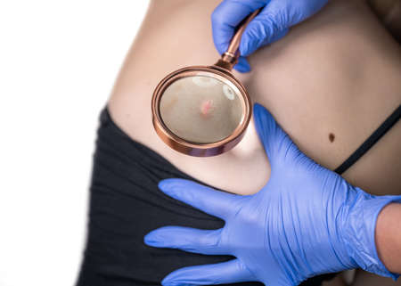
A man at a dermatologist appointment shows his birthmarks, moles and nevi. The doctor examines the patient with a dermatoscope. Benign and malignant birthmarks. Skin abnormalities care concept
Коллекция по умолчанию
Коллекция по умолчанию
Создать новую
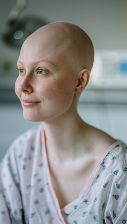
A cheerful woman with a shaved head smiles in a hospital setting, wearing a gown.
Коллекция по умолчанию
Коллекция по умолчанию
Создать новую
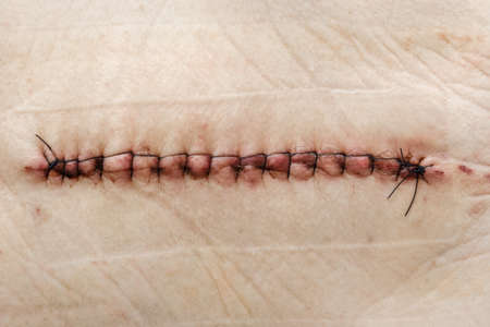
SuPostoperative healing suture on human skin with black medical threads rgery on the hip. Postoperative suture on the skin with iodine. Un Stitched up skin after an operation
Коллекция по умолчанию
Коллекция по умолчанию
Создать новую
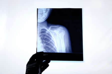
X-ray image of a man with a broken collarbone
Коллекция по умолчанию
Коллекция по умолчанию
Создать новую
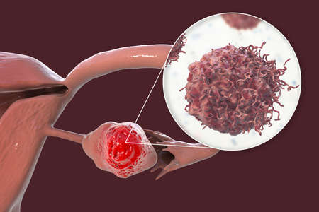
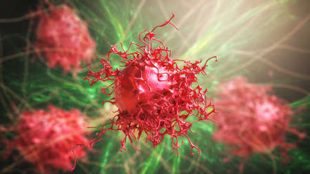
Ovarian cancer, 3D illustration showing malignant tumor in the left ovary and close-up view of cancer cells
Коллекция по умолчанию
Коллекция по умолчанию
Создать новую
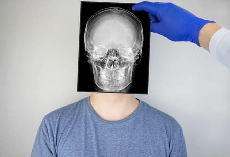
Survey radiography of the skull of a man. A doctor radiologist is studying an x-ray examination. A snapshot of the skull is placed on the patient’s head.
Коллекция по умолчанию
Коллекция по умолчанию
Создать новую
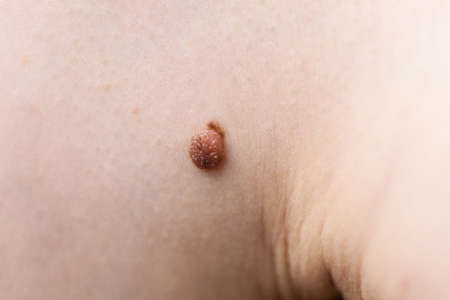
Unrecognizable person having dark fibroma, dermatology problem on skin. Close up of papilloma dermatosis
Коллекция по умолчанию
Коллекция по умолчанию
Создать новую
Chaos ink texture background, ink in water pattern frost. Crystal winter design
Коллекция по умолчанию
Коллекция по умолчанию
Создать новую

Tongue with taste buds Papilla across 100x
Коллекция по умолчанию
Коллекция по умолчанию
Создать новую
Soft fibroid in male under belly, macro photo. Dermatology problem concept. Selective focus
Коллекция по умолчанию
Коллекция по умолчанию
Создать новую
Bacillary dysentery, light micrograph, photo under microscope showing presence of bacteria and accumulation of inflammatory cells in intestinal epithelium
Коллекция по умолчанию
Коллекция по умолчанию
Создать новую
Endometriosis, a disorder in which cells similar to those in the endometrium grow outside the uterus. Light micrograph, photo under microscope
Коллекция по умолчанию
Коллекция по умолчанию
Создать новую

Close-up Medical Image: Lipoma Growth on Scalp - Documentary Photography Style
Коллекция по умолчанию
Коллекция по умолчанию
Создать новую
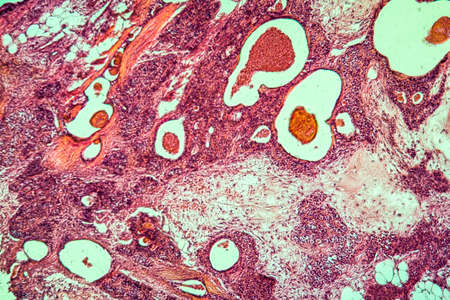
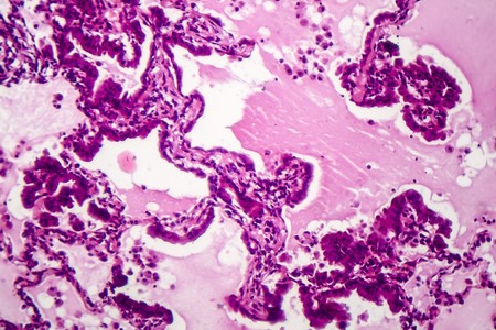
Intricate cells and structures are visible under magnification, displaying unique patterns and textures that reveal complex biological pathways.
Коллекция по умолчанию
Коллекция по умолчанию
Создать новую
Close up shooting of Doctor examines the growths on the skin of a female. Diagnosis of skin cancer. Hemangioma, angioma, papilloma, mole
Коллекция по умолчанию
Коллекция по умолчанию
Создать новую
Gastric carcinoma in tissue section 100x
Коллекция по умолчанию
Коллекция по умолчанию
Создать новую
wound on human skin
Коллекция по умолчанию
Коллекция по умолчанию
Создать новую
Testicular seminoma, light micrograph, photo under microscope. A most common germ cell tumor of the testis
Коллекция по умолчанию
Коллекция по умолчанию
Создать новую

Lacrimal gland tissue under the microscope 100x
Коллекция по умолчанию
Коллекция по умолчанию
Создать новую
Cervical cancer cells, 3D illustration. Malignant tumor of cervix uteri
Коллекция по умолчанию
Коллекция по умолчанию
Создать новую
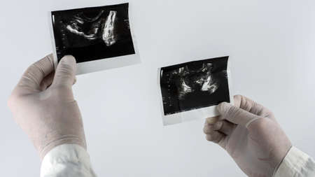
Prostate picture in the hand of a doctor, ultrasound of the prostate on a white background.
Коллекция по умолчанию
Коллекция по умолчанию
Создать новую
water flowing over boulders, rocks, and stones
Коллекция по умолчанию
Коллекция по умолчанию
Создать новую
Bladder cancer, light micrograph, photo under microscope
Коллекция по умолчанию
Коллекция по умолчанию
Создать новую

background of the skin on the head with a small hair
Коллекция по умолчанию
Коллекция по умолчанию
Создать новую

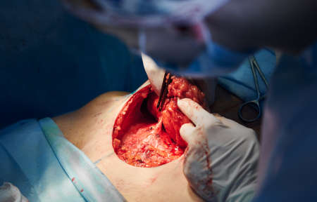
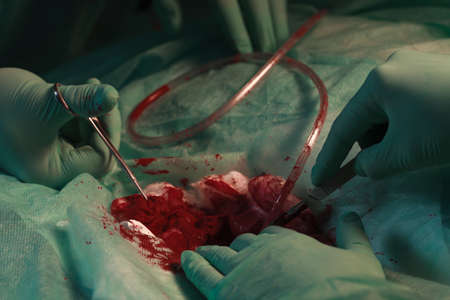
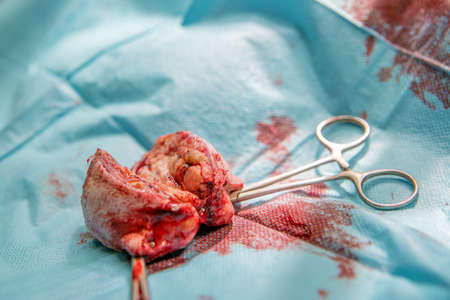
Rupture appendicitis explore laparotomy to appendectomy
Коллекция по умолчанию
Коллекция по умолчанию
Создать новую

Fibromyoma of the uterus diseased tissue 100x
Коллекция по умолчанию
Коллекция по умолчанию
Создать новую
Legion-Media
Создайте свои проекты на основе качественных стоковых фотографий и видео.


Copyright © Legion-Media.