
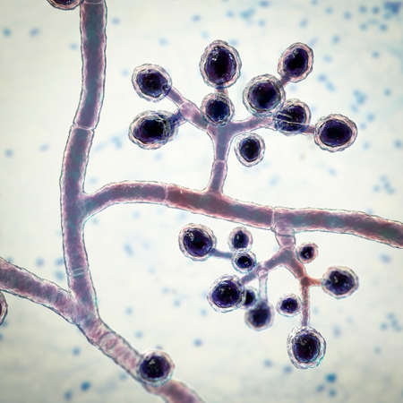
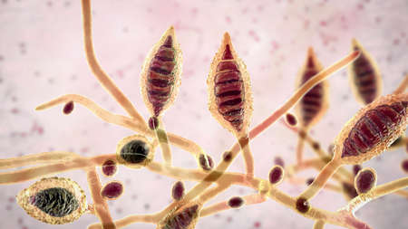
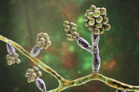
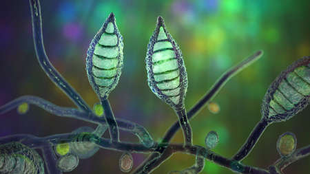
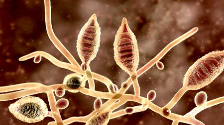
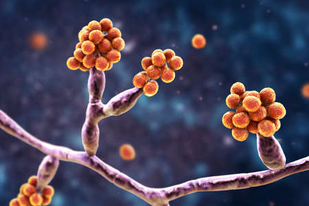
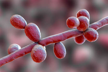
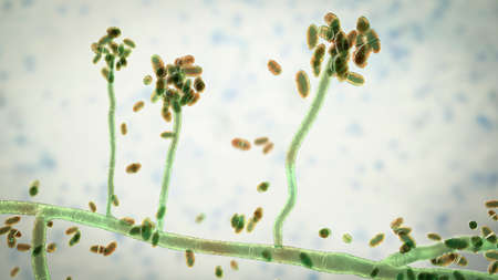
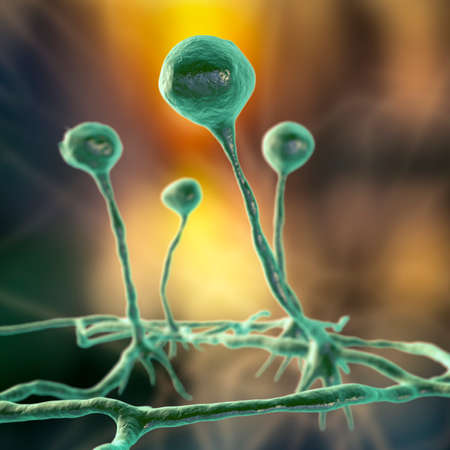
Fungi Trichophyton mentagrophytes, 3D illustration showing branched conidiophores bearing spherical microconidia. Causes skin infection (ringworm), hair and nail infections
Коллекция по умолчанию
Коллекция по умолчанию
Создать новую

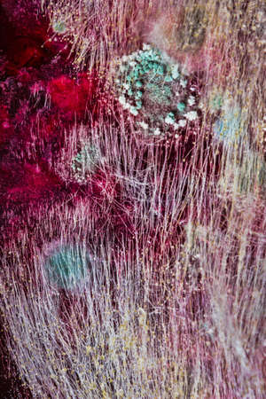
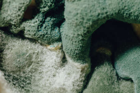
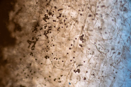
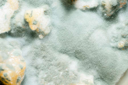
Fungus on old coffee ground for the background
Коллекция по умолчанию
Коллекция по умолчанию
Создать новую
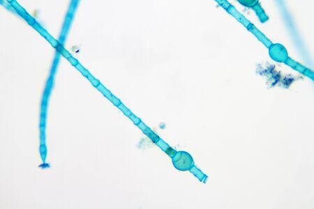
Aspergillus (mold) under the light microscopic view for education.
Коллекция по умолчанию
Коллекция по умолчанию
Создать новую
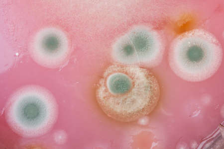
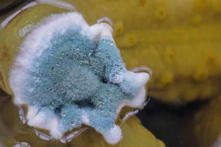
Mushroom cultivation. Macro. Fungi culture on petri dish plate, top view. Mycelium of mushrooms on agar in a petri dish
Коллекция по умолчанию
Коллекция по умолчанию
Создать новую
Fungi Trichophyton mentagrophytes, 3D illustration showing macroconidium, septate and spiral hyphae. Causes skin infection (ringworm, tinea capitis, tinea corporis and other), hair and nail infections
Коллекция по умолчанию
Коллекция по умолчанию
Создать новую
sour and moldy food, top view close-up
Коллекция по умолчанию
Коллекция по умолчанию
Создать новую
Microscopic fungi Microsporum canis, 3D illustration. Zoophilic dermatophyte fungus, causes infections of scalp (tinea capitis), body skin (tinea corporis) aquired from infected dogs and cats
Коллекция по умолчанию
Коллекция по умолчанию
Создать новую
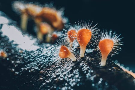
Orange mushroom ,Champagne mushroom or eyelash cup mushroom with sparkling droplets in the forest. Ecosystem or biological diversity concept.
Коллекция по умолчанию
Коллекция по умолчанию
Создать новую
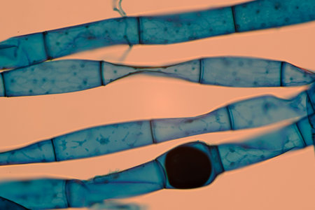
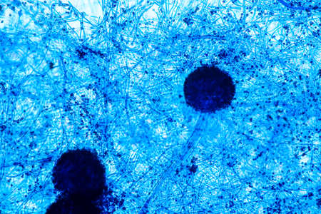
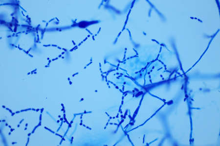
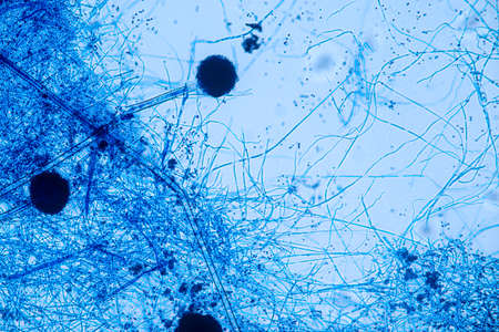
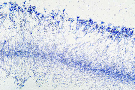
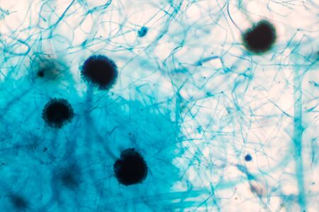
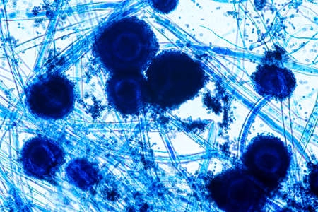
Microscopic view of blue-stained fungal hyphae with spores.
Коллекция по умолчанию
Коллекция по умолчанию
Создать новую
Microaneurysms, microscopic buldges in the artery walls filled with blood, 3D illustration. Found in the eye retina in diabetic retinopathy, and also in brain (Charcot-Bouchard aneurysms)
Коллекция по умолчанию
Коллекция по умолчанию
Создать новую
Microscopic fungi Trichosporon, 3D illustration shows septate hyphae, pseudohyphae, blastoconidia singly or in short chains, arthroconidia. Cause white piedra, superficial and invasive infections
Коллекция по умолчанию
Коллекция по умолчанию
Создать новую
abstract small Bokeh lights. Beautiful background pattern
Коллекция по умолчанию
Коллекция по умолчанию
Создать новую

Mixed of bacteria colonies in Petri dish
Коллекция по умолчанию
Коллекция по умолчанию
Создать новую
Mould fungi Madurella, 3D illustration. The microscopic fungus that causes black-grain mycetoma, or maduromycosis, an infection of human extremities and nervous system found in tropical areas
Коллекция по умолчанию
Коллекция по умолчанию
Создать новую

Fungus on old coffee ground for the background
Коллекция по умолчанию
Коллекция по умолчанию
Создать новую

Aspergillus niger and Aspergillus oryzae (mold) under microscope for Microbiology in Lab.
Коллекция по умолчанию
Коллекция по умолчанию
Создать новую
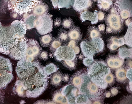
Mold colonies on the red fruit juice
Коллекция по умолчанию
Коллекция по умолчанию
Создать новую
Microscopic fungi Microsporum canis, 3D illustration. Zoophilic dermatophyte fungus, causes infections of scalp (tinea capitis), body skin (tinea corporis) acquired from infected dogs and cats
Коллекция по умолчанию
Коллекция по умолчанию
Создать новую
Aspergillus niger and Aspergillus oryzae (mold) under microscope for Microbiology in Lab.
Коллекция по умолчанию
Коллекция по умолчанию
Создать новую
Microscopic View of Septate Dematiaceous Fungal Hyphae and Possible Chlamydospores
Коллекция по умолчанию
Коллекция по умолчанию
Создать новую
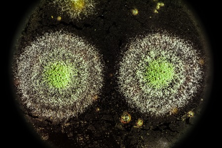
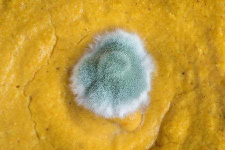
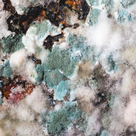
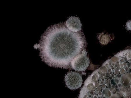
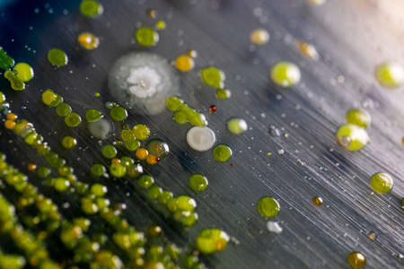
green yellow round fungal mold on a heterogeneous black surface, macro science abstract background
Коллекция по умолчанию
Коллекция по умолчанию
Создать новую
Microscope of black fungus spore strain with Lactophenol cotton blue, molds or yeasts with macro 40x lens, contamination in air room, pollution aerosol environmental. Microbiology laboratory concepts.
Коллекция по умолчанию
Коллекция по умолчанию
Создать новую
yellow mold spores macro shot with detail visible
Коллекция по умолчанию
Коллекция по умолчанию
Создать новую
Microscopic fungi Microsporum canis, 3D illustration. Zoophilic dermatophyte fungus, causes infections of scalp (tinea capitis), body skin (tinea corporis) aquired from infected dogs and cats
Коллекция по умолчанию
Коллекция по умолчанию
Создать новую

Mold on food magnification. texture of toxic green mold with dark green and white orange spots. Spoiled long-term storage product
Коллекция по умолчанию
Коллекция по умолчанию
Создать новую
Mould fungi Madurella, 3D illustration. The microscopic fungus that causes black-grain mycetoma, or maduromycosis, an infection of human extremities and nervous system found in tropical areas
Коллекция по умолчанию
Коллекция по умолчанию
Создать новую
blue mold background, macro view
Коллекция по умолчанию
Коллекция по умолчанию
Создать новую
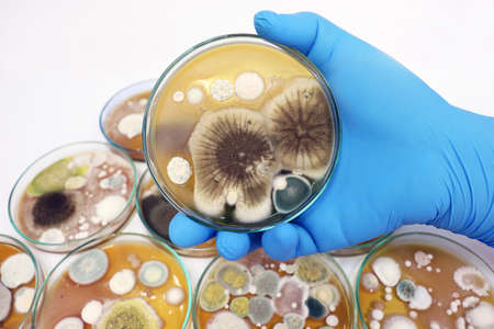
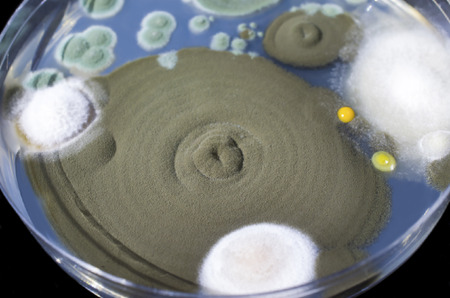
Backgrounds of Characteristics and Different shaped Colony of Bacteria and Mold growing on agar plates from Soil samples for education in Microbiology laboratory.
Коллекция по умолчанию
Коллекция по умолчанию
Создать новую

Fungus on old coffee ground for the background
Коллекция по умолчанию
Коллекция по умолчанию
Создать новую
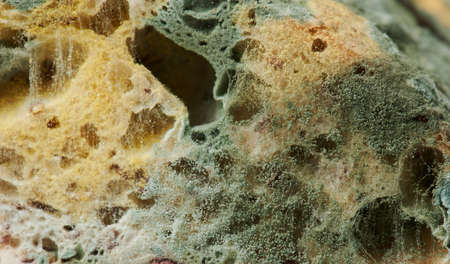
plum pieces fluttered with mould and mushrooms
Коллекция по умолчанию
Коллекция по умолчанию
Создать новую
Close-up of green, blue and white mold growing on a rotten pumpkin with seeds in the middle.
Коллекция по умолчанию
Коллекция по умолчанию
Создать новую

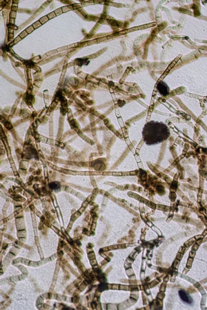
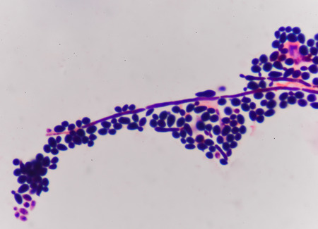
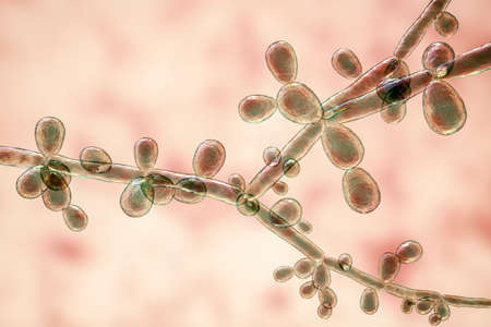
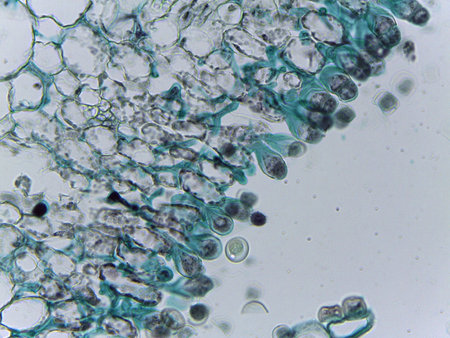
Rhizopus (bread mold) is a genus of common saprophytic fungi,Rhizopus (bread mold) under the microscope.
Коллекция по умолчанию
Коллекция по умолчанию
Создать новую
Aspergillus niger and Aspergillus oryzae (mold) under microscope for Microbiology in Lab.
Коллекция по умолчанию
Коллекция по умолчанию
Создать новую
Mould aka mold on red wine
Коллекция по умолчанию
Коллекция по умолчанию
Создать новую
growing magic mushrooms at home, legalizing psilocybin mushrooms in the world
Коллекция по умолчанию
Коллекция по умолчанию
Создать новую
budding yeast cell structure fine with microscope in laboratory.
Коллекция по умолчанию
Коллекция по умолчанию
Создать новую
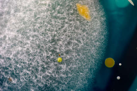
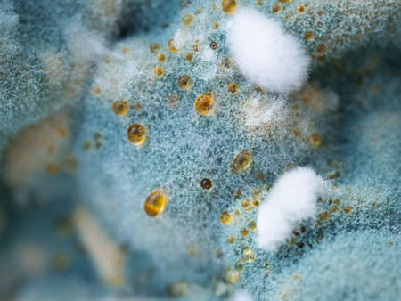
view of the microscope development of green mold on organic basis, macro abstract background
Коллекция по умолчанию
Коллекция по умолчанию
Создать новую
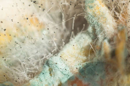
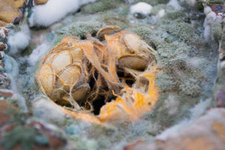
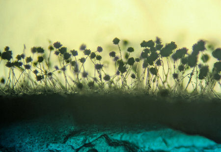
Tiny Slime mold growing on the forest floor macro photography close-up of penicillin, green mold, white fluff of tender Mold in the sun. Abstract closeup of penicillin Small mushrooms growing on a dried lemon peel. Fruit mold.
Коллекция по умолчанию
Коллекция по умолчанию
Создать новую
Candida tropicalis yeasts, microscopic fungi that cause infections in immunocompromised patients. Scientific 3D illustration showing pseudohyphae and blastoconidia formed singly or in small groups
Коллекция по умолчанию
Коллекция по умолчанию
Создать новую
Macro of mixed fungal and bacterial colonies cultured from dirty hands on malt agar Including Penecillium sp and Bacillus subtilis
Коллекция по умолчанию
Коллекция по умолчанию
Создать новую
Blastomyces dermatitidis fungi, the causative agent of the disease blastomycosis affecting lungs, more rarely skin, bones, other organs, 3D illustration. Filamentous form
Коллекция по умолчанию
Коллекция по умолчанию
Создать новую

Aspergillus niger and Aspergillus oryzae (mold) under microscope for Microbiology in Lab.
Коллекция по умолчанию
Коллекция по умолчанию
Создать новую
Aspergillus (mold) under the light microscopic view for education.
Коллекция по умолчанию
Коллекция по умолчанию
Создать новую
Candida tropicalis yeasts, microscopic fungi that cause infections in immunocompromised patients. Scientific 3D illustration showing pseudohyphae and blastoconidia formed singly or in small groups
Коллекция по умолчанию
Коллекция по умолчанию
Создать новую
Mold in food. Green fungus in bread spread.
Коллекция по умолчанию
Коллекция по умолчанию
Создать новую
Bacterial colonies mainly Bacillus subtilis probably from dirty hands cultured on malt agar in a petrie dish Macro
Коллекция по умолчанию
Коллекция по умолчанию
Создать новую
Mold fungi Acremonium (formerly Cephalosporium), 3D illustration. Etiologic agents of white grain mycetoma, nail infections, keratitis
Коллекция по умолчанию
Коллекция по умолчанию
Создать новую
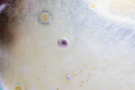
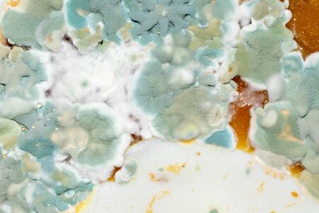
Mold close-up macro. Moldy fungus on food. Fluffy spores mold as a background or texture. Mold fungus. Abstract background.
Коллекция по умолчанию
Коллекция по умолчанию
Создать новую

Host cells with spores (mold) are inside wood under the microscope for education.
Коллекция по умолчанию
Коллекция по умолчанию
Создать новую
Backgrounds of Characteristics and Different shaped Colony of Bacteria and Mold growing on agar plates from Soil samples for education in Microbiology laboratory.
Коллекция по умолчанию
Коллекция по умолчанию
Создать новую

Characteristics and Different shaped Colony of Bacteria and Mold growing on agar plates from Soil samples for education in Microbiology laboratory.
Коллекция по умолчанию
Коллекция по умолчанию
Создать новую

Backgrounds of Characteristics and Different shaped Colony of Bacteria and Mold growing on agar plates from Soil samples for education in Microbiology laboratory.
Коллекция по умолчанию
Коллекция по умолчанию
Создать новую
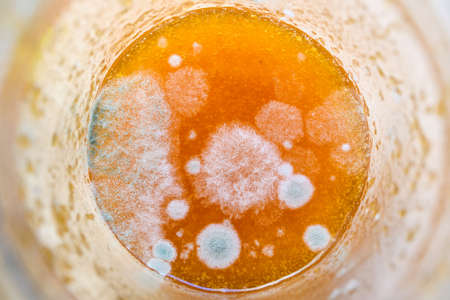
Close-up of mold on orange jam in a glass jar.
Коллекция по умолчанию
Коллекция по умолчанию
Создать новую
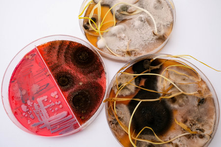
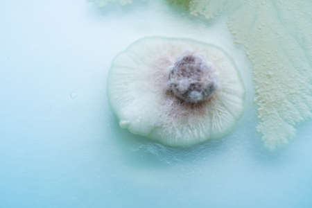
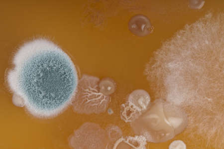
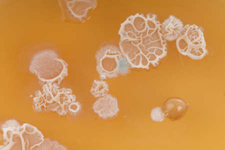
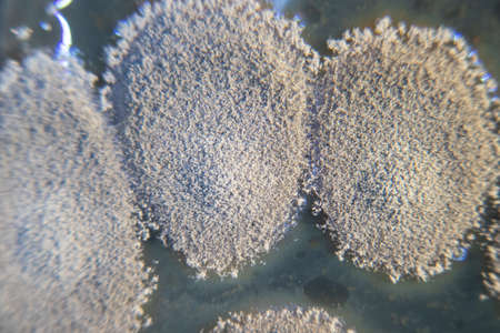
photo of fungi growth on agar media in petri dish
Коллекция по умолчанию
Коллекция по умолчанию
Создать новую
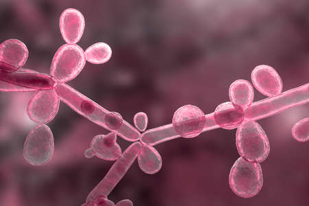
Candida tropicalis yeasts, microscopic fungi that cause infections in immunocompromised patients. Scientific 3D illustration showing pseudohyphae and blastoconidia formed singly or in small groups
Коллекция по умолчанию
Коллекция по умолчанию
Создать новую
High-Power Photomicrograph Revealing Multicellular Dematiaceous Fungal Spores Exhibiting Transverse and Longitudinal Septation
Коллекция по умолчанию
Коллекция по умолчанию
Создать новую

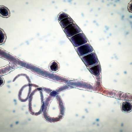
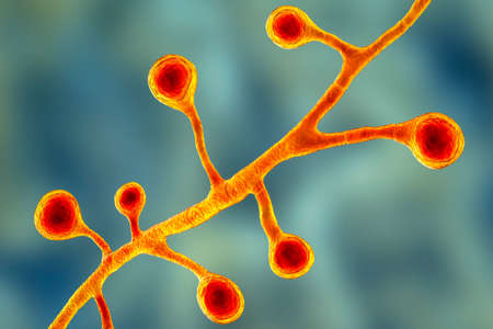
Fungus Trichophyton rubrum, 3D illustration showing macroconidia, microconidia and septate hyphae. Infects skin and nails causing dermatophytosis, especially on feet (tinea pedis), and onychomycosis
Коллекция по умолчанию
Коллекция по умолчанию
Создать новую
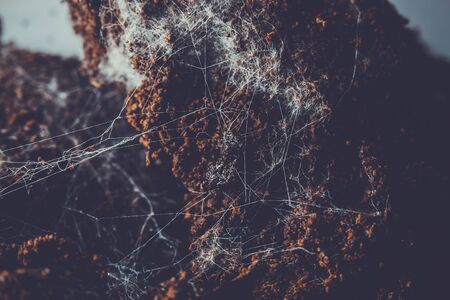
Fungus on old coffee ground for the background
Коллекция по умолчанию
Коллекция по умолчанию
Создать новую
Backgrounds of Characteristics and Different shaped Colony of Bacteria and Mold growing on agar plates from Soil samples for education in Microbiology laboratory.
Коллекция по умолчанию
Коллекция по умолчанию
Создать новую
Pumpkin covered with mold and fungus. Texture background for designers.
Коллекция по умолчанию
Коллекция по умолчанию
Создать новую
Mould aka mold on a glass of red wine - macro.
Коллекция по умолчанию
Коллекция по умолчанию
Создать новую
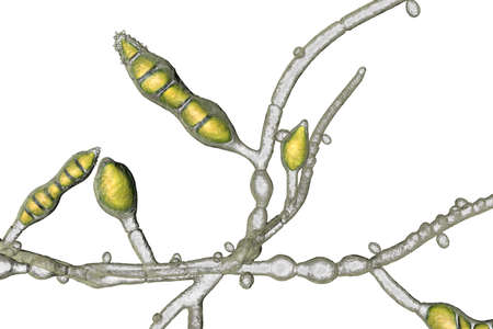
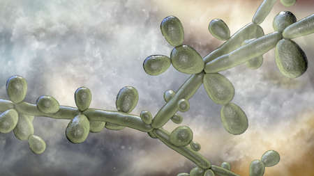
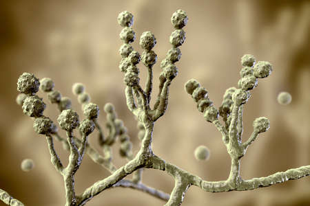
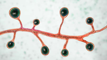
Penicillium, ascomycetous fungi, under a microscope. Mycelium, the vegetative part of the fungus, a mass of branching, thread-like hyphae. Used for antibiotics, fermenting, beverages and cheese. Photo
Коллекция по умолчанию
Коллекция по умолчанию
Создать новую
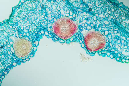
Rust fungus on milkweed plant, 200x
Коллекция по умолчанию
Коллекция по умолчанию
Создать новую
Rhizopus bread mold under the microscope. Rhizopus is a genus of common saprophytic fungi.
Коллекция по умолчанию
Коллекция по умолчанию
Создать новую
Backgrounds of Characteristics and Different shaped Colony of Bacteria and Mold growing on agar plates from Soil samples for education in Microbiology laboratory.
Коллекция по умолчанию
Коллекция по умолчанию
Создать новую
Aspergillus niger and Aspergillus oryzae (mold) under microscope for Microbiology in Lab.
Коллекция по умолчанию
Коллекция по умолчанию
Создать новую

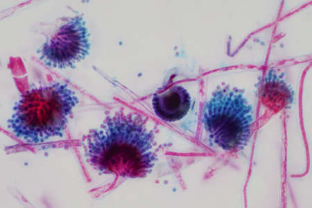
Microscopic fungi Cunninghamella, scientific 3D illustration. Pathogenic fungi from the order Mucorales, cause sinopulmonary and disseminated infections, one of the causative agents of mucormycosis
Коллекция по умолчанию
Коллекция по умолчанию
Создать новую

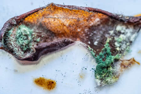
Old Green Fungus with White Fury Spots Growing on a Butter
Коллекция по умолчанию
Коллекция по умолчанию
Создать новую

Mould aka mold on a glass of red wine left open to the air - macro.
Коллекция по умолчанию
Коллекция по умолчанию
Создать новую
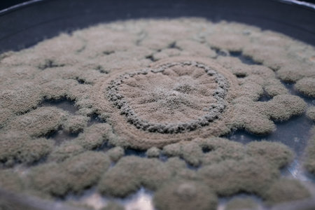
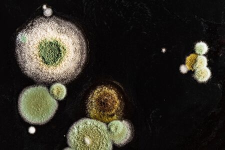
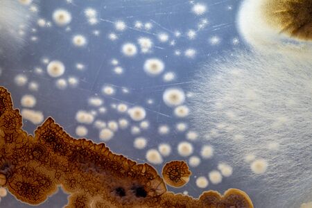
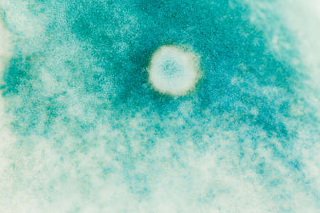
A close-up view of a circular pattern formed by mold or fungus on a surface, showcasing intricate textures and shapes
Коллекция по умолчанию
Коллекция по умолчанию
Создать новую
Malt Extract Agar in Petri dish use for growth media to isolate and cultivate yeasts, molds and fungal testing clinical samples, hold in scientist hands in medical health laboratory analysis disease.
Коллекция по умолчанию
Коллекция по умолчанию
Создать новую
Rhizopus mold, also known as bread mold and black fungus, 3D illustration. Opportunistic fungi that cause mucormycosis involving skin, nasal sinuses, brain and lungs. Complication of Covid-19
Коллекция по умолчанию
Коллекция по умолчанию
Создать новую

Slime molds, as a group, are polyphyletic under the microscope for education.
Коллекция по умолчанию
Коллекция по умолчанию
Создать новую
blue mold background, macro view
Коллекция по умолчанию
Коллекция по умолчанию
Создать новую
green and white mold macro photo. background
Коллекция по умолчанию
Коллекция по умолчанию
Создать новую

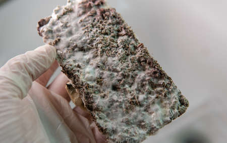
Extreme close-up of mold on the piece of bread
Коллекция по умолчанию
Коллекция по умолчанию
Создать новую
Microscopic fungi Microsporum audouinii, 3D illustration. Anthropophilic dermatophyte fungus, causes infections of scalp (tinea capitis), body skin (tinea corporis) mainly in children
Коллекция по умолчанию
Коллекция по умолчанию
Создать новую
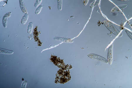
Plankton with microscopic ciliates
Коллекция по умолчанию
Коллекция по умолчанию
Создать новую
Backgrounds of Characteristics of Bacteria and Fungi for education in Microbiology laboratory.
Коллекция по умолчанию
Коллекция по умолчанию
Создать новую

Backgrounds of Characteristics of Bacteria and Fungi for education in Microbiology laboratory.
Коллекция по умолчанию
Коллекция по умолчанию
Создать новую
Candida tropicalis yeasts, microscopic fungi that cause infections in immunocompromised patients. Scientific 3D illustration showing pseudohyphae and blastoconidia formed singly or in small groups
Коллекция по умолчанию
Коллекция по умолчанию
Создать новую
Microscopic fungi Scopulariopsis brevicaulis, 3D illustration. Fungus that infects nails, causes subcutaneous and invasive infections, endocarditis, sinusitis, disseminated infection
Коллекция по умолчанию
Коллекция по умолчанию
Создать новую

mold on pickled cucumbers, botulism bacteria. natural texture
Коллекция по умолчанию
Коллекция по умолчанию
Создать новую
Green mold on food. Fungus on the apple jam
Коллекция по умолчанию
Коллекция по умолчанию
Создать новую
Spores of fungus on bread macro close up view
Коллекция по умолчанию
Коллекция по умолчанию
Создать новую
Microscopic Silhouette of Black Mold Fungi (Aspergillus niger or Rhizopus) with Sporangia
Коллекция по умолчанию
Коллекция по умолчанию
Создать новую

tea mold in a cup, black background
Коллекция по умолчанию
Коллекция по умолчанию
Создать новую

Microscopic fungi Microsporum canis, 3D illustration. Zoophilic dermatophyte fungus, causes infections of scalp (tinea capitis), body skin (tinea corporis) acquired from infected dogs and cats
Коллекция по умолчанию
Коллекция по умолчанию
Создать новую

Budding yeast cells with pseudohyphae Gram stain method
Коллекция по умолчанию
Коллекция по умолчанию
Создать новую
Blastomyces dermatitidis fungi, the causative agent of the disease blastomycosis affecting lungs, more rarely skin, bones, other organs, 3D illustration. Filamentous form
Коллекция по умолчанию
Коллекция по умолчанию
Создать новую
Microscope view of freshwater algae Oedogonium.
Коллекция по умолчанию
Коллекция по умолчанию
Создать новую

Microscopic fungi Scopulariopsis brevicaulis, 3D illustration. Fungus that infects nails, causes subcutaneous and invasive infections, endocarditis, sinusitis, disseminated infection
Коллекция по умолчанию
Коллекция по умолчанию
Создать новую
Close-up photo of a blue mold pattern on a pickle in a jar.the concept of violation of storage conditions, the danger of poisoning with spoiled products.
Коллекция по умолчанию
Коллекция по умолчанию
Создать новую
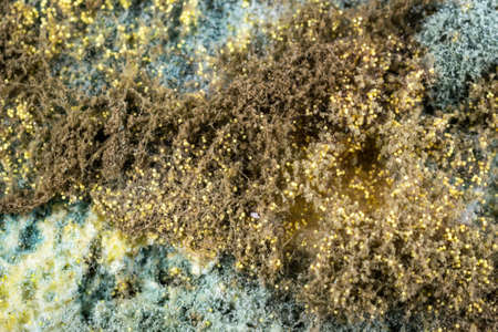
Colonies of mold fungi cultivated from indoor air on Petri dish with Sabourad dextrose agar
Коллекция по умолчанию
Коллекция по умолчанию
Создать новую
Microscopic Visualization of Cystopus Candidus Albugo White Rust Fungal Pathogen
Коллекция по умолчанию
Коллекция по умолчанию
Создать новую

Fungus on old coffee ground for the background
Коллекция по умолчанию
Коллекция по умолчанию
Создать новую
Dematiaceous Fungal Spores Exhibiting Transverse and Longitudinal Septation, Likely of the Genus Alternaria.
Коллекция по умолчанию
Коллекция по умолчанию
Создать новую
view of growing mold on the surface of a rotten tomato.
Коллекция по умолчанию
Коллекция по умолчанию
Создать новую
3D illustration of fungi Trichophyton rubrum which cause tinea, athlete's foot, ringworm, jock itch and similar infections of the skin, nail, beard and scalp on colorful background
Коллекция по умолчанию
Коллекция по умолчанию
Создать новую
Fungi Coccidioides immitis, saprophytic stage, 3D illustration showing fungal arthroconidia. Pathogenic fungi that reside in soil and can cause infection coccidioidomycosis, or Valley fever
Коллекция по умолчанию
Коллекция по умолчанию
Создать новую
Legion-Media
Создайте свои проекты на основе качественных стоковых фотографий и видео.


Copyright © Legion-Media.