

This x-ray image depicts a human hand, revealing a clear view of a long bone, Detailed view of the ulna and radius bones in X-ray, AI Generated
Коллекция по умолчанию
Коллекция по умолчанию
Создать новую

X-Ray Of Carpal And Metacarpal Bones In The Human Hand
Коллекция по умолчанию
Коллекция по умолчанию
Создать новую

Bone fractures vector illustration. Educational labeled broken leg, arm scheme. Various damaged injury types with titles. Trauma description with transverse, linear, greenstick, comminuted and spiral.
Коллекция по умолчанию
Коллекция по умолчанию
Создать новую

Human Foot X-Ray On Black Background
Коллекция по умолчанию
Коллекция по умолчанию
Создать новую

Hand x-ray
Коллекция по умолчанию
Коллекция по умолчанию
Создать новую

x-ray fixation fracture wrist joint
Коллекция по умолчанию
Коллекция по умолчанию
Создать новую

X-ray of the wrist and human radius
Коллекция по умолчанию
Коллекция по умолчанию
Создать новую

Film x-ray show fracture plate of arm for fix arm’s bone
Коллекция по умолчанию
Коллекция по умолчанию
Создать новую

X-ray of both human knee
Коллекция по умолчанию
Коллекция по умолчанию
Создать новую

X-ray image of wrist joint, PA and lateral view, Showing ulna fracture.
Коллекция по умолчанию
Коллекция по умолчанию
Создать новую

X-ray of human knees, closeup
Коллекция по умолчанию
Коллекция по умолчанию
Создать новую

X-ray from broken leg of dog
Коллекция по умолчанию
Коллекция по умолчанию
Создать новую

film x-ray forearm AP : show fracture shaft of ulnar(forearm's bone)
Коллекция по умолчанию
Коллекция по умолчанию
Создать новую

X-rays image of leg fracture patients
Коллекция по умолчанию
Коллекция по умолчанию
Создать новую

xray of an arm with elbow joint visible
Коллекция по умолчанию
Коллекция по умолчанию
Создать новую

Human Knee Joint Inflammation Anatomical Close-Up, Highlighted Pain, Blue Background
Коллекция по умолчанию
Коллекция по умолчанию
Создать новую

X-ray image of tibia and fibula fracture. AP and lateral view.
Коллекция по умолчанию
Коллекция по умолчанию
Создать новую

Intermediate Cuneiform Foot bone Anatomy For Medical Concept 3D Illustration
Коллекция по умолчанию
Коллекция по умолчанию
Создать новую

collection x-ray OA knee
Коллекция по умолчанию
Коллекция по умолчанию
Создать новую

Xray foot heel ankle test scan results showing orthopedic titanium metal implant screws after injury surgical operation.
Коллекция по умолчанию
Коллекция по умолчанию
Создать новую

X-ray of the broken leg
Коллекция по умолчанию
Коллекция по умолчанию
Создать новую
X-ray view of a vertebral column in a dog, highlighting spinal structure and disc health
Коллекция по умолчанию
Коллекция по умолчанию
Создать новую

Ankle feet & knee joint X-ray human photo film
Коллекция по умолчанию
Коллекция по умолчанию
Создать новую

Close up bone x-ray medical science background
Коллекция по умолчанию
Коллекция по умолчанию
Создать новую

X-ray of dogs paw fracture. Radiograph of the broken paw of a dog.
Коллекция по умолчанию
Коллекция по умолчанию
Создать новую

fracture shaft of radius ulnar bone
Коллекция по умолчанию
Коллекция по умолчанию
Создать новую

X-ray image of a human foot highlighting the detailed bone structure and metatarsal bones. Concept of medical diagnostics, skeletal analysis, foot anatomy, and healthcare visualization.
Коллекция по умолчанию
Коллекция по умолчанию
Создать новую

film leg AP/lateral : show fracture shaft of tibia and fibular (leg's bone). patient was operated and insert plate and screw for fix leg's bone
Коллекция по умолчанию
Коллекция по умолчанию
Создать новую

Knees
Коллекция по умолчанию
Коллекция по умолчанию
Создать новую

Fractures to the radius born with displacement of the right woman's arm we see on an X-ray
Коллекция по умолчанию
Коллекция по умолчанию
Создать новую

X ray film of tibia leg fracture
Коллекция по умолчанию
Коллекция по умолчанию
Создать новую

High-resolution X-ray image of a human shoulder joint. Concept of medical imaging, bone structure, orthopedic diagnosis, and skeletal anatomy.
Коллекция по умолчанию
Коллекция по умолчанию
Создать новую

X-ray of both human arms and hands
Коллекция по умолчанию
Коллекция по умолчанию
Создать новую

black hand of death, the walking dead, zombie theme, halloween theme, zombie hands, black background, isolated, hand of death, mummy hands, the hands of the devil, black nails, hands monster
Коллекция по умолчанию
Коллекция по умолчанию
Создать новую

X-ray of human knee. Problems with bone or joint.
Коллекция по умолчанию
Коллекция по умолчанию
Создать новую

X-Ray image of human foot with highlighted ankle joint. 3D illustration.
Коллекция по умолчанию
Коллекция по умолчанию
Создать новую

A modern veterinary X-ray display showing an animal skeleton
Коллекция по умолчанию
Коллекция по умолчанию
Создать новую

Flexible white 3d hand. Next generation futuristic prosthetic articulated hand. Generate ai
Коллекция по умолчанию
Коллекция по умолчанию
Создать новую

Fracture shaft of ulnar bone ( forearm bone ) : ( front and side view )
Коллекция по умолчанию
Коллекция по умолчанию
Создать новую

x-ray of the hands, detail of the phalanges and joints,trapeze,scaphoid,Pyramidal,Pisiform,capitate, distal, frontal, proximal, interflaginian, lunate, intercapitate
Коллекция по умолчанию
Коллекция по умолчанию
Создать новую

X-rays of leg fracture patients
Коллекция по умолчанию
Коллекция по умолчанию
Создать новую

collection image of leg fracture and surgical treatment by internal fixation with plate and screw break tibia and fibula bone
Коллекция по умолчанию
Коллекция по умолчанию
Создать новую

This x-ray image captures the intricate skeletal structure of a human hand, highlighting the bones and joints in detail, An x-ray image of a stitched wound, AI Generated
Коллекция по умолчанию
Коллекция по умолчанию
Создать новую

3d rendered medically accurate illustration of the ankle joint
Коллекция по умолчанию
Коллекция по умолчанию
Создать новую

Closeup of x-ray of human knees
Коллекция по умолчанию
Коллекция по умолчанию
Создать новую

Bone tissue human skeleton under microscope cells structure medical science biology background texture magnification research structure health microbiology internal materials organs surgical study
Коллекция по умолчанию
Коллекция по умолчанию
Создать новую

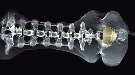
detail of a x-ray of a human spine
Коллекция по умолчанию
Коллекция по умолчанию
Создать новую

human forearm skeleton anatomy bone
Коллекция по умолчанию
Коллекция по умолчанию
Создать новую

x-ray image of human foot joint , side view
Коллекция по умолчанию
Коллекция по умолчанию
Создать новую

Film right elbow including forearm
Коллекция по умолчанию
Коллекция по умолчанию
Создать новую

X-ray orthopedic medical CAT scan of painful knee meniscus injury leg in Traumatology hospital clinic with prosthetics Trauma implant.
Коллекция по умолчанию
Коллекция по умолчанию
Создать новую

X-ray film of finger fracture
Коллекция по умолчанию
Коллекция по умолчанию
Создать новую

Knee joint implant screw xray showing in medical orthpodedic traumatology scan.
Коллекция по умолчанию
Коллекция по умолчанию
Создать новую

X-rays of the human foot, highlighted in red
Коллекция по умолчанию
Коллекция по умолчанию
Создать новую

knee with total replacement x-ray image on black background
Коллекция по умолчанию
Коллекция по умолчанию
Создать новую

X-ray orthopedic medical CAT scan of painful knee meniscus injury leg in Traumatology hospital clinic with prosthetics Trauma implant.
Коллекция по умолчанию
Коллекция по умолчанию
Создать новую

detail of a x-ray of a human spine
Коллекция по умолчанию
Коллекция по умолчанию
Создать новую

Knee joint meniscus x-ray test scan results photo showing injury and pain and orthopedic surgery and Traumatology surgical titanium metal implant
Коллекция по умолчанию
Коллекция по умолчанию
Создать новую

Femur bone affected by Legg-Calve-Perthes Disease, a childhood hip disorder that affects the blood supply to the femoral head, 3D illustration shows affected left femur bone (right side of the image)
Коллекция по умолчанию
Коллекция по умолчанию
Создать новую

Knee joint implant screw xray showing in medical orthpodedic traumatology scan.
Коллекция по умолчанию
Коллекция по умолчанию
Создать новую

X-ray of both human legs
Коллекция по умолчанию
Коллекция по умолчанию
Создать новую

Types of bone fractures medical skeleton anatomy educational vector illustration. Medical science
Коллекция по умолчанию
Коллекция по умолчанию
Создать новую

X-ray Knee Joint Fracture proximal tibia and Poat fix fracture proximal tibia with plate and screws. Normal joint space. Minimal joint effusion.
Коллекция по умолчанию
Коллекция по умолчанию
Создать новую

X-ray image of fracture forearm with wooden splint.
Коллекция по умолчанию
Коллекция по умолчанию
Создать новую

X-ray of human hands injury
Коллекция по умолчанию
Коллекция по умолчанию
Создать новую

X-ray from dog in negative
Коллекция по умолчанию
Коллекция по умолчанию
Создать новую

x ray of fractures bone
Коллекция по умолчанию
Коллекция по умолчанию
Создать новую

Cervical X-ray film
Коллекция по умолчанию
Коллекция по умолчанию
Создать новую

the xray of a human neck
Коллекция по умолчанию
Коллекция по умолчанию
Создать новую

X-Ray Of Carpal And Metacarpal Bones In The Human Hand
Коллекция по умолчанию
Коллекция по умолчанию
Создать новую

X-ray of human bones on dark background. Vector illustration.
Коллекция по умолчанию
Коллекция по умолчанию
Создать новую

Medical hospital x-ray feet traumatology scan.
Коллекция по умолчанию
Коллекция по умолчанию
Создать новую

Human foot ankle and leg in x-ray, on gray background. The foot ankle is highlighted by red colour.
Коллекция по умолчанию
Коллекция по умолчанию
Создать новую

Intertrochanteric femur fracture (right) and normal femur (left), 3D illustration showing a break between the greater and lesser trochanters of the femur.
Коллекция по умолчанию
Коллекция по умолчанию
Создать новую

X-ray image of broken foot, AP and obliqe view
Коллекция по умолчанию
Коллекция по умолчанию
Создать новую

Snake skeleton of King Cobra (Ophiophagus hannah), the world's longest venomous snake.
Коллекция по умолчанию
Коллекция по умолчанию
Создать новую

hand x-ray
Коллекция по умолчанию
Коллекция по умолчанию
Создать новую

Human Skeleton Foot Anatomy 3D Rendering on Blue Background
Коллекция по умолчанию
Коллекция по умолчанию
Создать новую

Foot and toes injury x-ray scan orthopedics and Traumatology radiology test results photo.
Коллекция по умолчанию
Коллекция по умолчанию
Создать новую

X-ray of a knee
Коллекция по умолчанию
Коллекция по умолчанию
Создать новую

X-rays of leg fracture patients
Коллекция по умолчанию
Коллекция по умолчанию
Создать новую

Medical hospital x-ray feet traumatology scan.
Коллекция по умолчанию
Коллекция по умолчанию
Создать новую

X-ray photo of human knee joint isolated on white background
Коллекция по умолчанию
Коллекция по умолчанию
Создать новую

Fracture at 3rd and 4th metacarpal bone . Film x-ray of adult hands . Oblique view .
Коллекция по умолчанию
Коллекция по умолчанию
Создать новую

fracture shaft of radius ulnar bone, x-ray film
Коллекция по умолчанию
Коллекция по умолчанию
Создать новую

broken arm
Коллекция по умолчанию
Коллекция по умолчанию
Создать новую

A close-up display of a human hand skeleton showcases the intricate arrangement of bones and joints. The natural light accentuates the fine details, providing insights into human anatomy and skeletal composition.
Коллекция по умолчанию
Коллекция по умолчанию
Создать новую

x-ray image of human foot joint , back view
Коллекция по умолчанию
Коллекция по умолчанию
Создать новую

X-ray image of wrist joint for diagnosis rheumatoid arthritis .
Коллекция по умолчанию
Коллекция по умолчанию
Создать новую

Knee joint replacement orthopedic titanium metal Traaumatology ball and socket implant x-ray image of old age patient.
Коллекция по умолчанию
Коллекция по умолчанию
Создать новую

film x-ray elbow AP show normal human
Коллекция по умолчанию
Коллекция по умолчанию
Создать новую

X-ray image of knee joint, AP and lateral view.
Коллекция по умолчанию
Коллекция по умолчанию
Создать новую

Xray foot heel ankle test scan results showing orthopedic titanium metal implant screws after injury surgical operation.
Коллекция по умолчанию
Коллекция по умолчанию
Создать новую

X-ray image of forearm, lateral view, Showing ulnar and radius fractues.
Коллекция по умолчанию
Коллекция по умолчанию
Создать новую

Broken leg xray image showing tibia and fibula fracture.
Коллекция по умолчанию
Коллекция по умолчанию
Создать новую

Broken arm with plaster
Коллекция по умолчанию
Коллекция по умолчанию
Создать новую

This x-ray image reveals the detailed structure of a mans back and shoulder, Skeletal view of a human's scapula through 3D X-ray, AI Generated
Коллекция по умолчанию
Коллекция по умолчанию
Создать новую

fiilm x-ray wrist show fracture distal radius forearm bone
Коллекция по умолчанию
Коллекция по умолчанию
Создать новую

Bone tissue human skeleton under microscope cells structure medical science biology background texture magnification research structure health microbiology internal materials organs surgical study
Коллекция по умолчанию
Коллекция по умолчанию
Создать новую

X-ray from broken leg of dog in negative
Коллекция по умолчанию
Коллекция по умолчанию
Создать новую
Legion-Media
Создайте свои проекты на основе качественных стоковых фотографий и видео.


Copyright © Legion-Media.