
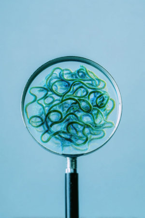
Close up of a magnifying glass revealing a complex network of intertwined green fibers, highlighting the intricate details and textures
Коллекция по умолчанию
Коллекция по умолчанию
Создать новую

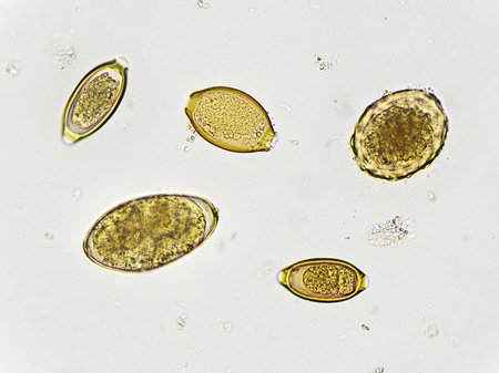
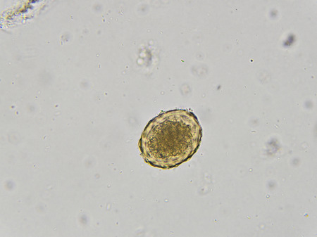
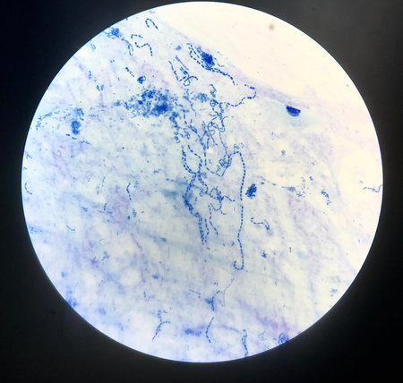
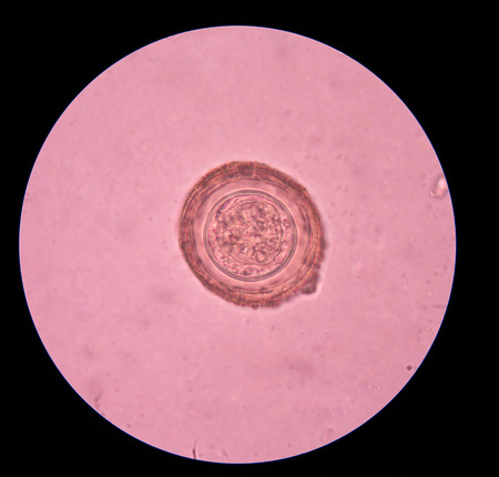
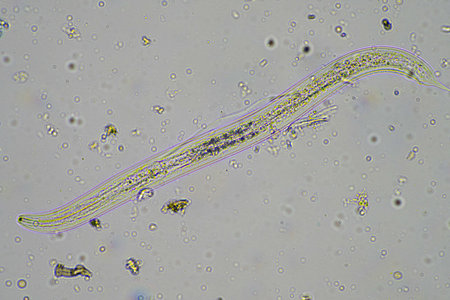
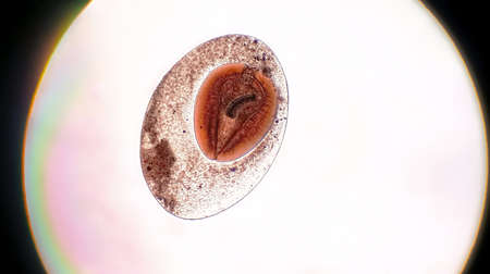
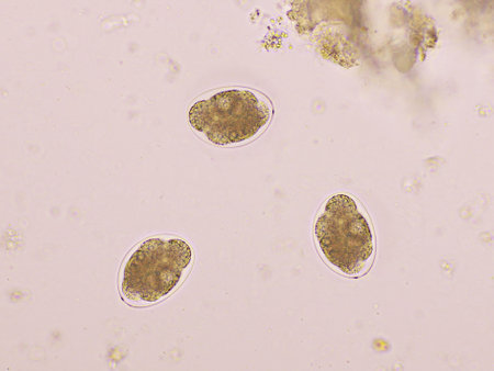
Egg of Ascaris lumbricoides (roundworm) in human stool, analyze by microscope, 400x
Коллекция по умолчанию
Коллекция по умолчанию
Создать новую

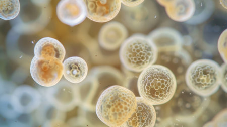
Apple pollen from a blossom in spring under the microscope
Коллекция по умолчанию
Коллекция по умолчанию
Создать новую

A close up of a purple and blue jellyfish with bubbles, AI
Коллекция по умолчанию
Коллекция по умолчанию
Создать новую
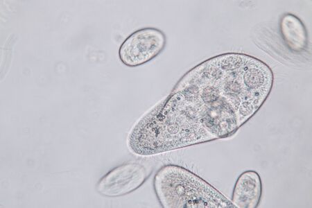
Paramecium caudatum is a genus of unicellular ciliated protozoan and Bacterium under the microscope.
Коллекция по умолчанию
Коллекция по умолчанию
Создать новую
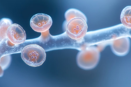
Molluscum contagiosum virus, 3D illustration. A virus from Poxvirus family, causes skin infection with numerous small raised lesions
Коллекция по умолчанию
Коллекция по умолчанию
Создать новую

Leuconostoc bacteria, 3D illustration. Coccoid lactic acid bacteria, found on plants, used for production of fermented milk, can cause meningitis, bacteremia, urinary tract and pulmonary infections
Коллекция по умолчанию
Коллекция по умолчанию
Создать новую
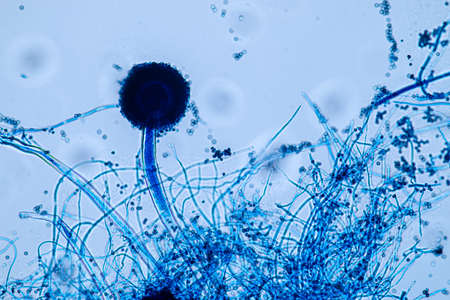
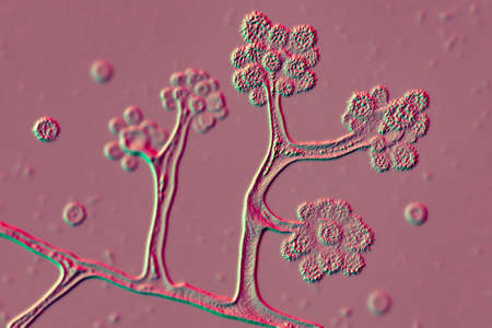
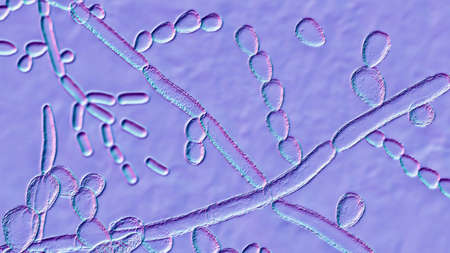
Aspergillus niger and Aspergillus oryzae (mold) under microscope for Microbiology in Lab.
Коллекция по умолчанию
Коллекция по умолчанию
Создать новую
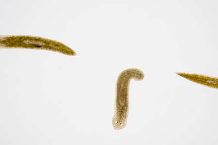
Planarian parasite (flatworm) under microscope view.
Коллекция по умолчанию
Коллекция по умолчанию
Создать новую

Ascaris lumbricoides, a large roundworm, fertilized egg, 3D illustration
Коллекция по умолчанию
Коллекция по умолчанию
Создать новую
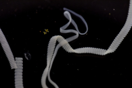
The study of Tapeworm infection is caused by ingesting food or water contaminated with tapeworm eggs or larvae in laboratory.
Коллекция по умолчанию
Коллекция по умолчанию
Создать новую
Borrelia bacteria in blood, 3D illustration. The causative agent of Lyme disease and relapsing fever. Borrelia recurrentis, B. burgdorferi in blood smear under microscope
Коллекция по умолчанию
Коллекция по умолчанию
Создать новую

Blood vessel with flowing blood cells, 3D illustration. Small blood vessels, capillaries
Коллекция по умолчанию
Коллекция по умолчанию
Создать новую
An intricate digital representation of bacteria displaying vibrant colors and glowing elements, highlighting the fascinating world of microscopic life in a stunning abstract setting.
Коллекция по умолчанию
Коллекция по умолчанию
Создать новую

Detailed macro image reveals microorganisms with thin cell walls in linear arrangement, Ai Generated.
Коллекция по умолчанию
Коллекция по умолчанию
Создать новую
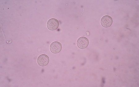
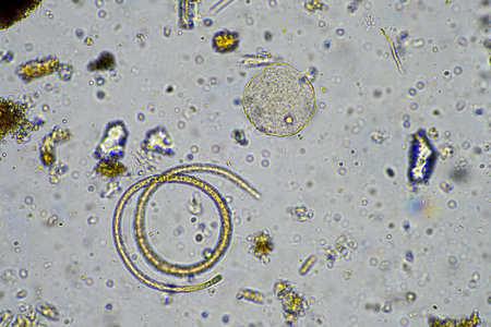
Eggs of helminth in stool
Коллекция по умолчанию
Коллекция по умолчанию
Создать новую

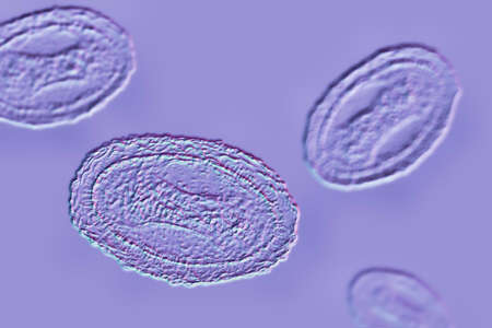
Opisthorchis viverrini, common name Southeast Asian liver fluke, is a trematode parasite.
Коллекция по умолчанию
Коллекция по умолчанию
Создать новую

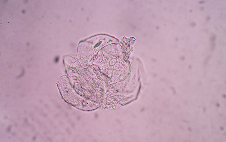
parasite eggs.
Коллекция по умолчанию
Коллекция по умолчанию
Создать новую

Leuconostoc bacteria, 3D illustration. Coccoid lactic acid bacteria, found on plants, used for production of fermented milk, can cause meningitis, bacteremia, urinary tract and pulmonary infections
Коллекция по умолчанию
Коллекция по умолчанию
Создать новую
A stunning macro image captures a network of interconnected, glowing blue cells against a deep black backdrop, revealing intricate details.
Коллекция по умолчанию
Коллекция по умолчанию
Создать новую

Yersinia enterocolitica bacteria, 3D illustration. Gram-negative bacteria of Enterobacteriaceae family, the causative agent of yersiniosis, mesenteric lymphadenitis
Коллекция по умолчанию
Коллекция по умолчанию
Создать новую
bladder epithelial cells in urine.
Коллекция по умолчанию
Коллекция по умолчанию
Создать новую

Egg of Parasitic nematode worm (roundworm) Ascaris lumbricoides which inhabits human intestine and causes disease ascariasis
Коллекция по умолчанию
Коллекция по умолчанию
Создать новую
Close-up view of glowing bacteria and viruses with spiky exteriors, floating in a dark blue, luminous, and abstract background.
Коллекция по умолчанию
Коллекция по умолчанию
Создать новую

The study of Acanthocephala is a phylum of parasitic worms known as acanthocephalans, thorny-headed worms or spiny-headed worms in laboratory.
Коллекция по умолчанию
Коллекция по умолчанию
Создать новую
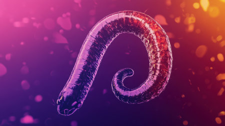
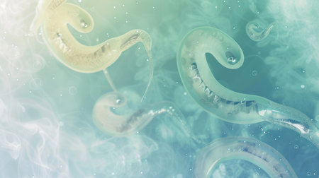
This vibrant abstract composition features a stylized worm design, showcasing smooth curves and a colorful gradient that captivates the viewer's attention.
Коллекция по умолчанию
Коллекция по умолчанию
Создать новую

Egg of Taenia in stool
Коллекция по умолчанию
Коллекция по умолчанию
Создать новую
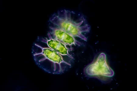
Volvox in drop of water under the microscope for classroom education.
Коллекция по умолчанию
Коллекция по умолчанию
Создать новую
Numerous small blue bubbles are seen floating on the surface of the water, creating a mesmerizing pattern in the sunlight. The bubbles gently rise and fall with the movement of the water.
Коллекция по умолчанию
Коллекция по умолчанию
Создать новую
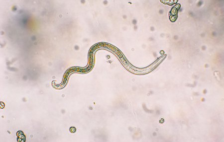
Toxocara canis second stage larvae hatch from eggs
Коллекция по умолчанию
Коллекция по умолчанию
Создать новую
Discover a captivating microscopic world with colorful microorganisms floating gracefully in a liquid environment, showcasing intricate shapes and unique textures.
Коллекция по умолчанию
Коллекция по умолчанию
Создать новую

Trichinella spiralis larvae in muscle tissue under the microscope. Trichinella spiralis is a nematode parasite responsible for trichosis and affecting mammals.
Коллекция по умолчанию
Коллекция по умолчанию
Создать новую
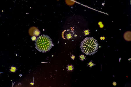
Protozoa and Green Algae in waste water under the microscope.
Коллекция по умолчанию
Коллекция по умолчанию
Создать новую
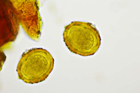
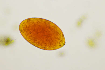
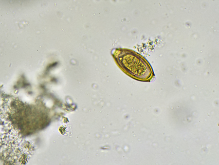
Egg of Ascaris lumbricoides (roundworm) in stool, analyze by microscope
Коллекция по умолчанию
Коллекция по умолчанию
Создать новую
Egg of Ascaris lumbricoides (roundworm) in human stool, analyze by microscope, 400x
Коллекция по умолчанию
Коллекция по умолчанию
Создать новую
Close up blue bacteria cells with microscope.
Коллекция по умолчанию
Коллекция по умолчанию
Создать новую
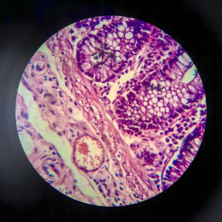
Histopathology of human liver under microscope view for medical education.
Коллекция по умолчанию
Коллекция по умолчанию
Создать новую
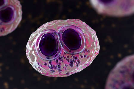
Cytomegalovirus CMV in a human cell, owl's eye inclusion in nucleus, multinucleated cell, 3D illustration. It is herpes virus, causes diseases in fetus, organ transplant patients, HIV infected people
Коллекция по умолчанию
Коллекция по умолчанию
Создать новую

Proglottid (body unit) of tapeworm Taenia saginata, 3D illustration. A flatworm parasitizing animal and human intestine. Proglottid contains uterus with 12-30 primary lateral branches filled with eggs
Коллекция по умолчанию
Коллекция по умолчанию
Создать новую

Ascaris lumbricoides life cycle. Silhouette of a man with internal organs. The arrows indicate the direction of worm migration in the human body and environment. Eggs, larva and adult specimens of ascarids
Коллекция по умолчанию
Коллекция по умолчанию
Создать новую
close-up photograph of small, green spheres with a textured surface, floating gracefully against a blurred background of green leaves.
Коллекция по умолчанию
Коллекция по умолчанию
Создать новую
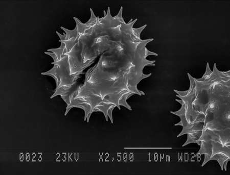
Scanning electron micrograph of one daisy pollen grain. Nottingham, UK
Коллекция по умолчанию
Коллекция по умолчанию
Создать новую
Numerous tiny white bubbles float gracefully on the surface of the water. The bubbles appear to be delicate and light, moving with the gentle current in a mesmerizing manner.
Коллекция по умолчанию
Коллекция по умолчанию
Создать новую

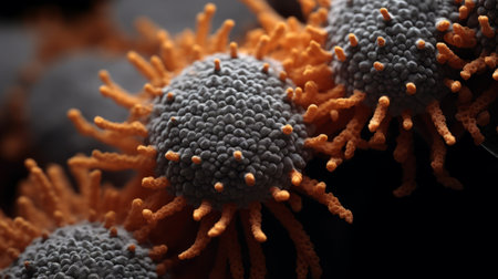
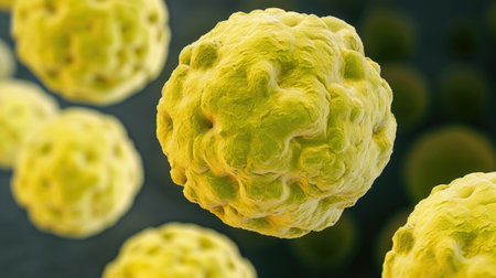
Monkeypox virus model. Concept of searching for antibodies to Monkeypox virus. Anti-Monkeypox Virus VACV-5C7 antibodies. Mutated monkey fever. Model Monkey pox bacteria on blue background.
Коллекция по умолчанию
Коллекция по умолчанию
Создать новую
Hymenolepis diminuta parasite egg in stool exam.
Коллекция по умолчанию
Коллекция по умолчанию
Создать новую
Bacillary dysentery, light micrograph, photo under microscope showing presence of bacteria and accumulation of inflammatory cells in intestinal epithelium
Коллекция по умолчанию
Коллекция по умолчанию
Создать новую
Characteristics of Lichen, hyphae and Symbiotic algae under the microscope for education.
Коллекция по умолчанию
Коллекция по умолчанию
Создать новую
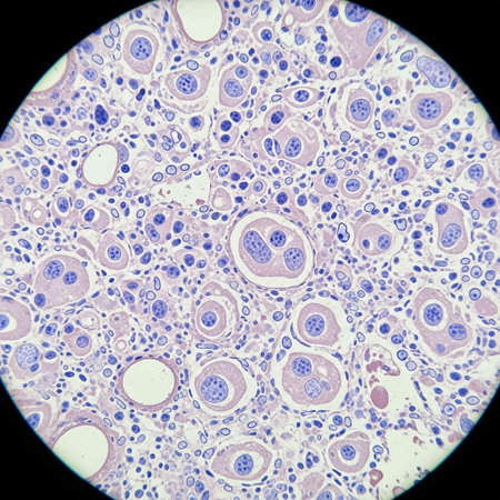
Worms in infected liver 100x
Коллекция по умолчанию
Коллекция по умолчанию
Создать новую
Bacteria Ebola virus under a microscope
Коллекция по умолчанию
Коллекция по умолчанию
Создать новую
soil sample containing soil biology, with bacteria, fungi, amoeba, flagellate, and arcella, on a sustainable agricultural farm in australia
Коллекция по умолчанию
Коллекция по умолчанию
Создать новую
microorganisms and soil biology, with nematodes and fungi under the microscope. in a soil and compost sample in australia
Коллекция по умолчанию
Коллекция по умолчанию
Создать новую
Detailed macro image reveals microorganisms with thin cell walls in linear arrangement, Ai Generated.
Коллекция по умолчанию
Коллекция по умолчанию
Создать новую
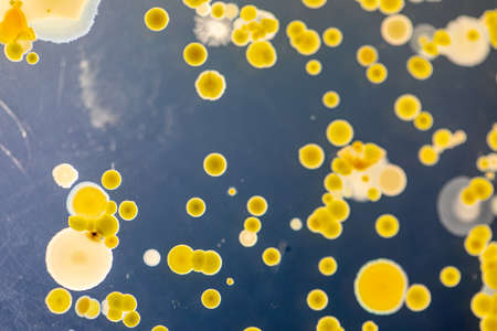
Backgrounds of Characteristics and Different shaped Colony of Bacteria and Mold growing on agar plates from Soil samples for education in Microbiology laboratory.
Коллекция по умолчанию
Коллекция по умолчанию
Создать новую

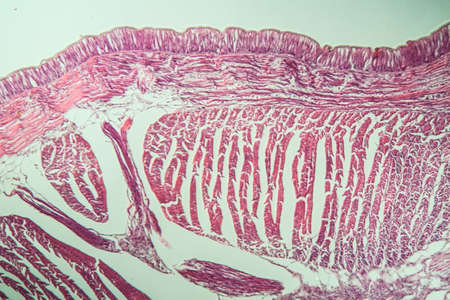
Stomach tissue under the microscope 100x
Коллекция по умолчанию
Коллекция по умолчанию
Создать новую

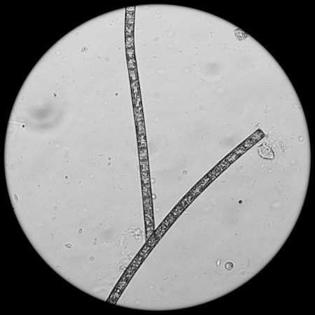
Eggs of Trichuris trichiura in stool
Коллекция по умолчанию
Коллекция по умолчанию
Создать новую

Close up Plant epidermis with stomata or Leaf Epidermis (Stomata) under microscope.
Коллекция по умолчанию
Коллекция по умолчанию
Создать новую
Streptococcus pyogenes bacteria. 3D computer illustration of Streptococcus pyogenes, or group-A Streptococcus, bacteria. S. pyogenes is a gram-positive spherical (coccus) bacteria
Коллекция по умолчанию
Коллекция по умолчанию
Создать новую
Moniliformis dubius in the Intestine of rat, intermediate host
Коллекция по умолчанию
Коллекция по умолчанию
Создать новую
Protozoa and Green Algae in waste water under the microscope.
Коллекция по умолчанию
Коллекция по умолчанию
Создать новую
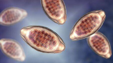
Egg of intestinal fluke in human stool, analyze by microscope, original magnification 400x
Коллекция по умолчанию
Коллекция по умолчанию
Создать новую
Close-up visualization of cell division under a microscope, showcasing glowing blue cells in the process of mitosis. Ideal for biology, science, and medical research themes.
Коллекция по умолчанию
Коллекция по умолчанию
Создать новую
a close-up portrait photograph showcasing the intricate details of unknown staphylococcus aureus biological virus alien flowers. the photo captures the brittle and dry nature of the flowers, while also highlighting their elegant and beautiful features. the rich and vivid contrast, along with the depth of field and black tones, adds a crisp and realistic touch to the image. shot on a 100mm lens with an
Коллекция по умолчанию
Коллекция по умолчанию
Создать новую
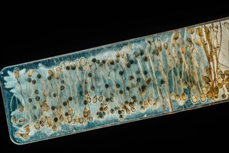
sporangia of a microscopic organism, microbiology concept
Коллекция по умолчанию
Коллекция по умолчанию
Создать новую
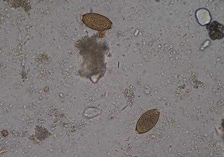
Egg of Hookworm in human stool, analyze by microscope
Коллекция по умолчанию
Коллекция по умолчанию
Создать новую
microscope slide with detailed view of plant stem, complete with cells and minutiae, created with generative ai
Коллекция по умолчанию
Коллекция по умолчанию
Создать новую

Ovarian cancer, light micrograph, photo under microscope. Photograph shows a fragment of a cancerous tumor in the female ovary. Selective focus
Коллекция по умолчанию
Коллекция по умолчанию
Создать новую
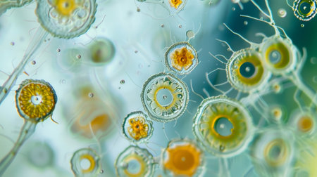
A microscope slide containing a sample of plankton viewed under high magnification to study its composition
Коллекция по умолчанию
Коллекция по умолчанию
Создать новую
Various types of bacteria in a petri dish under a microscope, scientific medical laboratory research
Коллекция по умолчанию
Коллекция по умолчанию
Создать новую
Protozoa and Green Algae in waste water under the microscope.
Коллекция по умолчанию
Коллекция по умолчанию
Создать новую

Egg of Ascaris lumbricoides (roundworm) in human stool, analyze by microscope, 400x
Коллекция по умолчанию
Коллекция по умолчанию
Создать новую
Egg of parasite in stool examition testing finding with microscope.
Коллекция по умолчанию
Коллекция по умолчанию
Создать новую

Opisthorchis viverrini egg in stool under microscopic
Коллекция по умолчанию
Коллекция по умолчанию
Создать новую
Background of a group of worms for medical uses
Коллекция по умолчанию
Коллекция по умолчанию
Создать новую

Bacteria methicillin-resistant Staphylococcus aureus MRSA, multidrug resistant bacteria, 3D illustration
Коллекция по умолчанию
Коллекция по умолчанию
Создать новую
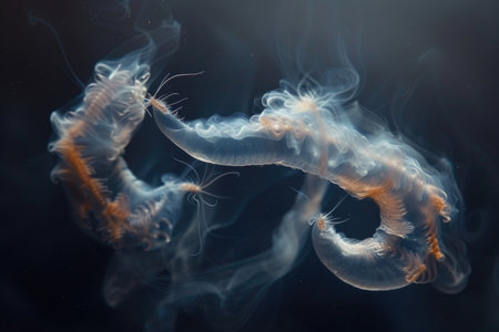
An intriguing scene of deep-sea smoldering dark worms releasing mysterious smoky trails
Коллекция по умолчанию
Коллекция по умолчанию
Создать новую

Protozoa and Green Algae in waste water under the microscope.
Коллекция по умолчанию
Коллекция по умолчанию
Создать новую
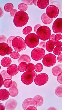
Blood cells in human body under microscope view for education in laboratory.
Коллекция по умолчанию
Коллекция по умолчанию
Создать новую

Thyroid follicular carcinoma, light micrograph, photo under microscope
Коллекция по умолчанию
Коллекция по умолчанию
Создать новую
water algae cell macro, micrograph.
Коллекция по умолчанию
Коллекция по умолчанию
Создать новую
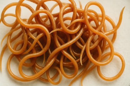
Tangled intestinal roundworms exhibiting complex structure on neutral background. concept of parasitology, intricate biology, and complex worm formations in close-up view.
Коллекция по умолчанию
Коллекция по умолчанию
Создать новую
Earthworm histology cross section 10th segment 100x
Коллекция по умолчанию
Коллекция по умолчанию
Создать новую
Eggs of parasitic roundworm Trichuris trichiura, or whipworm, the causative agent of trichuriasis, disease of a human large intestine, 3D illustration
Коллекция по умолчанию
Коллекция по умолчанию
Создать новую
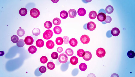
Red blood cells are visibly floating and interacting under a microscope showcasing their circular shape and vibrant red color in detail.
Коллекция по умолчанию
Коллекция по умолчанию
Создать новую
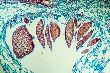
Nestwurz orchid root cross 100x
Коллекция по умолчанию
Коллекция по умолчанию
Создать новую
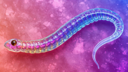
A stunning close-up of a colorful, transparent snake-like creature set against a vibrant pink and purple background. This image highlights the fascinating anatomy and beauty of wildlife.
Коллекция по умолчанию
Коллекция по умолчанию
Создать новую

Bacteria, virus, cell 3d illustration
Коллекция по умолчанию
Коллекция по умолчанию
Создать новую
squamous cell
Коллекция по умолчанию
Коллекция по умолчанию
Создать новую

Parasitic protozoans Toxoplasma gondii, the causative agent of toxoplasmosis in tachyzoite stage, 3D illustration
Коллекция по умолчанию
Коллекция по умолчанию
Создать новую
Egg of Trichuris trichiura in stool
Коллекция по умолчанию
Коллекция по умолчанию
Создать новую
Microscopic fungi Cunninghamella, scientific 3D illustration. Pathogenic fungi from the order Mucorales, cause sinopulmonary and disseminated infections, one of the causative agents of mucormycosis
Коллекция по умолчанию
Коллекция по умолчанию
Создать новую
Streptococcus pyogenes bacteria. 3D computer illustration of Streptococcus pyogenes, or group-A Streptococcus, bacteria. S. pyogenes is a gram-positive spherical (coccus) bacteria
Коллекция по умолчанию
Коллекция по умолчанию
Создать новую
this is a close up of a bubble snails, eggs of snails
Коллекция по умолчанию
Коллекция по умолчанию
Создать новую

Euglena is a genus of single-celled flagellate Eukaryotes under microscopic view for education.
Коллекция по умолчанию
Коллекция по умолчанию
Создать новую
Volvox in drop of water under the microscope for classroom education.
Коллекция по умолчанию
Коллекция по умолчанию
Создать новую
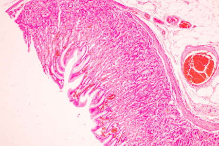
Tissue of Stomach Human under the microscope in Lab.
Коллекция по умолчанию
Коллекция по умолчанию
Создать новую
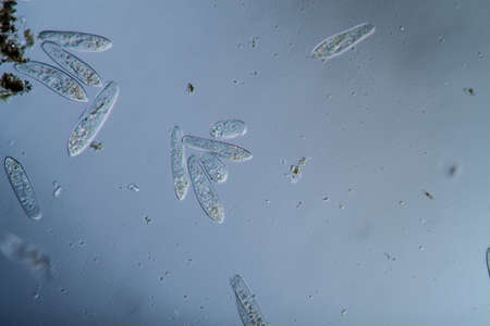
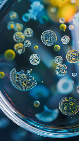
Plankton with microscopic ciliates
Коллекция по умолчанию
Коллекция по умолчанию
Создать новую

Histopathology of human under microscope view for education in laboratory.
Коллекция по умолчанию
Коллекция по умолчанию
Создать новую
living soil life in a soil sample under the microscope in a lab
Коллекция по умолчанию
Коллекция по умолчанию
Создать новую
Microscopic fungi Trichosporon, 3D illustration shows septate hyphae, pseudohyphae, blastoconidia singly or in short chains, arthroconidia. Cause white piedra, superficial and invasive infections
Коллекция по умолчанию
Коллекция по умолчанию
Создать новую

Microbiota, Probiotic Streptococcus bacteria on mucosa 3d illustration
Коллекция по умолчанию
Коллекция по умолчанию
Создать новую
Legion-Media
Создайте свои проекты на основе качественных стоковых фотографий и видео.


Copyright © Legion-Media.