
Microbes of different shapes, 3D illustration. Group of microorganisms. Digital art. Picture render by neural network.
Коллекция по умолчанию
Коллекция по умолчанию
Создать новую

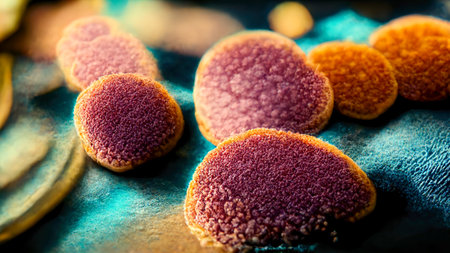
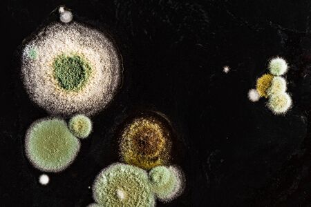
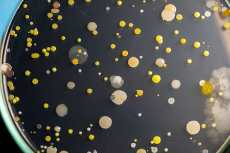
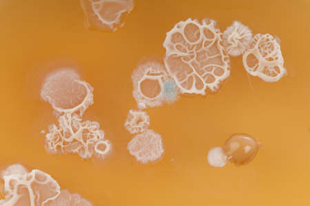
Backgrounds of Characteristics and Different shaped Colony of Bacteria and Mold growing on agar plates from Soil samples for education in Microbiology laboratory.
Коллекция по умолчанию
Коллекция по умолчанию
Создать новую
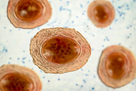
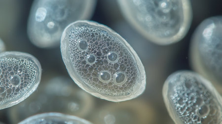
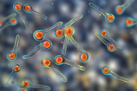
Ascaris lumbricoides, a large roundworm, unfertilized egg, 3D illustration
Коллекция по умолчанию
Коллекция по умолчанию
Создать новую
Backgrounds of Characteristics and Different shaped Colony of Bacteria and Mold growing on agar plates from Soil samples for education in Microbiology laboratory.
Коллекция по умолчанию
Коллекция по умолчанию
Создать новую

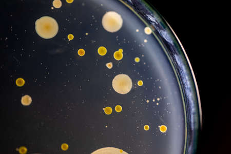
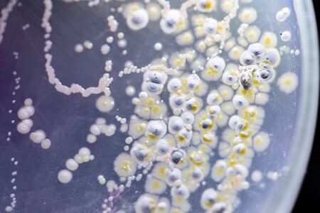
Mixed of bacteria colonies in Petri dish
Коллекция по умолчанию
Коллекция по умолчанию
Создать новую
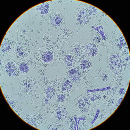
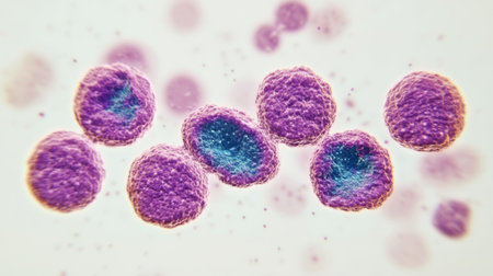
Close up of purple cells under a microscope reveals intricate structures typical of biological lab research.
Коллекция по умолчанию
Коллекция по умолчанию
Создать новую
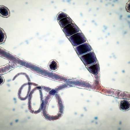
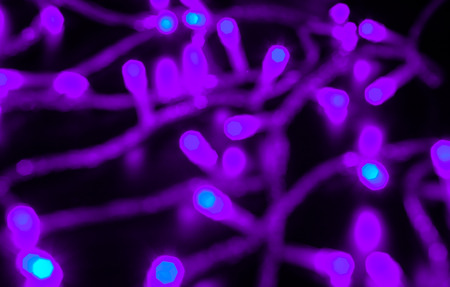
Fungi Trichophyton mentagrophytes, 3D illustration showing macroconidium, septate and spiral hyphae. Causes skin infection (ringworm, tinea capitis, tinea corporis and other), hair and nail infections
Коллекция по умолчанию
Коллекция по умолчанию
Создать новую
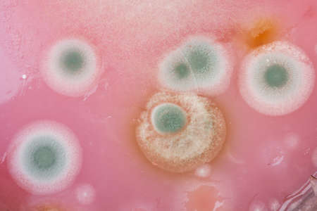
Macro View of Aspergillus terreus Colony Growth in a Petri Dish, Highlighting Fungal Morphology for Mycology Studies.
Коллекция по умолчанию
Коллекция по умолчанию
Создать новую
A highly detailed 3D illustration showing various bacteria and microbes in different colors, providing a close-up microscopic view of microorganisms
Коллекция по умолчанию
Коллекция по умолчанию
Создать новую
Cytomegalovirus CMV in a human cell, owl's eye inclusion in nucleus, multinucleated cell, 3D illustration. It is herpes virus, causes diseases in fetus, organ transplant patients, HIV infected people
Коллекция по умолчанию
Коллекция по умолчанию
Создать новую
Volvox in drop of water under the microscope for classroom education.
Коллекция по умолчанию
Коллекция по умолчанию
Создать новую
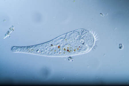
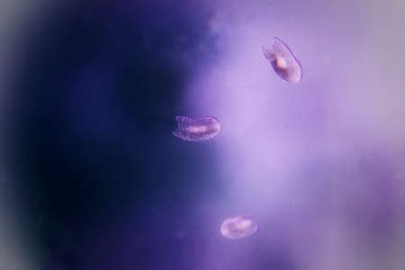
Trumpet animal as a microscopic plankton animal in drops of water
Коллекция по умолчанию
Коллекция по умолчанию
Создать новую
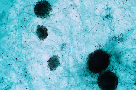
view of the microscope development of green mold on organic basis, macro abstract background
Коллекция по умолчанию
Коллекция по умолчанию
Создать новую
Close-up of a single Petri dish with visible microorganism colonies, on a reflective lab surface, representing research in microbiology.
Коллекция по умолчанию
Коллекция по умолчанию
Создать новую
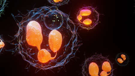
Parasite eggs, worm eggs, ai illustration in 3D style. Close-up view of worm eggs with highly detailed texture. Scientific background
Коллекция по умолчанию
Коллекция по умолчанию
Создать новую
abstract small Bokeh lights. Beautiful background pattern
Коллекция по умолчанию
Коллекция по умолчанию
Создать новую
Rare image of Ghost flatworm - Maricola (Planarian) triclad flatworms in reef aquarium glass
Коллекция по умолчанию
Коллекция по умолчанию
Создать новую
blue mold background, macro view
Коллекция по умолчанию
Коллекция по умолчанию
Создать новую
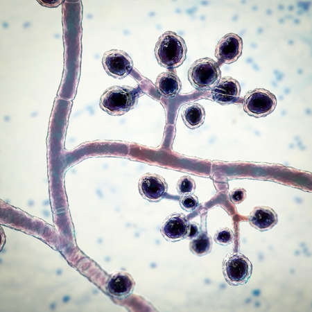
Fungi Trichophyton mentagrophytes, 3D illustration showing branched conidiophores bearing spherical microconidia. Causes skin infection (ringworm), hair and nail infections
Коллекция по умолчанию
Коллекция по умолчанию
Создать новую
budding yeast cell structure fine with microscope in laboratory.
Коллекция по умолчанию
Коллекция по умолчанию
Создать новую
Ice texture background, ink in water pattern frost. Crystal winter design
Коллекция по умолчанию
Коллекция по умолчанию
Создать новую

Host cells with spores (mold) are inside wood under the microscope for education.
Коллекция по умолчанию
Коллекция по умолчанию
Создать новую
Microaneurysms, microscopic buldges in the artery walls filled with blood, 3D illustration. Found in the eye retina in diabetic retinopathy, and also in brain (Charcot-Bouchard aneurysms)
Коллекция по умолчанию
Коллекция по умолчанию
Создать новую

Aspergillus niger and Aspergillus oryzae (mold) under microscope for Microbiology in Lab.
Коллекция по умолчанию
Коллекция по умолчанию
Создать новую
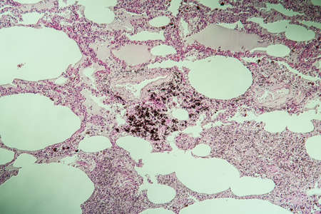
Lung tissue as dust lung under the microscope 100x
Коллекция по умолчанию
Коллекция по умолчанию
Создать новую
Bacteria methicillin-resistant Staphylococcus aureus MRSA, multidrug resistant bacteria, 3D illustration
Коллекция по умолчанию
Коллекция по умолчанию
Создать новую
sour and moldy food, top view close-up
Коллекция по умолчанию
Коллекция по умолчанию
Создать новую
Backgrounds of Characteristics and Different shaped Colony of Bacteria and Mold growing on agar plates from Soil samples for education in Microbiology laboratory.
Коллекция по умолчанию
Коллекция по умолчанию
Создать новую
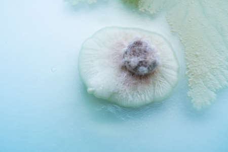
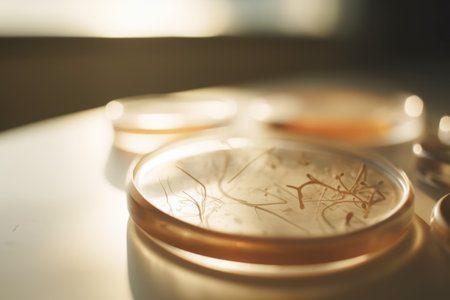
Mushroom cultivation. Macro. Fungi culture on petri dish plate, top view. Mycelium of mushrooms on agar in a petri dish
Коллекция по умолчанию
Коллекция по умолчанию
Создать новую

3D illustration of abstract light background with bokeh defocused lights
Коллекция по умолчанию
Коллекция по умолчанию
Создать новую
Backgrounds of Characteristics and Different shaped Colony of Bacteria and Mold growing on agar plates from Soil samples for education in Microbiology laboratory.
Коллекция по умолчанию
Коллекция по умолчанию
Создать новую

Backgrounds of Colony Characteristics of Fungus and algae in petri dish for education.
Коллекция по умолчанию
Коллекция по умолчанию
Создать новую

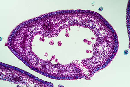
Histological Uterus human, Uterine tube human, Placenta human and Umbilical cord Human under the microscope for education
Коллекция по умолчанию
Коллекция по умолчанию
Создать новую
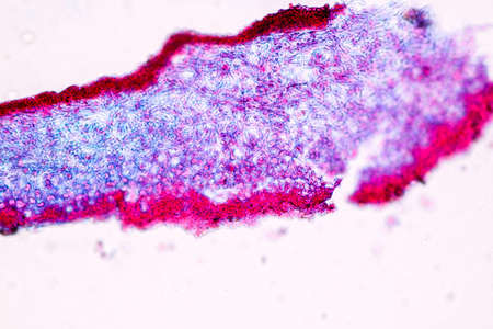
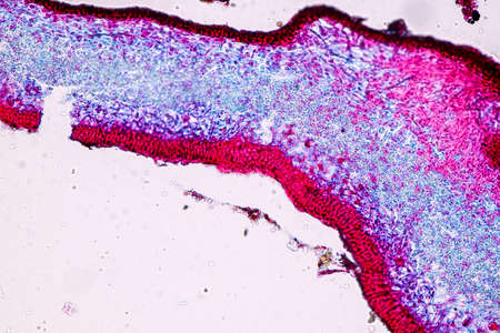
Heather leaf cross section under the microscope, 200x
Коллекция по умолчанию
Коллекция по умолчанию
Создать новую
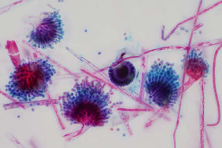
Characteristics of Lichen, hyphae and Symbiotic algae under the microscope for education.
Коллекция по умолчанию
Коллекция по умолчанию
Создать новую
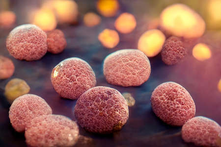
A close up of many small, round, clear objects with a lot of holes in them. The objects are all different sizes and are scattered throughout the image. Scene is one of curiosity and wonder
Коллекция по умолчанию
Коллекция по умолчанию
Создать новую
3D illustration of Phagocytosis. Neutrophe that uses its plasma membrane to engulf bacteria. From endocytosis to exocytosis. Digestion process in phagocytes. immune system, 3d render
Коллекция по умолчанию
Коллекция по умолчанию
Создать новую

Activated and non-activated platelets, thrombocytes, 3D illustration. Activated thrombocytes have cell membrane projections on the surface, unactivated platelets are biconvex discoid, or lens-shaped
Коллекция по умолчанию
Коллекция по умолчанию
Создать новую
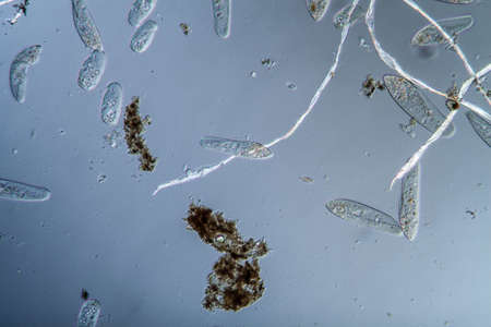
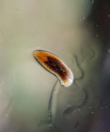
Plankton with microscopic ciliates
Коллекция по умолчанию
Коллекция по умолчанию
Создать новую
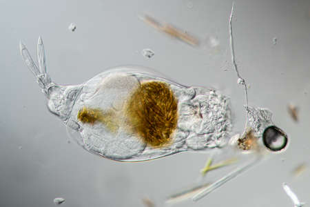
Rotifer foraging in the stream 200x
Коллекция по умолчанию
Коллекция по умолчанию
Создать новую
Microscopic fungi Trichosporon, 3D illustration shows septate hyphae, pseudohyphae, blastoconidia singly or in short chains, arthroconidia. Cause white piedra, superficial and invasive infections
Коллекция по умолчанию
Коллекция по умолчанию
Создать новую
Backgrounds of Characteristics and Different shaped Colony of Bacteria and Mold growing on agar plates from Soil samples for education in Microbiology laboratory.
Коллекция по умолчанию
Коллекция по умолчанию
Создать новую
chemist wearing gloves at laboratory. testing process with glass plate and sample. viruses and health care concept
Коллекция по умолчанию
Коллекция по умолчанию
Создать новую
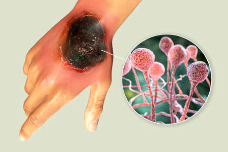
Cutaneous mucormycosis, a disease caused by fungi Mucor, also known as black fungus, 3D illustration showing skin leasion and closep view of Mucor fungus. Covid-19 complication
Коллекция по умолчанию
Коллекция по умолчанию
Создать новую
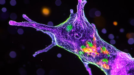
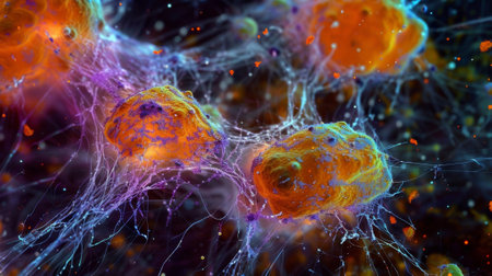
This striking microscopic image showcases a neuron illuminated by fluorescent markers against a dark backdrop, highlighting its intricate structure and connectivity.
Коллекция по умолчанию
Коллекция по умолчанию
Создать новую
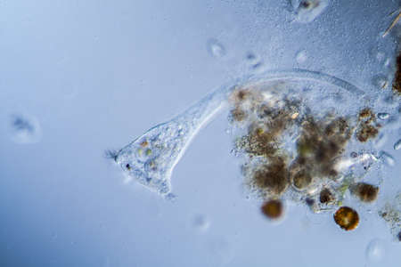
Trumpet animal as a microscopic plankton animal in drops of water
Коллекция по умолчанию
Коллекция по умолчанию
Создать новую
Aspergillus (mold) under the light microscopic view for education.
Коллекция по умолчанию
Коллекция по умолчанию
Создать новую
diseased ear tissue infected with Aspergillus 200x
Коллекция по умолчанию
Коллекция по умолчанию
Создать новую
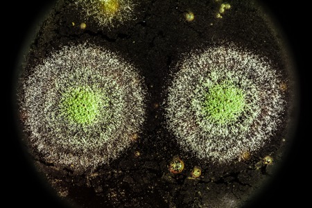
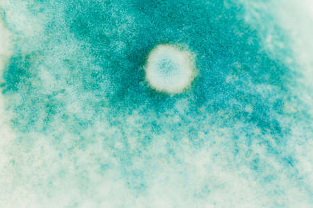
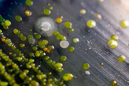
green yellow round fungal mold on a heterogeneous black surface, macro science abstract background
Коллекция по умолчанию
Коллекция по умолчанию
Создать новую

Cytomegalovirus CMV in a human cell, owl's eye inclusion in nucleus, multinucleated cell, 3D illustration. It is herpes virus, causes diseases in fetus, organ transplant patients, HIV infected people
Коллекция по умолчанию
Коллекция по умолчанию
Создать новую
Backgrounds of Characteristics and Different shaped Colony of Bacteria and Mold growing on agar plates from Soil samples for education in Microbiology laboratory.
Коллекция по умолчанию
Коллекция по умолчанию
Создать новую

Meningococcal meningitis, cerebrospinal fluid smear containing neutrophils with and without bacteria Neisseria meningitidis, 3D illustration
Коллекция по умолчанию
Коллекция по умолчанию
Создать новую

Abstract macro image of particles looking like bacteria, macro shot, microbiology theme
Коллекция по умолчанию
Коллекция по умолчанию
Создать новую

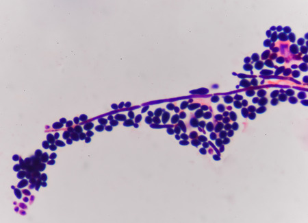
Microscopic Fungi samples over white background
Коллекция по умолчанию
Коллекция по умолчанию
Создать новую
Aspergillus (mold) under the light microscopic view for education.
Коллекция по умолчанию
Коллекция по умолчанию
Создать новую

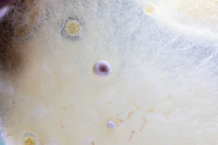
Characteristics and Different shaped Colony of Bacteria and Mold growing on agar plates from Soil samples for education in Microbiology laboratory.
Коллекция по умолчанию
Коллекция по умолчанию
Создать новую
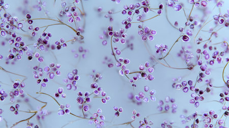
This image captures delicate purple flowers with fine stems set against a soft blue background, evoking a tranquil and ethereal feeling perfect for decor.
Коллекция по умолчанию
Коллекция по умолчанию
Создать новую
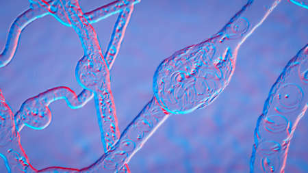
Cross-section through the lichen symbiote body 100x
Коллекция по умолчанию
Коллекция по умолчанию
Создать новую
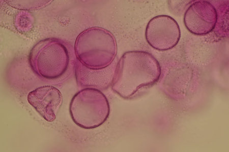
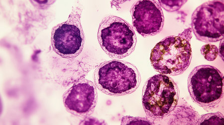
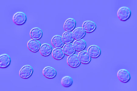
Close-up of pink circular cells under a microscope
Коллекция по умолчанию
Коллекция по умолчанию
Создать новую
A microscope slide containing a sample of plankton viewed under high magnification to study its composition
Коллекция по умолчанию
Коллекция по умолчанию
Создать новую
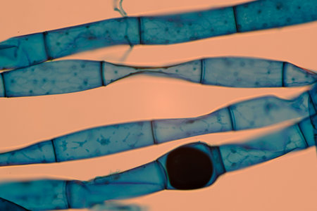
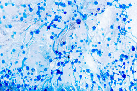
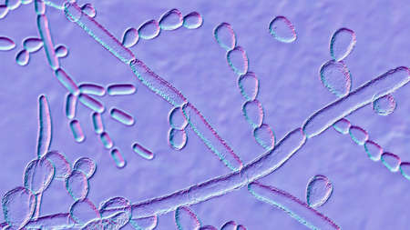
Microscopic view of blue-stained fungal hyphae with spores.
Коллекция по умолчанию
Коллекция по умолчанию
Создать новую
Colorful microorganism cells close up. illustration in high details for medical education and science. ai generative.
Коллекция по умолчанию
Коллекция по умолчанию
Создать новую

Blue spots from the dye in the white tub dissolves in water
Коллекция по умолчанию
Коллекция по умолчанию
Создать новую
Microscopic fungi Scopulariopsis brevicaulis, 3D illustration. Fungus that infects nails, causes subcutaneous and invasive infections, endocarditis, sinusitis, disseminated infection
Коллекция по умолчанию
Коллекция по умолчанию
Создать новую
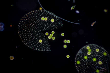
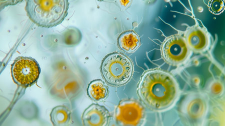
Dive into a stunning microscopic view showcasing vibrant microorganisms in an aquatic setting. This image reveals the intricacies of diverse life forms through scientific observation.
Коллекция по умолчанию
Коллекция по умолчанию
Создать новую

Cancer Cell in blood cells human showing abnormal cells.
Коллекция по умолчанию
Коллекция по умолчанию
Создать новую
Mould fungi Madurella, 3D illustration. The microscopic fungus that causes black-grain mycetoma, or maduromycosis, an infection of human extremities and nervous system found in tropical areas
Коллекция по умолчанию
Коллекция по умолчанию
Создать новую
Backgrounds of Characteristics and Different shaped Colony of Bacteria and Mold growing on agar plates from Soil samples for education in Microbiology laboratory.
Коллекция по умолчанию
Коллекция по умолчанию
Создать новую
Pancreas cancer cells, light micrograph for medical education.
Коллекция по умолчанию
Коллекция по умолчанию
Создать новую
Backgrounds of Characteristics and Different shaped Colony of Bacteria and Mold growing on agar plates from Soil samples for education in Microbiology laboratory.
Коллекция по умолчанию
Коллекция по умолчанию
Создать новую
An intricate, illuminated plant structure thrives within a circular transparent vessel against a dark background. The detailed design and unique form are captivating.
Коллекция по умолчанию
Коллекция по умолчанию
Создать новую

Backgrounds of Characteristics and Different shaped Colony of Bacteria and Mold growing on agar plates from Soil samples for education in Microbiology laboratory.
Коллекция по умолчанию
Коллекция по умолчанию
Создать новую
Characteristics of Lichen, hyphae and Symbiotic algae under the microscope for education.
Коллекция по умолчанию
Коллекция по умолчанию
Создать новую
In the human brain there are approximately 1.5 trillion astrocytes making them the most abundant cell type in the central nervous system
Коллекция по умолчанию
Коллекция по умолчанию
Создать новую
Characteristics of Hair cell of human under microscope view for education in laboratory.
Коллекция по умолчанию
Коллекция по умолчанию
Создать новую
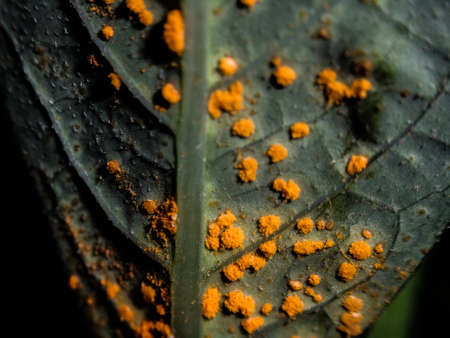
yellow fungus on green leaf
Коллекция по умолчанию
Коллекция по умолчанию
Создать новую
Clostridium tetani bacteria, the causative agent of tetanus, 3D illustration
Коллекция по умолчанию
Коллекция по умолчанию
Создать новую
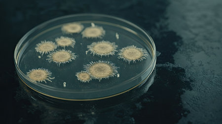
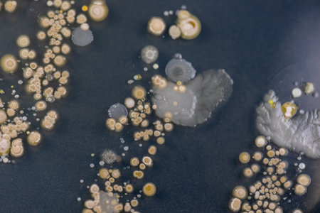
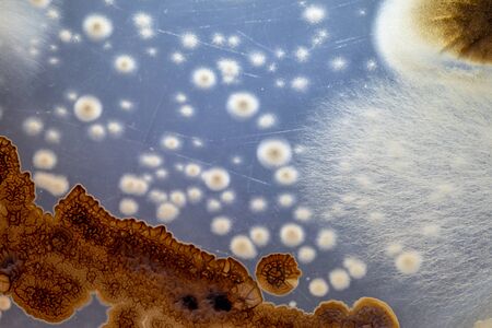
Bacterial colonies mainly Bacillus subtilis probably from dirty hands cultured on malt agar in a petrie dish Macro
Коллекция по умолчанию
Коллекция по умолчанию
Создать новую
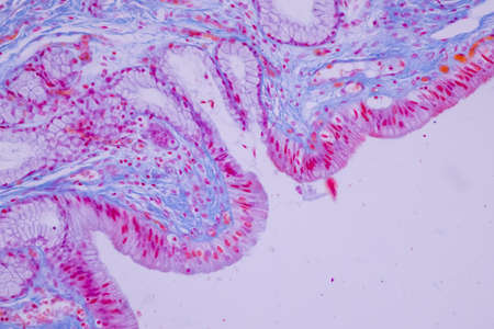
Columnar epithelium of human gall bladder under the microscope in Lab.
Коллекция по умолчанию
Коллекция по умолчанию
Создать новую
Bacterial culture growth on MacConkey agar (Gram Negative Bacilli)contains small light grains. Focus on all agar surface.
Коллекция по умолчанию
Коллекция по умолчанию
Создать новую
Backgrounds of Characteristics and Different shaped Colony of Bacteria and Mold growing on agar plates from Soil samples for education in Microbiology laboratory.
Коллекция по умолчанию
Коллекция по умолчанию
Создать новую
Leech on the glass. Bloodsucking animal. subclass of ringworms from the belt-type class. Hirudotherapy.
Коллекция по умолчанию
Коллекция по умолчанию
Создать новую
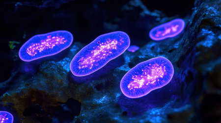
Bacteria emit a brilliant glow under UV light in a dark setting, showing vibrant colors and intricate patterns.
Коллекция по умолчанию
Коллекция по умолчанию
Создать новую
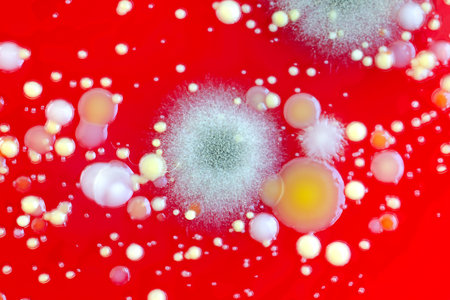
Bacterial and mold fungi colonies grown from indoor air on blood sheep agar, photograph showing microbial diversity and air contamination.
Коллекция по умолчанию
Коллекция по умолчанию
Создать новую
microbiology laboratory test-tubes in hand of biochemist
Коллекция по умолчанию
Коллекция по умолчанию
Создать новую

Backgrounds of Characteristics and Different shaped Colony of Bacteria and Mold growing on agar plates from Soil samples for education in Microbiology laboratory.
Коллекция по умолчанию
Коллекция по умолчанию
Создать новую
petri dish with microbe culture growing on plate, under microscope, created with generative ai
Коллекция по умолчанию
Коллекция по умолчанию
Создать новую
Chromosomes Human under the microscope for education.
Коллекция по умолчанию
Коллекция по умолчанию
Создать новую

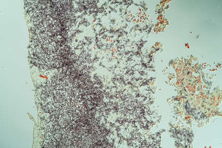
Slime molds, as a group, are polyphyletic under the microscope for education.
Коллекция по умолчанию
Коллекция по умолчанию
Создать новую
Colonies of bacteria growth on agar plate medium in microbiology laboratory.
Коллекция по умолчанию
Коллекция по умолчанию
Создать новую
Mould fungi Madurella, 3D illustration. The microscopic fungus that causes black-grain mycetoma, or maduromycosis, an infection of human extremities and nervous system found in tropical areas
Коллекция по умолчанию
Коллекция по умолчанию
Создать новую

Parasites on a sheet of paper. Extruded from the skin parasites. Acari parasites
Коллекция по умолчанию
Коллекция по умолчанию
Создать новую

Backgrounds of Colony Characteristics of Mold under the microscope.
Коллекция по умолчанию
Коллекция по умолчанию
Создать новую
Thyroid follicular carcinoma, light micrograph, photo under microscope
Коллекция по умолчанию
Коллекция по умолчанию
Создать новую
Copepod crab swims through the water
Коллекция по умолчанию
Коллекция по умолчанию
Создать новую
Beautiful 3d rendering of VIRUS microscope.
Коллекция по умолчанию
Коллекция по умолчанию
Создать новую
Microscopic View Rendered Image of Abnormal, Diseased Cells in Biology and Medicine Illustration
Коллекция по умолчанию
Коллекция по умолчанию
Создать новую

Blood smear of leukemia patient under microscope
Коллекция по умолчанию
Коллекция по умолчанию
Создать новую
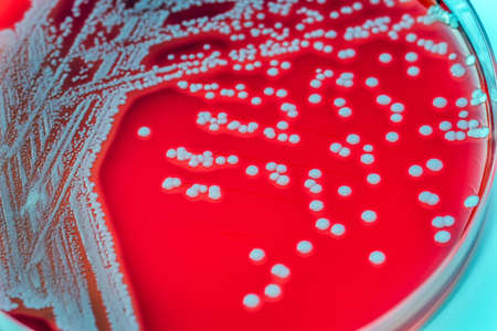
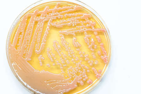
Staphylococcus aureus: Gram-positive, to Gram-variable, nonmotile, Coccus,beta haemolysis, saprotrophic bacterium that belongs to the family Staphylococcus growth on blood agar.
Коллекция по умолчанию
Коллекция по умолчанию
Создать новую
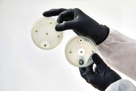
Petri plates with bacteria cells on table in lab, created using generative ai technology
Коллекция по умолчанию
Коллекция по умолчанию
Создать новую
Legion-Media
Создайте свои проекты на основе качественных стоковых фотографий и видео.


Copyright © Legion-Media.