
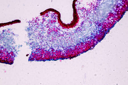
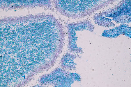
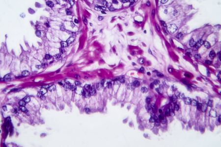
Columnar epithelium of human gall bladder under the microscope in Lab.
Коллекция по умолчанию
Коллекция по умолчанию
Создать новую

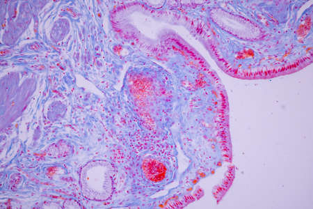
Ovarian cancer, light micrograph, photo under microscope. Photograph shows a fragment of a cancerous tumor in the female ovary. Selective focus
Коллекция по умолчанию
Коллекция по умолчанию
Создать новую

Stomach tissue under the microscope 100x
Коллекция по умолчанию
Коллекция по умолчанию
Создать новую

Bladder cancer, light micrograph, photo under microscope
Коллекция по умолчанию
Коллекция по умолчанию
Создать новую
Education anatomy and Histological sample of Human under the microscope.
Коллекция по умолчанию
Коллекция по умолчанию
Создать новую
A detailed photograph of a vibrant orange sea anemone, showcasing its complex structure against a stark black backdrop.
Коллекция по умолчанию
Коллекция по умолчанию
Создать новую
Nestwurz orchid root cross 100x
Коллекция по умолчанию
Коллекция по умолчанию
Создать новую
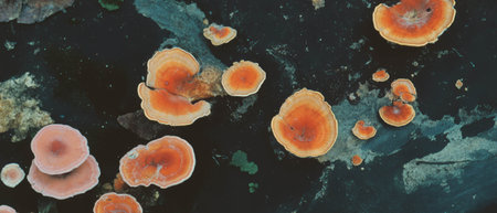
destructive mushroom in wood fabric 100x
Коллекция по умолчанию
Коллекция по умолчанию
Создать новую
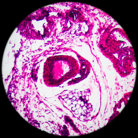
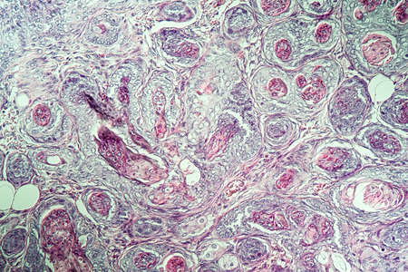
Inguinal testicles gonadically diseased tissue 100x
Коллекция по умолчанию
Коллекция по умолчанию
Создать новую
Characteristics of Lichen, hyphae and Symbiotic algae under the microscope for education.
Коллекция по умолчанию
Коллекция по умолчанию
Создать новую

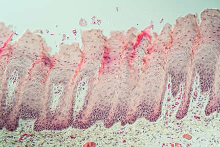
Tongue with taste buds Papilla across 100x
Коллекция по умолчанию
Коллекция по умолчанию
Создать новую

Thyroid follicular carcinoma, light micrograph, photo under microscope
Коллекция по умолчанию
Коллекция по умолчанию
Создать новую
Tongue Tissue with taste buds across 200x
Коллекция по умолчанию
Коллекция по умолчанию
Создать новую
Characteristics of Lichen, hyphae and Symbiotic algae under the microscope for education.
Коллекция по умолчанию
Коллекция по умолчанию
Создать новую
Vibrant cross-section of a developing seed under UV light, highlighting the embryo and endosperm in bright colors
Коллекция по умолчанию
Коллекция по умолчанию
Создать новую
A highresolution image of tight junctions in epithelial cells displaying the various types and arrangements of proteins that make up these junctions
Коллекция по умолчанию
Коллекция по умолчанию
Создать новую
Tissue of Stomach Human under the microscope in Lab.
Коллекция по умолчанию
Коллекция по умолчанию
Создать новую
a close-up photo of an eye showcasing vibrant blue and red colors, with intricate threads running through it. this captivating image, created in the style of unreal engine, resembles the stunning nature-inspired imagery often seen in national geographic. the eye's mesmerizing combination of dark orange and light navy hues adds a touch of schizocore aesthetics to this photo-realistic still life. ai generated
Коллекция по умолчанию
Коллекция по умолчанию
Создать новую

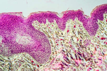
Scalp and hair follicles of human under the microscope in Lab.
Коллекция по умолчанию
Коллекция по умолчанию
Создать новую

Characteristics of Lichen, hyphae and Symbiotic algae under the microscope for education.
Коллекция по умолчанию
Коллекция по умолчанию
Создать новую
A close up of a pink and blue cell with a tree inside of it. The cell is surrounded by a pink and blue background
Коллекция по умолчанию
Коллекция по умолчанию
Создать новую
Histopathology of acute nephritis, light micrograph, photo under microscope
Коллекция по умолчанию
Коллекция по умолчанию
Создать новую
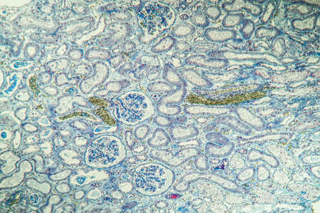
Atrophy kidney tissue under the microscope 100x
Коллекция по умолчанию
Коллекция по умолчанию
Создать новую
serous gland tissue under the microscope 100x
Коллекция по умолчанию
Коллекция по умолчанию
Создать новую
Squamous cell carcinoma of the uterus, light micrograph, photo under microscope
Коллекция по умолчанию
Коллекция по умолчанию
Создать новую
a vibrant 3d animation showcases a cell adorned with intricate embellishments. the cell is beautifully colored in light indigo and pink hues, reminiscent of a petzval 85mm f22 lens. the addition of light cyan and orange accents adds depth to the image. this captivating artwork resembles tilt-shift photography and resembles detailed miniatures found in medical imaging films. ai generated
Коллекция по умолчанию
Коллекция по умолчанию
Создать новую
Characteristics of Lichen, hyphae and Symbiotic algae under the microscope for education.
Коллекция по умолчанию
Коллекция по умолчанию
Создать новую
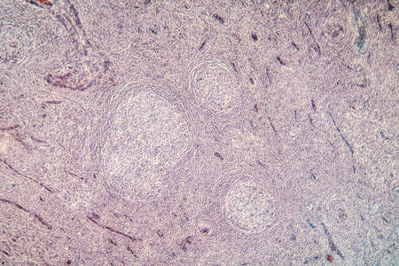
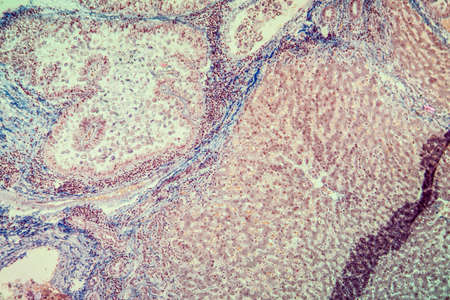
science medical anthropotomy physiology microscopic section of lymph gland tissue background
Коллекция по умолчанию
Коллекция по умолчанию
Создать новую
This colorful coral structure features unique shapes and textures thriving in a clear ocean environment under gentle sunlight.
Коллекция по умолчанию
Коллекция по умолчанию
Создать новую
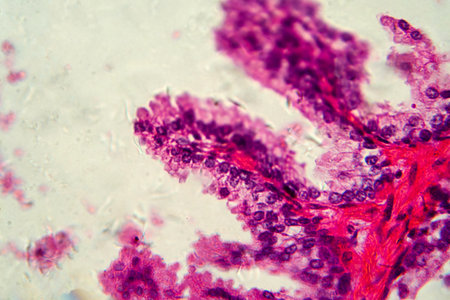
squamous cell
Коллекция по умолчанию
Коллекция по умолчанию
Создать новую
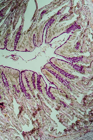
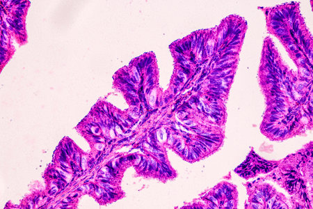
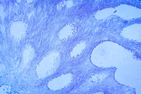
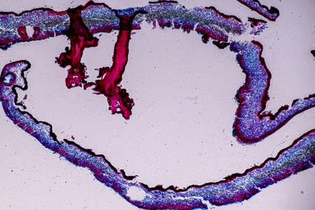
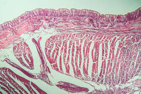
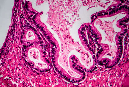
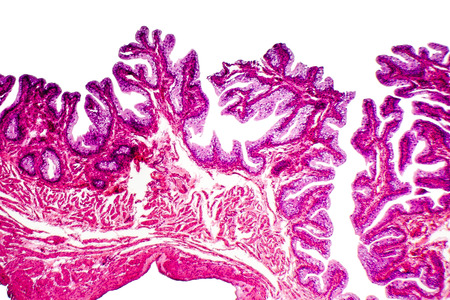
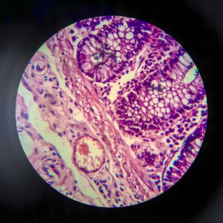
Cross-section through the intestine with glands 200x
Коллекция по умолчанию
Коллекция по умолчанию
Создать новую
Uterine cancer, light micrograph, photo under microscope
Коллекция по умолчанию
Коллекция по умолчанию
Создать новую
close up of red cabbage leaf with water droplets, abstract background
Коллекция по умолчанию
Коллекция по умолчанию
Создать новую
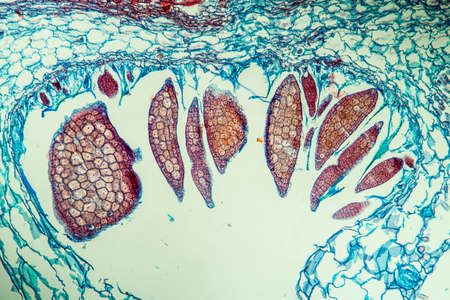
Earthworm histology cross section 10th segment 100x
Коллекция по умолчанию
Коллекция по умолчанию
Создать новую
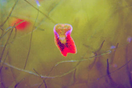
Red planaria flatworms - Convolutriloba retrogemma
Коллекция по умолчанию
Коллекция по умолчанию
Создать новую

Histopathology of human under microscope view for education in laboratory.
Коллекция по умолчанию
Коллекция по умолчанию
Создать новую
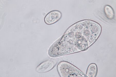
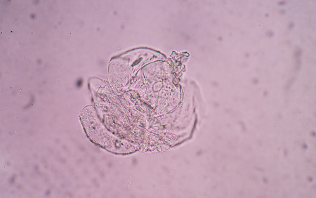
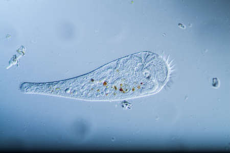
Paramecium caudatum is a genus of unicellular ciliated protozoan and Bacterium under the microscope.
Коллекция по умолчанию
Коллекция по умолчанию
Создать новую
Microscopic view of human cells under microscope.
Коллекция по умолчанию
Коллекция по умолчанию
Создать новую
Ovarian cancer, light micrograph, photo under microscope. Photograph shows a fragment of a cancerous tumor in the female ovary. Selective focus
Коллекция по умолчанию
Коллекция по умолчанию
Создать новую
Characteristics of Lichen, hyphae and Symbiotic algae under the microscope for education.
Коллекция по умолчанию
Коллекция по умолчанию
Создать новую
Mushrooms growing on textured tree bark showcasing varied colors and patterns in nature
Коллекция по умолчанию
Коллекция по умолчанию
Создать новую
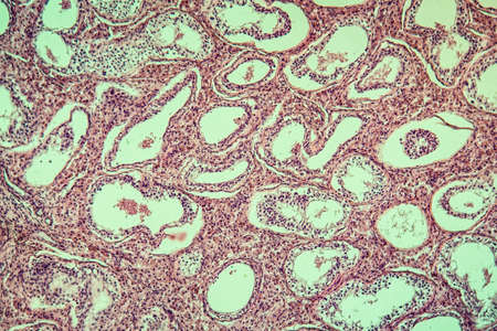
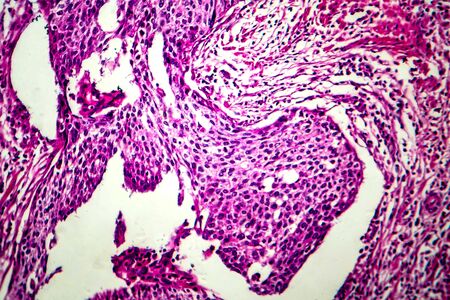
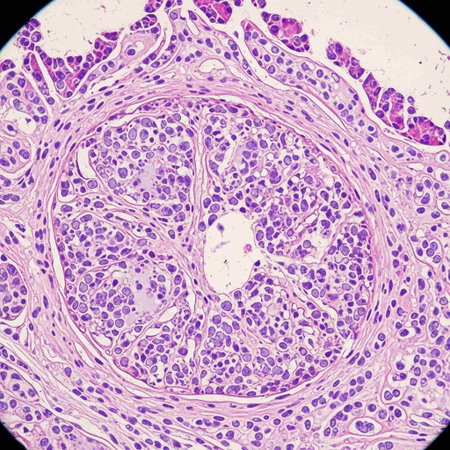
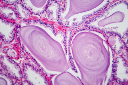
Photomicrograph showing histological features of benign prostatic hyperplasia. Enlarged prostate gland with nodular proliferation of glandular and stromal components. High-resolution histology image.
Коллекция по умолчанию
Коллекция по умолчанию
Создать новую
Colon tissue with diverticulum 100x
Коллекция по умолчанию
Коллекция по умолчанию
Создать новую
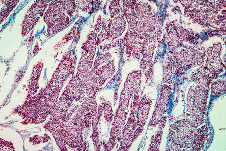
Histopathology of cirrhosis, light micrograph, photo under microscope
Коллекция по умолчанию
Коллекция по умолчанию
Создать новую
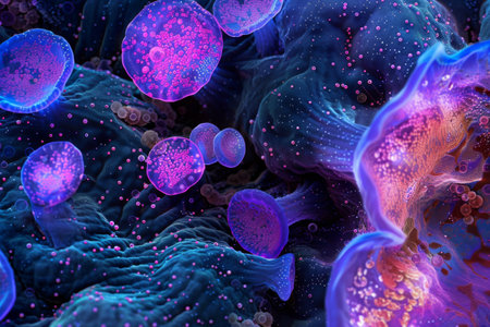
Cellular structures are magnified to reveal their complex shapes and vibrant colors, emphasizing the beauty of biology at a microscopic level.
Коллекция по умолчанию
Коллекция по умолчанию
Создать новую

Photo Picture of Some Jellyfish Dangerous Poisonous Medusa
Коллекция по умолчанию
Коллекция по умолчанию
Создать новую

Condyloma acuminatum, also known as genital warts. Light micrograph, photo under microscope
Коллекция по умолчанию
Коллекция по умолчанию
Создать новую
Atrophy kidney tissue under the microscope 100x
Коллекция по умолчанию
Коллекция по умолчанию
Создать новую
Numerous small blue bubbles are seen floating on the surface of the water, creating a mesmerizing pattern in the sunlight. The bubbles gently rise and fall with the movement of the water.
Коллекция по умолчанию
Коллекция по умолчанию
Создать новую
Embryo with endosperm across 100x
Коллекция по умолчанию
Коллекция по умолчанию
Создать новую
Actinomyces in the jaw diseased tissue 200x
Коллекция по умолчанию
Коллекция по умолчанию
Создать новую
Tissue of Stomach Human under the microscope in Lab.
Коллекция по умолчанию
Коллекция по умолчанию
Создать новую
Wonderful aquatic life in ocean seabed. anemones. Coral reef design. Generative AI. Illustration for banner, poster, cover, brochure or presentation.
Коллекция по умолчанию
Коллекция по умолчанию
Создать новую
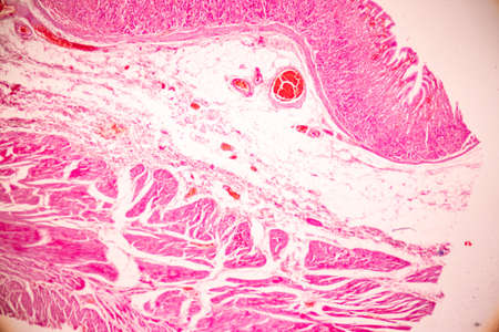
Fibroepithelium Diseased tissue 100x
Коллекция по умолчанию
Коллекция по умолчанию
Создать новую
Transitional epithelium tissue of the urinary bladder under microscope, light micrograph, hematoxylin eosin staining
Коллекция по умолчанию
Коллекция по умолчанию
Создать новую
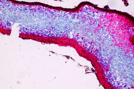
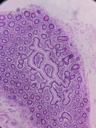
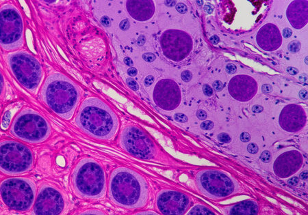
Palatal tonsils transverse 100x under a microscope
Коллекция по умолчанию
Коллекция по умолчанию
Создать новую
Microscopic fungi Cunninghamella, scientific 3D illustration. Pathogenic fungi from the order Mucorales, cause sinopulmonary and disseminated infections, one of the causative agents of mucormycosis
Коллекция по умолчанию
Коллекция по умолчанию
Создать новую
Trumpet animal as a microscopic plankton animal in drops of water
Коллекция по умолчанию
Коллекция по умолчанию
Создать новую
A group of chaotically surreal bacteria with a face. cartoon picture
Коллекция по умолчанию
Коллекция по умолчанию
Создать новую
Tongue Tissue with taste buds across 200x
Коллекция по умолчанию
Коллекция по умолчанию
Создать новую
Photomicrograph showing histological features of benign prostatic hyperplasia. Enlarged prostate gland with nodular proliferation of glandular and stromal components.
Коллекция по умолчанию
Коллекция по умолчанию
Создать новую
Pancreas cancer cell under microscope view for medical education.
Коллекция по умолчанию
Коллекция по умолчанию
Создать новую
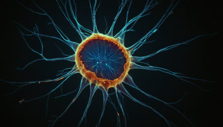
Radiant Neuron Structure
Коллекция по умолчанию
Коллекция по умолчанию
Создать новую
Exploring the microscopic world of cells and tissues, AI generated
Коллекция по умолчанию
Коллекция по умолчанию
Создать новую
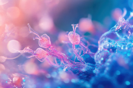
Close-up of a colorful seaweed in blue and pink colors
Коллекция по умолчанию
Коллекция по умолчанию
Создать новую
Characteristics of fungi living in wood as a group, are polyphyletic under the microscope for education.
Коллекция по умолчанию
Коллекция по умолчанию
Создать новую
Fibrin deposits in the kidney, microscopy 100x
Коллекция по умолчанию
Коллекция по умолчанию
Создать новую

A close up of a purple and blue jellyfish with bubbles, AI
Коллекция по умолчанию
Коллекция по умолчанию
Создать новую
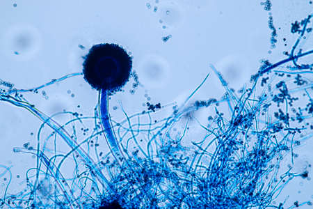
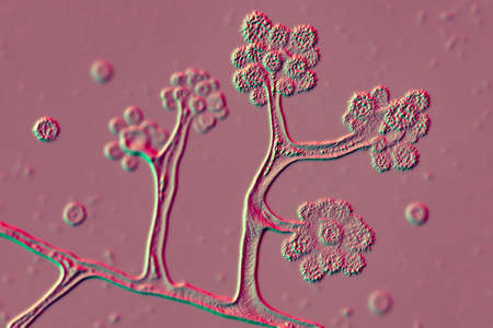
Aspergillus niger and Aspergillus oryzae (mold) under microscope for Microbiology in Lab.
Коллекция по умолчанию
Коллекция по умолчанию
Создать новую
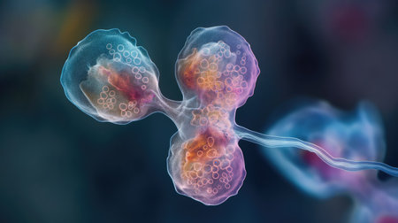
Cell division process, micro
Коллекция по умолчанию
Коллекция по умолчанию
Создать новую
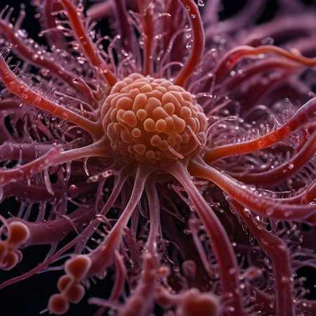
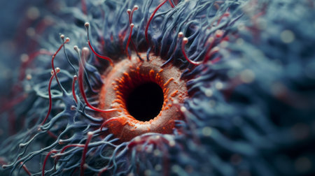
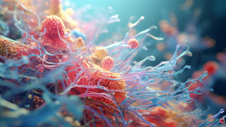
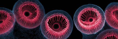
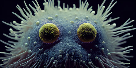
This image shows a close-up of an object that appears to be organic in nature, resembling a microscopic view of a cellular structure. The central focus is an orb-like formation with clusters of smaller, bead-like structures on its surface. Surrounding this are numerous filament-like protrusions, some with branching extensions, all against a dark background. The object and its protrusions are in various shades of pink and red, and theres a translucent quality to some of the extensions, giving a sense of depth and complexity. Water droplets are visible across the surfaces, suggesting a wet environment and adding a reflective quality to the image. The overall appearance is reminiscent of an image you might see in biology or microbiology related to cellular organisms, perhaps a virus, a sea anemone, or a stylized depiction of a cancer cell.
Коллекция по умолчанию
Коллекция по умолчанию
Создать новую
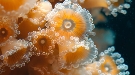
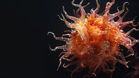
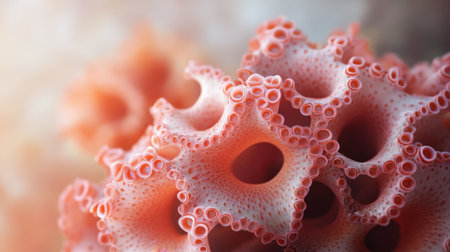
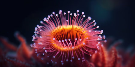
A dense cluster of orange marine polyps, each with a ribbed oral disc and a crown of translucent, white, bubble-tipped tentacles.
Коллекция по умолчанию
Коллекция по умолчанию
Создать новую

Tissue of Small intestine (Duodenum), Large intestine Human and Stomach Human under the microscope in Lab.
Коллекция по умолчанию
Коллекция по умолчанию
Создать новую
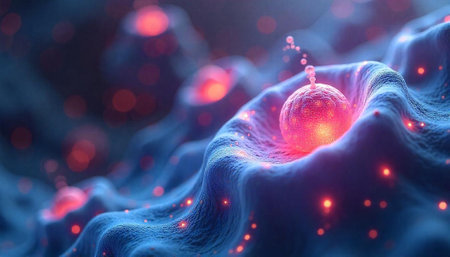
Abstract cell structure with vibrant lighting and blue background
Коллекция по умолчанию
Коллекция по умолчанию
Создать новую
Detailed macro image reveals microorganisms with thin cell walls in linear arrangement, Ai Generated.
Коллекция по умолчанию
Коллекция по умолчанию
Создать новую
Neuron cells with synapse connections viewed under microscope with glowing abstract background in blue and pink. Medical and science background concept
Коллекция по умолчанию
Коллекция по умолчанию
Создать новую

Papillary thyroid carcinoma, light micrograph, photo under microscope. The most common type of thyroid cancer
Коллекция по умолчанию
Коллекция по умолчанию
Создать новую

Growing cancer cell concept image. 3d rendering
Коллекция по умолчанию
Коллекция по умолчанию
Создать новую
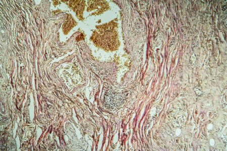
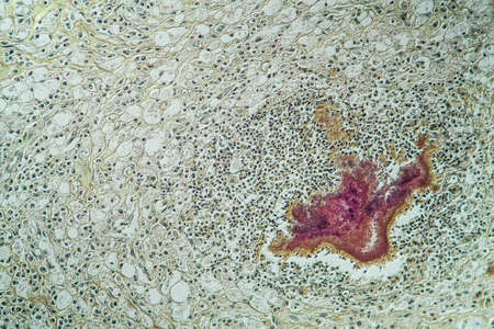
Carcinoma in guinea pigs, tissue 100x
Коллекция по умолчанию
Коллекция по умолчанию
Создать новую

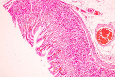
Characteristics Tissue of Olfactory epithelium Human under the microscope in Lab.
Коллекция по умолчанию
Коллекция по умолчанию
Создать новую
A colorful image of a cell with a purple and blue blob in the center. The image is abstract and has a mood of curiosity and wonder
Коллекция по умолчанию
Коллекция по умолчанию
Создать новую
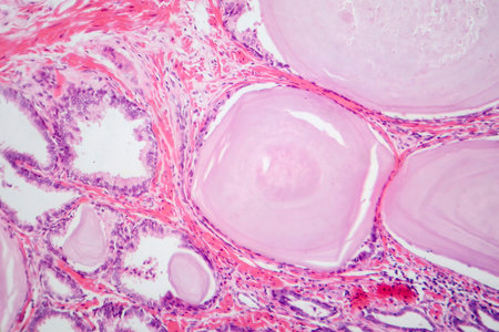
Photomicrograph showing histological features of benign prostatic hyperplasia. Enlarged prostate gland with nodular proliferation of glandular and stromal components.
Коллекция по умолчанию
Коллекция по умолчанию
Создать новую
Bacillary dysentery, light micrograph, photo under microscope showing presence of bacteria and accumulation of inflammatory cells in intestinal epithelium
Коллекция по умолчанию
Коллекция по умолчанию
Создать новую
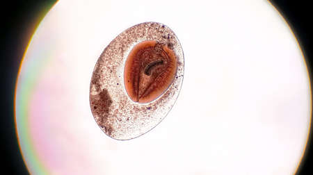
Moniliformis dubius in the Intestine of rat, intermediate host
Коллекция по умолчанию
Коллекция по умолчанию
Создать новую
This detailed microscopic image showcases various cellular structures, highlighted in striking purple tones. The intricate patterns and textures reveal the complexity of biological tissues, making it a valuable resource for educational and scientific purposes
Коллекция по умолчанию
Коллекция по умолчанию
Создать новую
Coccidiosis of liver tissue under the microscope 100x
Коллекция по умолчанию
Коллекция по умолчанию
Создать новую
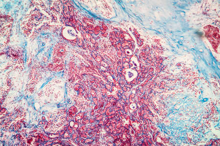
Basal cell cancer Diseased tissue 100x
Коллекция по умолчанию
Коллекция по умолчанию
Создать новую

diseased liver with cirrhosis 100x under the microscope
Коллекция по умолчанию
Коллекция по умолчанию
Создать новую
Prostate cancer, light micrograph, photo under microscope
Коллекция по умолчанию
Коллекция по умолчанию
Создать новую
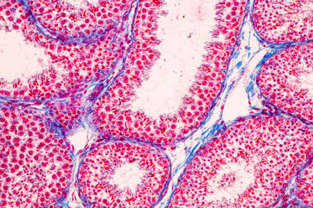
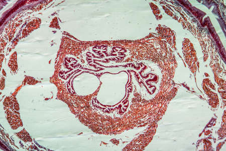
Anatomy and Histological Epididymis and Testis human cells under microscope.
Коллекция по умолчанию
Коллекция по умолчанию
Создать новую

Marine creatures, Medusozoa, jellyfish with jelly-like body and bell shape.
Коллекция по умолчанию
Коллекция по умолчанию
Создать новую
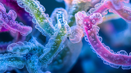
A close-up of a colorful cellular structure featuring fine filaments and textures. This detailed representation highlights the complexity of microscopic life forms in a scientific setting.
Коллекция по умолчанию
Коллекция по умолчанию
Создать новую
An AI generated illustration of a macro photograph of a colony of bacteria, forming a visible biofilm
Коллекция по умолчанию
Коллекция по умолчанию
Создать новую
Education anatomy and Histological sample of Human under the microscope.
Коллекция по умолчанию
Коллекция по умолчанию
Создать новую
Stunning microscopic image of colorful cellular structures demonstrates the intricate details and vibrant colors found in the world of microbiology and life forms.
Коллекция по умолчанию
Коллекция по умолчанию
Создать новую
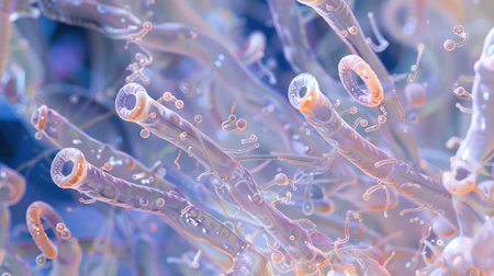
A vibrant marine animal swims gracefully through deep blue ocean waters showcasing its unique shape and colors.
Коллекция по умолчанию
Коллекция по умолчанию
Создать новую
The activation of TLRs leading to the production of proinflammatory cytokines as seen in a micrograph of immune cells
Коллекция по умолчанию
Коллекция по умолчанию
Создать новую
Uterine cancer, light micrograph, photo under microscope
Коллекция по умолчанию
Коллекция по умолчанию
Создать новую
Bowel with goblet cells in the dark field 100x
Коллекция по умолчанию
Коллекция по умолчанию
Создать новую
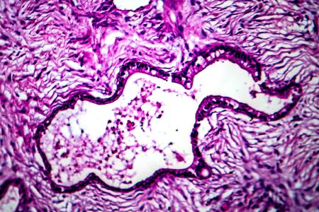
Lungworm under the microscope 100x
Коллекция по умолчанию
Коллекция по умолчанию
Создать новую
Legion-Media
Создайте свои проекты на основе качественных стоковых фотографий и видео.


Copyright © Legion-Media.