

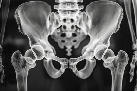
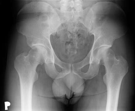
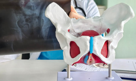
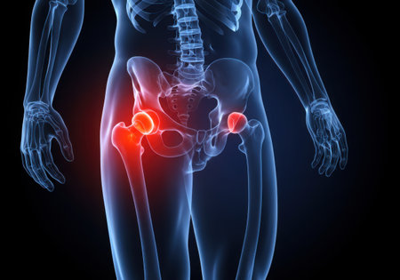
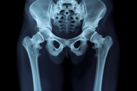
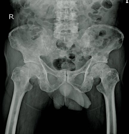
X-ray of the pelvic bones of a man. A doctor radiologist is studying an x-ray examination. A hip joint is placed on the patient’s body
Коллекция по умолчанию
Коллекция по умолчанию
Создать новую
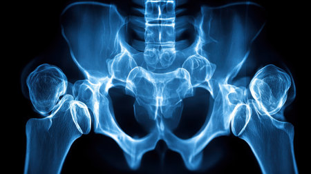
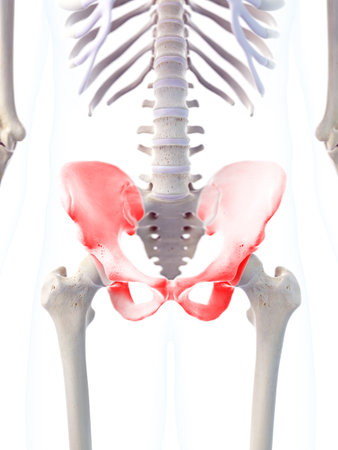
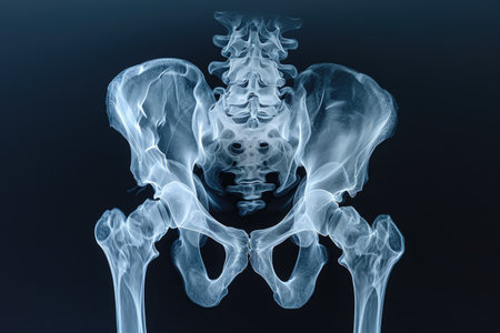
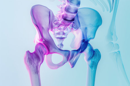
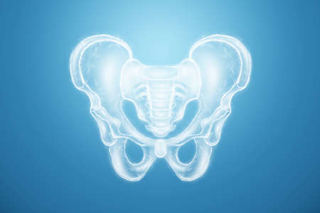
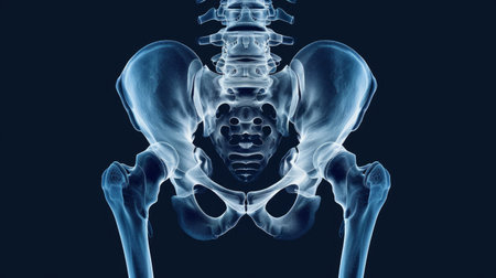
This high-resolution X-ray image of the human pelvis and hip joint displays detailed anatomical structures in stunning blue tones. Ideal for educational and medical purposes.
Коллекция по умолчанию
Коллекция по умолчанию
Создать новую
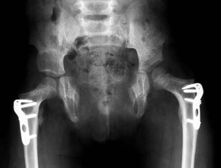
Detailed X-ray image showing a pelvis with an endoprosthesis, specifically designed for hip joint support in rheumatic diseases like Rheumatoid Arthritis.
Коллекция по умолчанию
Коллекция по умолчанию
Создать новую
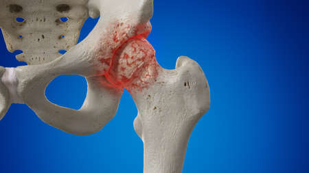
3d rendered illustration of an arthritic hip joint
Коллекция по умолчанию
Коллекция по умолчанию
Создать новую
some parts of human body
Коллекция по умолчанию
Коллекция по умолчанию
Создать новую
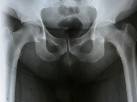
Rontgen picture of male pelvis
Коллекция по умолчанию
Коллекция по умолчанию
Создать новую
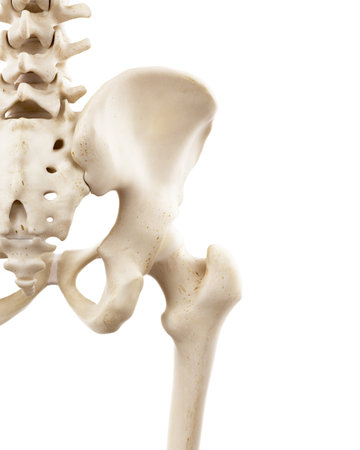
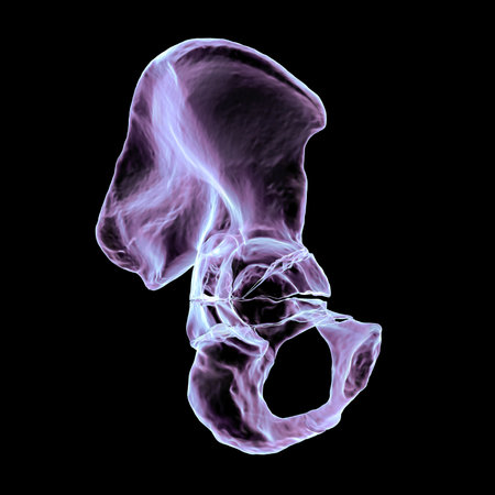
medically accurate 3d illustration of the skeletal hip
Коллекция по умолчанию
Коллекция по умолчанию
Создать новую
x-ray film broken bone at hip install plate with screw
Коллекция по умолчанию
Коллекция по умолчанию
Создать новую
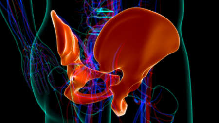
3D medical illustration of the pelvis bone
Коллекция по умолчанию
Коллекция по умолчанию
Создать новую

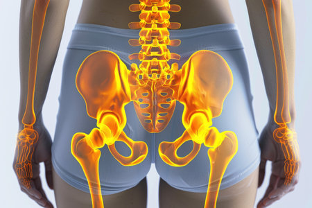
medically accurate 3d illustration of the highlighted human hip
Коллекция по умолчанию
Коллекция по умолчанию
Создать новую
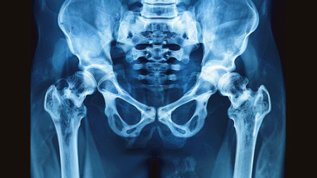
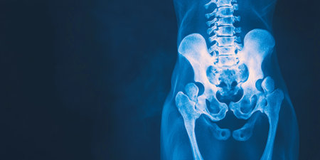
This X-ray reveals a detailed view of the human pelvis, highlighting the hip joints and surrounding bone structure in a clinical environment, showcasing alignment and health.
Коллекция по умолчанию
Коллекция по умолчанию
Создать новую
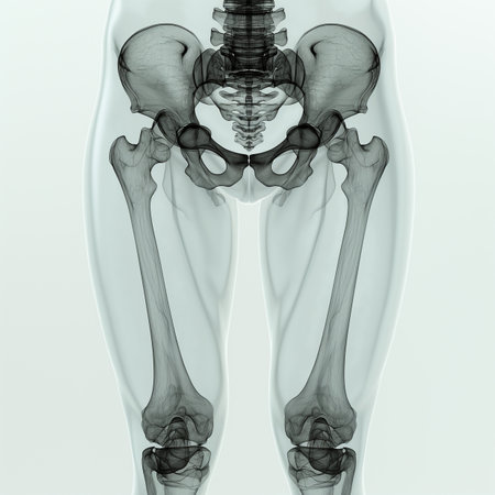
Human skeleton anatomy - Bone x-ray. Medical concept. 3D Rendering
Коллекция по умолчанию
Коллекция по умолчанию
Создать новую

3d rendered medically accurate illustration of the hip joint
Коллекция по умолчанию
Коллекция по умолчанию
Создать новую
Detailed white human spine model on black background
Коллекция по умолчанию
Коллекция по умолчанию
Создать новую
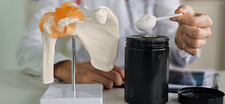
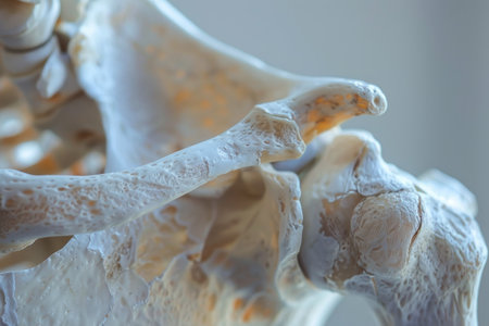
Doctor demonstrates the use of supplement powder next to a shoulder anatomy model in a clinical setting
Коллекция по умолчанию
Коллекция по умолчанию
Создать новую

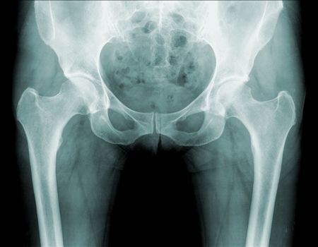
hip xray
Коллекция по умолчанию
Коллекция по умолчанию
Создать новую
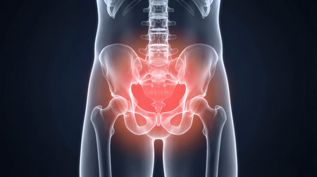
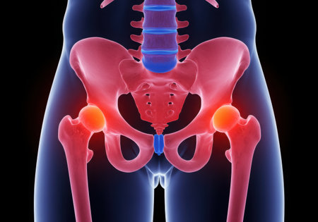
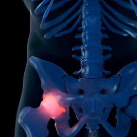
Detailed translucent rendering of human pelvic bones lower spine and hip joints concentrated area of bright red glowing pain
Коллекция по умолчанию
Коллекция по умолчанию
Создать новую

medically accurate illustration of the inferior gemellus
Коллекция по умолчанию
Коллекция по умолчанию
Создать новую
This x-ray image provides a detailed view of the human skeleton, revealing the intricate structure and positioning of bones, Three-dimensional X-ray film of human pelvic bones, AI Generated
Коллекция по умолчанию
Коллекция по умолчанию
Создать новую
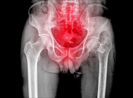
Preventive and control medical examination. X-ray of the Ilium and hip bones.
Коллекция по умолчанию
Коллекция по умолчанию
Создать новую
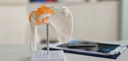
Detailed model of a human shoulder joint displayed on a desk with educational materials in the background
Коллекция по умолчанию
Коллекция по умолчанию
Создать новую
3d rendered illustration of the human bladder
Коллекция по умолчанию
Коллекция по умолчанию
Создать новую
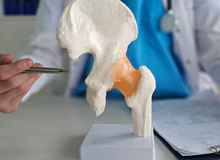
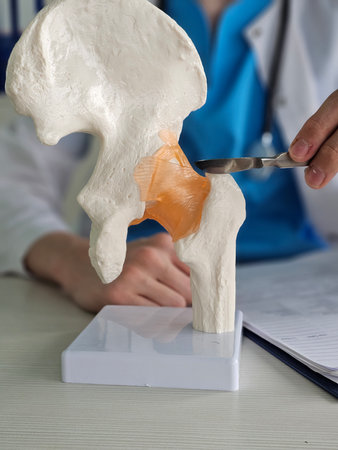
Model of hip joint anatomy displayed in medical office during patient consultation
Коллекция по умолчанию
Коллекция по умолчанию
Создать новую

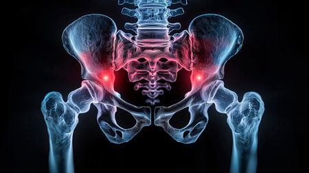
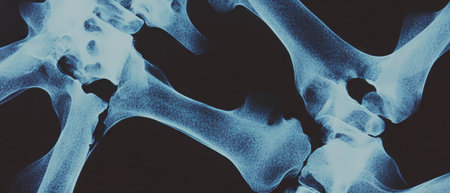
X-ray scan image of hip joints human skeleton in blue gray tones. Scanned in orthopedics traumatology surgery hospital clinic.
Коллекция по умолчанию
Коллекция по умолчанию
Создать новую
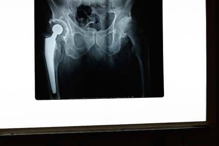
A x-ray of a hip and prosthesis on a x-ray screen
Коллекция по умолчанию
Коллекция по умолчанию
Создать новую
Radiography of an spectacular pelvis fracture
Коллекция по умолчанию
Коллекция по умолчанию
Создать новую
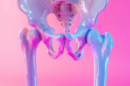
A close up of a skeleton's hip bone with a pink and purple hue. The bone is shown in a 3D image, and the colors give it a unique and artistic appearance
Коллекция по умолчанию
Коллекция по умолчанию
Создать новую
Lumbar Spine and Pelvis XRay Highlighting Skeletal Structure and Medical Imaging Techniques.
Коллекция по умолчанию
Коллекция по умолчанию
Создать новую
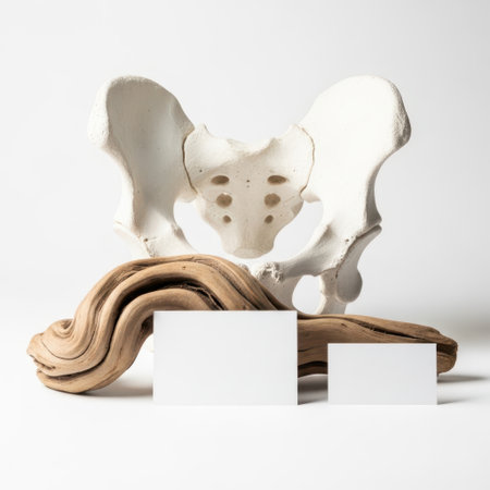
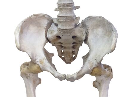
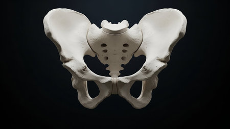
Pelvic bone model isolated on white background
Коллекция по умолчанию
Коллекция по умолчанию
Создать новую
Medical professional examining a hip joint model during a consultation in a clinic setting
Коллекция по умолчанию
Коллекция по умолчанию
Создать новую
Hologram, ultrasound image, anterior view of the male pelvis, sacrum isolated on a blue background. Anatomy, medicine, scientific concepts. 3D rendering, 3D illustration
Коллекция по умолчанию
Коллекция по умолчанию
Создать новую
Hip replacement on medical background. 3d illustration
Коллекция по умолчанию
Коллекция по умолчанию
Создать новую
X-Ray human skeleton anatomy, 3D
Коллекция по умолчанию
Коллекция по умолчанию
Создать новую
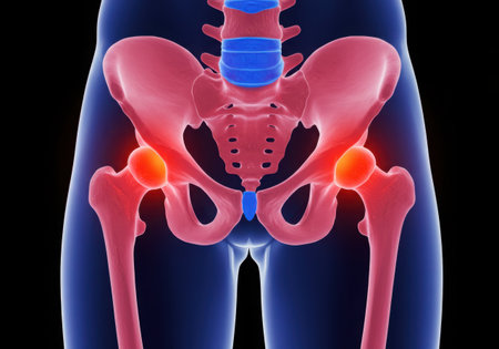
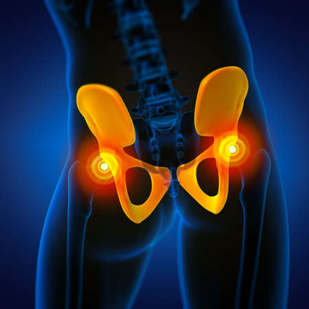
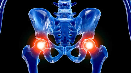
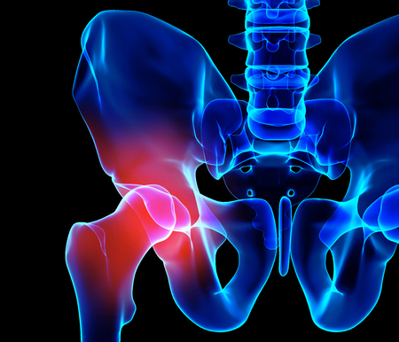
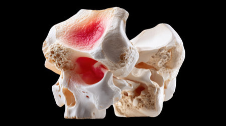
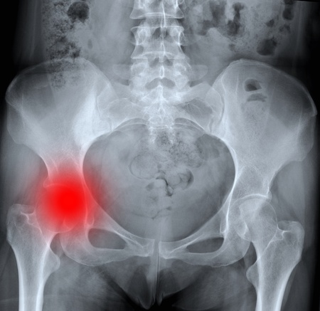
An anatomical illustration of the human pelvis and hip joints, highlighting areas of pain in red, against a dark background.
Коллекция по умолчанию
Коллекция по умолчанию
Создать новую
pelvis, uterus, skeleton, anatomy, location, female, closeup, small bone canal bounded anteriorly pubic bones symphysis pelvic
Коллекция по умолчанию
Коллекция по умолчанию
Создать новую
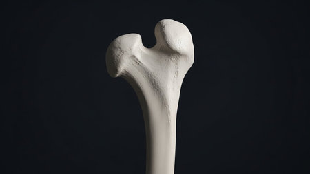
Detailed view of a human femur bone against a dark background, highlighting its anatomy and structure.
Коллекция по умолчанию
Коллекция по умолчанию
Создать новую
Artificial skeleton such as skull, bone and teeth in colleges and universities laboratory for teaching, learning, research forensic, anatomy, biology and ancient science
Коллекция по умолчанию
Коллекция по умолчанию
Создать новую

medically accurate 3d illustration of the human hip
Коллекция по умолчанию
Коллекция по умолчанию
Создать новую
A colorful sports stadium with a central circular platform, surrounded by a multi-colored running track and empty seating areas.
Коллекция по умолчанию
Коллекция по умолчанию
Создать новую
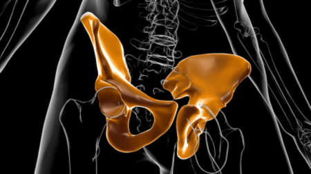
3d rendered medically accurate illustration of the human skeletal hip
Коллекция по умолчанию
Коллекция по умолчанию
Создать новую
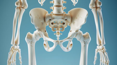
Human Skeleton Hip or Pelvic bone Anatomy For Medical Concept 3D Illustration
Коллекция по умолчанию
Коллекция по умолчанию
Создать новую

hip dysplasia of an 14 month old dog
Коллекция по умолчанию
Коллекция по умолчанию
Создать новую

Hand holding shoulder, clavicle X-ray image. Acromion, acromial end fracture. Arm trauma. Health care, injury diagnostics concept. High quality photo
Коллекция по умолчанию
Коллекция по умолчанию
Создать новую

Close up of male doctors hand showing ischial tuberosity or sits bones on skeleton spine model
Коллекция по умолчанию
Коллекция по умолчанию
Создать новую

Rontgen picture of male pelvis
Коллекция по умолчанию
Коллекция по умолчанию
Создать новую
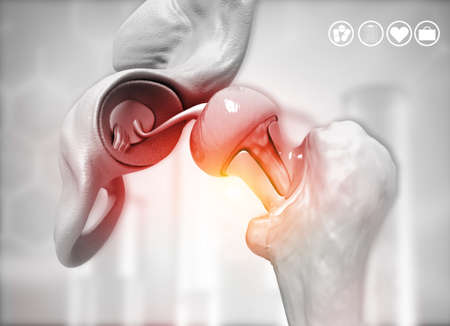
Anatomical visualization highlighting femoral head inflammation and acetabulum socket with red glowing pain indicator
Коллекция по умолчанию
Коллекция по умолчанию
Создать новую
Undernourished Teenager with Visible Ribs and Sunken Stomach
Коллекция по умолчанию
Коллекция по умолчанию
Создать новую
A pink background with a blue and purple skeleton. The skeleton is in the middle of the image and is the main focus
Коллекция по умолчанию
Коллекция по умолчанию
Создать новую
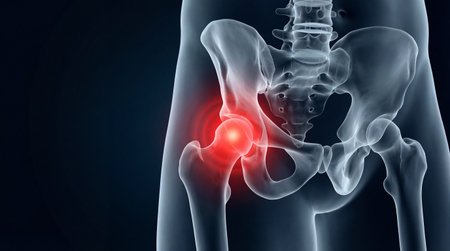
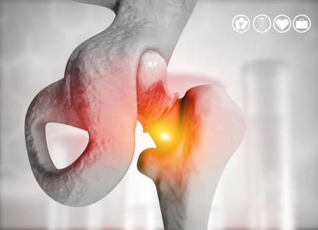
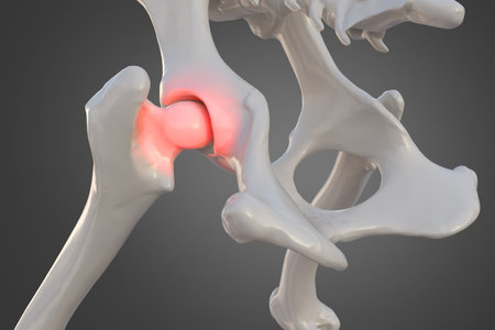
3d rendered illustration of a painful hip joint
Коллекция по умолчанию
Коллекция по умолчанию
Создать новую
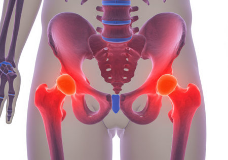
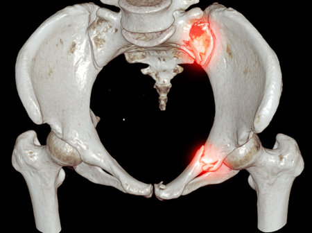
An anatomical illustration of the human pelvis and hip joints, highlighting areas of inflammation and pain in red and orange.
Коллекция по умолчанию
Коллекция по умолчанию
Создать новую
Human Skeleton Hip or Pelvic bone Anatomy For Medical Concept 3D Illustration
Коллекция по умолчанию
Коллекция по умолчанию
Создать новую

3d rendered illustration of the skeletal hip
Коллекция по умолчанию
Коллекция по умолчанию
Создать новую

Close-up of a woman in jeans and a sweater holding a stick
Коллекция по умолчанию
Коллекция по умолчанию
Создать новую
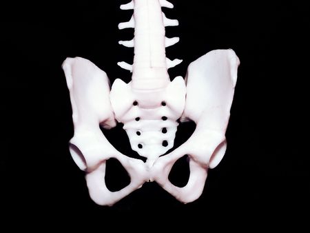
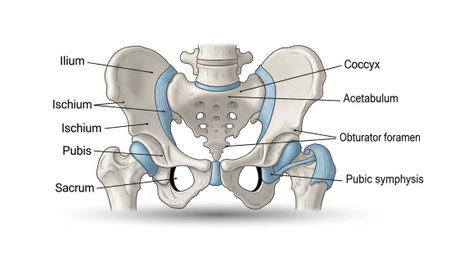
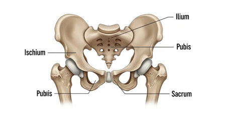
Human pelvic bones annotated
Коллекция по умолчанию
Коллекция по умолчанию
Создать новую
Osteoarthritis of the knee and hand. Healthcare concept image
Коллекция по умолчанию
Коллекция по умолчанию
Создать новую
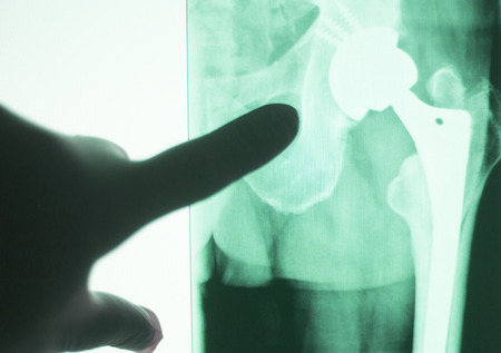
Hip joint replacement impant x-ray test scan results of old aged person with arthritis and joints pain.
Коллекция по умолчанию
Коллекция по умолчанию
Создать новую

bones and skull of an unknown animal in the Spitalfields Market
Коллекция по умолчанию
Коллекция по умолчанию
Создать новую
Rontgen picture of male pelvis
Коллекция по умолчанию
Коллекция по умолчанию
Создать новую

medically accurate illustration of the gluteus minimus
Коллекция по умолчанию
Коллекция по умолчанию
Создать новую
Pelvic bone with femoral neck joint isolated on white background
Коллекция по умолчанию
Коллекция по умолчанию
Создать новую

medically accurate 3d illustration of the human hip
Коллекция по умолчанию
Коллекция по умолчанию
Создать новую
Hip replacement on medical background. 3d illustration
Коллекция по умолчанию
Коллекция по умолчанию
Создать новую

X-ray of pelvic bones
Коллекция по умолчанию
Коллекция по умолчанию
Создать новую
CT Scan pelvic bone with both hip joint 3D rendering showign fracture of sacrum and superior pubic rumus.
Коллекция по умолчанию
Коллекция по умолчанию
Создать новую
3D illustration, hip painful skeleton x-ray, medical concept.
Коллекция по умолчанию
Коллекция по умолчанию
Создать новую
A translucent human skeleton displays glowing red areas indicating pain and inflammation in both hip joints and the surrounding pelvic region.
Коллекция по умолчанию
Коллекция по умолчанию
Создать новую
Several spoiled rotten apples on a gray background, close-up, copy space. Improper storage of fruits. Moldy texture on the apple. Dangerous food
Коллекция по умолчанию
Коллекция по умолчанию
Создать новую
Detailed view of human pelvis bone anatomy isolated on black
Коллекция по умолчанию
Коллекция по умолчанию
Создать новую

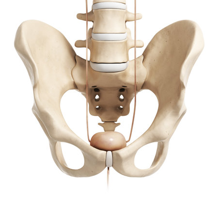
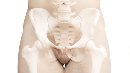
Human skeleton: pelvis and sacrum. Isolated on white. Medically accurate 3D illustration
Коллекция по умолчанию
Коллекция по умолчанию
Создать новую

Hip replacement implant installed in the pelvis bone. X-ray view. Medically accurate 3D illustration
Коллекция по умолчанию
Коллекция по умолчанию
Создать новую
An anatomical illustration of the human pelvis and hip joints, with areas of inflammation or pain glowing red.
Коллекция по умолчанию
Коллекция по умолчанию
Создать новую
An x-ray image showcasing the intricate details of a human skeleton, Human hip bones showing in X-ray film, AI Generated
Коллекция по умолчанию
Коллекция по умолчанию
Создать новую
Human skeleton anatomy. Anatomy of the human bones. Realistic illustration.
Коллекция по умолчанию
Коллекция по умолчанию
Создать новую
A large, empty arena stage is bathed in blue and white lights, creating a dramatic and inviting atmosphere. The center of the stage is illuminated with a circular ring of lights, ready for a performance or event.
Коллекция по умолчанию
Коллекция по умолчанию
Создать новую
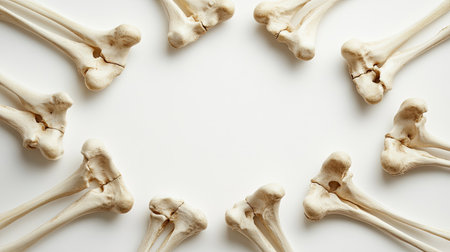
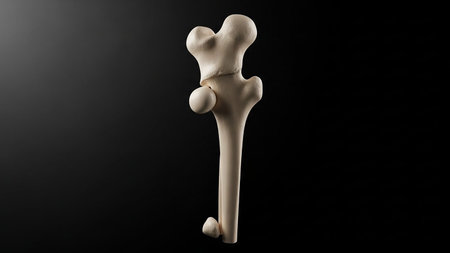
This image displays crossed femur bones arranged elegantly on a smooth white background. Ideal for medical illustrations, educational content, and anatomical studies.
Коллекция по умолчанию
Коллекция по умолчанию
Создать новую

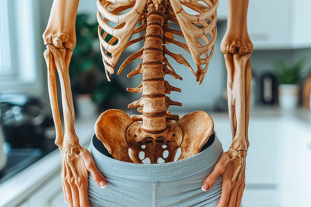
Human skeleton pelvis. Skeletal system anatomy, body structure, medical education concept. Reproductive, urinary or digestive systems. High quality photo
Коллекция по умолчанию
Коллекция по умолчанию
Создать новую
Human Hip Bones and Pelvic Region X-Ray Visualization
Коллекция по умолчанию
Коллекция по умолчанию
Создать новую
3d rendered illustration of the hip bone
Коллекция по умолчанию
Коллекция по умолчанию
Создать новую
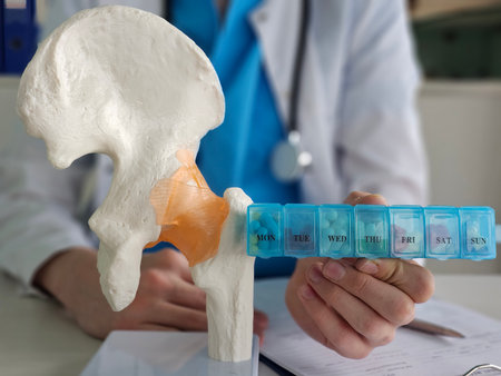
Doctor demonstrates hip joint model while holding a weekly pill organizer during patient consultation
Коллекция по умолчанию
Коллекция по умолчанию
Создать новую
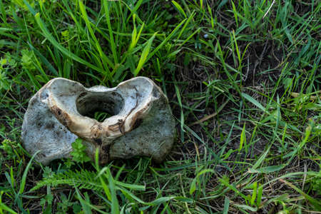
Animal bone lies on the ground
Коллекция по умолчанию
Коллекция по умолчанию
Создать новую
Canine dysplasia, dog bone with visible hip joint and femur affected by inflammation due to dysplasia, 3d illustration
Коллекция по умолчанию
Коллекция по умолчанию
Создать новую
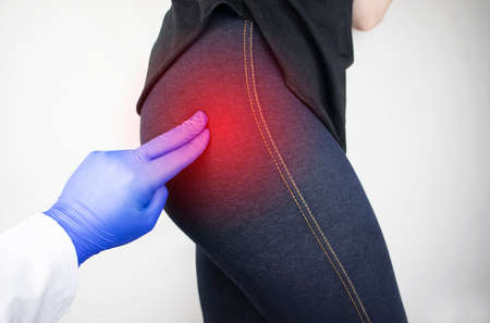
A woman suffers from pain in the buttock. The doctor diagnoses the patient piriformis syndrome, pinch of the sciatic nerve, lumbar osteochondrosis or sciatica
Коллекция по умолчанию
Коллекция по умолчанию
Создать новую
Detailed X-ray image of human pelvis and lower spine
Коллекция по умолчанию
Коллекция по умолчанию
Создать новую
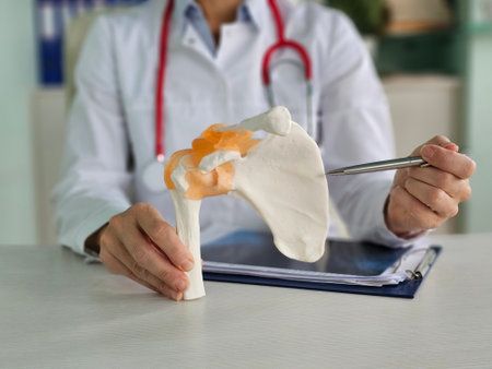
Medical professional explains shoulder anatomy using model in clinic during consultation
Коллекция по умолчанию
Коллекция по умолчанию
Создать новую
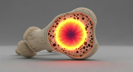
A detailed 3D illustration showing a cross-section of a bone with its spongy trabecular structure and glowing...
Коллекция по умолчанию
Коллекция по умолчанию
Создать новую

Roasted whole peeled chestnut over wooden background
Коллекция по умолчанию
Коллекция по умолчанию
Создать новую
Detailed view of a human femur bone against a dark background, highlighting its structure
Коллекция по умолчанию
Коллекция по умолчанию
Создать новую
X-ray Both hip finding Multiple hyperdensity and hypodensity bone lesions at both iliac bones, and both femur.
Prominent osteolytic lesions at Lt femur. impression: Multiple bone metastasis.
Коллекция по умолчанию
Коллекция по умолчанию
Создать новую
Cartilage degeneration in vertebrae showing osteoarthritis on transparent background
Коллекция по умолчанию
Коллекция по умолчанию
Создать новую
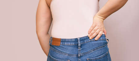
Woman lower back pain. Hold hand near body. Self chiropractic massage. jeans and top shirt. Sport muscle nerve displacement
Коллекция по умолчанию
Коллекция по умолчанию
Создать новую
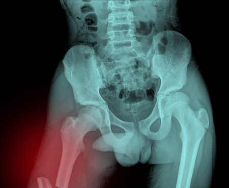
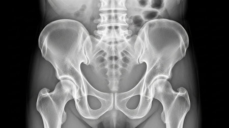
X-ray of the pelvis and spinal column
Коллекция по умолчанию
Коллекция по умолчанию
Создать новую
Detailed view of the shoulder bones of a horse, showing intricate details and structure, A close-up view of the shoulder bones with visible signs of degeneration and arthritis
Коллекция по умолчанию
Коллекция по умолчанию
Создать новую
High contrast X-ray texture showcasing coarse grain and an industrial aesthetic
Коллекция по умолчанию
Коллекция по умолчанию
Создать новую
High-resolution X-ray image of a human pelvis and hip bones. Concept of medical imaging, skeletal anatomy, diagnostic tool, healthcare illustration.
Коллекция по умолчанию
Коллекция по умолчанию
Создать новую
Pelvic bones with acetabulum fracture, a break in the hip socket where the femoral head meets the pelvis, 3D illustration. Often caused by high-impact trauma like car accidents or falls.
Коллекция по умолчанию
Коллекция по умолчанию
Создать новую
x-ray of a human hip , black background
Коллекция по умолчанию
Коллекция по умолчанию
Создать новую
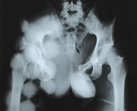
X-ray of pelvis
Коллекция по умолчанию
Коллекция по умолчанию
Создать новую
3d rendered medically accurate illustration of a painful hip joint
Коллекция по умолчанию
Коллекция по умолчанию
Создать новую

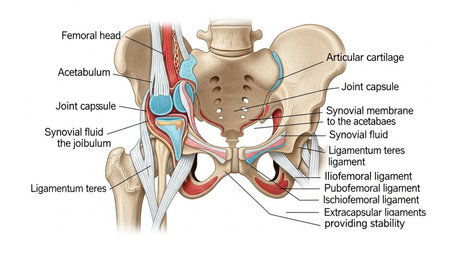
3d rendered medically accurate illustration of the ligaments of the hip
Коллекция по умолчанию
Коллекция по умолчанию
Создать новую
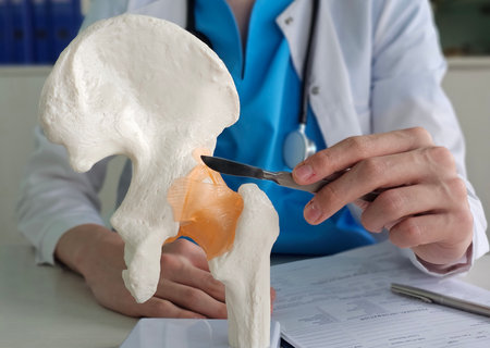
Hip replacement surgery. A doctor holds a scalpel. Hip arthroplasty concept
Коллекция по умолчанию
Коллекция по умолчанию
Создать новую
Legion-Media
Создайте свои проекты на основе качественных стоковых фотографий и видео.


Copyright © Legion-Media.